Direct Cardiac Segmentation from Undersampled K-space Using Transformers

0
Sign in to get full access
Overview
- This paper presents a novel approach for direct cardiac segmentation from undersampled k-space data using transformers.
- The proposed method aims to bypass the traditional image reconstruction step and perform segmentation directly from the undersampled k-space data.
- The authors leverage the powerful representation learning capabilities of transformers to effectively extract relevant features from the k-space data.
- The approach demonstrates improved segmentation accuracy and efficiency compared to traditional methods that first reconstruct the image and then perform segmentation.
Plain English Explanation
In medical imaging, such as cardiac MRI, the process of capturing and analyzing the images involves several steps. One crucial step is image reconstruction, where the raw data collected by the imaging device (known as k-space data) is transformed into a clear, usable image. After this reconstruction, the image can then be analyzed and segmented to identify specific structures, like the heart.
The researchers in this paper propose a new approach that bypasses the image reconstruction step and instead performs the segmentation directly on the k-space data. This is achieved by leveraging the powerful capabilities of transformers, a type of deep learning model that has shown remarkable success in various tasks, including natural language processing and computer vision.
The key idea is that the transformers can learn to extract the relevant features from the k-space data, without the need for a separate image reconstruction step. This has several potential benefits, such as improved accuracy and increased efficiency, as the segmentation can be performed directly on the raw data.
By bypassing the image reconstruction step, the proposed method aims to streamline the overall cardiac imaging workflow and potentially lead to faster and more accurate diagnoses for patients.
Technical Explanation
The paper presents a novel transformer-based approach for direct cardiac segmentation from undersampled k-space data. The key components of the proposed method include:
-
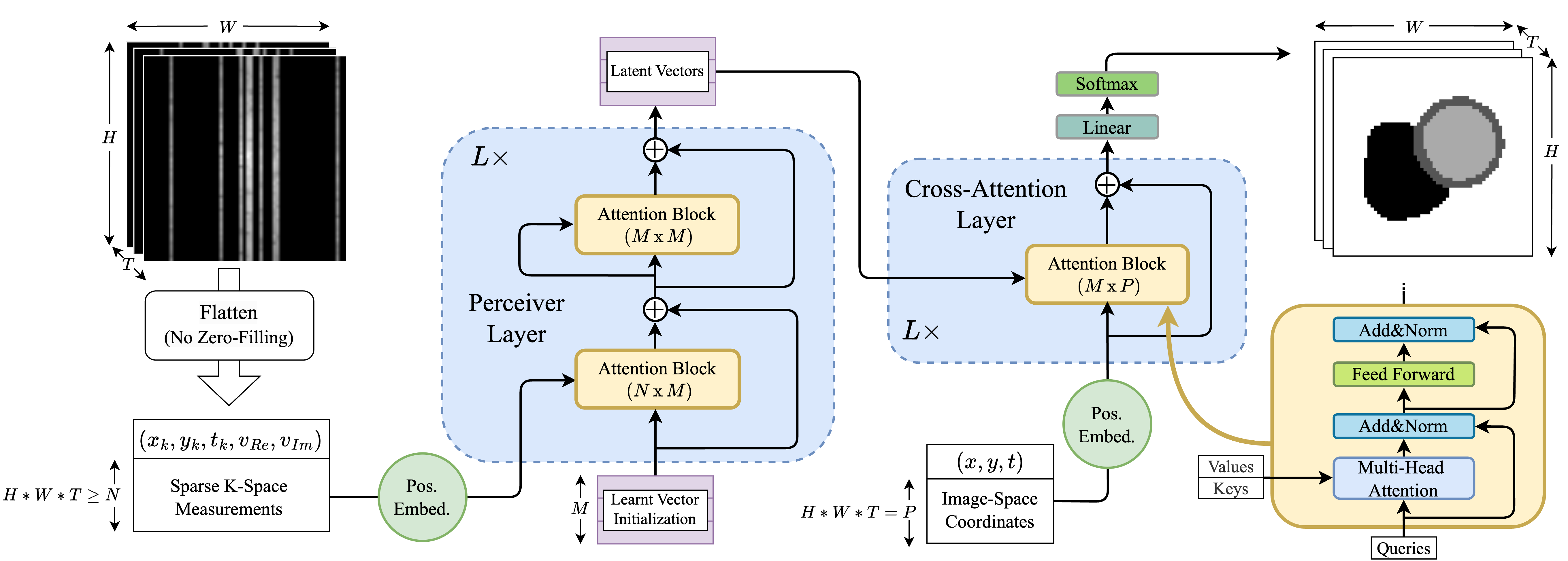
K-space Transformer Encoder: The authors design a transformer-based encoder that takes the undersampled k-space data as input and learns to extract relevant features. This is in contrast to traditional methods that first reconstruct the image and then perform segmentation.
-
Segmentation Head: The extracted features from the transformer encoder are passed to a segmentation head, which produces the final cardiac segmentation maps. The segmentation head is designed to be robust to the undersampled nature of the input k-space data.
-
Training and Optimization: The model is trained end-to-end using a combination of reconstruction and segmentation loss functions. This allows the transformer encoder to learn features that are directly relevant for the segmentation task, without the need for a separate image reconstruction step.
The authors evaluate the proposed method on a cardiac MRI dataset and compare its performance to traditional approaches that first reconstruct the image and then perform segmentation. The results demonstrate that the direct k-space segmentation method achieves superior segmentation accuracy and efficiency, highlighting the benefits of the transformer-based approach.
Critical Analysis
The paper presents a promising approach for direct cardiac segmentation from undersampled k-space data, leveraging the powerful representation learning capabilities of transformers. However, the authors acknowledge several limitations and areas for further research:
-
Generalization to different imaging modalities: The evaluation in the paper is limited to cardiac MRI data. Further research is needed to assess the generalizability of the approach to other imaging modalities, such as whole-heart 3D T1 representation learning or multi-view cardiac image segmentation.
-
Robustness to different undersampling patterns: The experiments in the paper focus on a specific undersampling pattern. Additional research is required to evaluate the method's performance under various undersampling scenarios, as real-world applications may involve different k-space sampling strategies.
-
Computational efficiency: While the proposed method demonstrates improved efficiency compared to traditional approaches, the computational requirements of the transformer-based architecture may still be a concern, especially for real-time applications. Further optimization and architectural refinements could help address this issue.
Overall, the paper presents a novel and promising approach that showcases the potential of transformers for direct cardiac segmentation from undersampled k-space data. Continued research in this direction may lead to significant advancements in the field of medical image analysis and clinical decision support systems.
Conclusion
This paper introduces a transformer-based approach for direct cardiac segmentation from undersampled k-space data, bypassing the traditional image reconstruction step. The proposed method leverages the powerful representation learning capabilities of transformers to effectively extract relevant features from the raw k-space data, leading to improved segmentation accuracy and efficiency compared to traditional methods.
The authors demonstrate the effectiveness of their approach on a cardiac MRI dataset, highlighting the potential benefits of this direct k-space segmentation technique. While the paper presents promising results, further research is needed to address the generalization, robustness, and computational efficiency concerns highlighted in the critical analysis.
Overall, the work contributes to the ongoing efforts in the medical imaging community to develop more efficient and accurate segmentation algorithms, which can ultimately lead to better clinical decision-making and patient care.
This summary was produced with help from an AI and may contain inaccuracies - check out the links to read the original source documents!
Related Papers
0
Direct Cardiac Segmentation from Undersampled K-space Using Transformers
Yundi Zhang, Nil Stolt-Ans'o, Jiazhen Pan, Wenqi Huang, Kerstin Hammernik, Daniel Rueckert
The prevailing deep learning-based methods of predicting cardiac segmentation involve reconstructed magnetic resonance (MR) images. The heavy dependency of segmentation approaches on image quality significantly limits the acceleration rate in fast MR reconstruction. Moreover, the practice of treating reconstruction and segmentation as separate sequential processes leads to artifact generation and information loss in the intermediate stage. These issues pose a great risk to achieving high-quality outcomes. To leverage the redundant k-space information overlooked in this dual-step pipeline, we introduce a novel approach to directly deriving segmentations from sparse k-space samples using a transformer (DiSK). DiSK operates by globally extracting latent features from 2D+time k-space data with attention blocks and subsequently predicting the segmentation label of query points. We evaluate our model under various acceleration factors (ranging from 4 to 64) and compare against two image-based segmentation baselines. Our model consistently outperforms the baselines in Dice and Hausdorff distances across foreground classes for all presented sampling rates.
Read more6/4/2024

0
Classification, Regression and Segmentation directly from k-Space in Cardiac MRI
Ruochen Li, Jiazhen Pan, Youxiang Zhu, Juncheng Ni, Daniel Rueckert
Cardiac Magnetic Resonance Imaging (CMR) is the gold standard for diagnosing cardiovascular diseases. Clinical diagnoses predominantly rely on magnitude-only Digital Imaging and Communications in Medicine (DICOM) images, omitting crucial phase information that might provide additional diagnostic benefits. In contrast, k-space is complex-valued and encompasses both magnitude and phase information, while humans cannot directly perceive. In this work, we propose KMAE, a Transformer-based model specifically designed to process k-space data directly, eliminating conventional intermediary conversion steps to the image domain. KMAE can handle critical cardiac disease classification, relevant phenotype regression, and cardiac morphology segmentation tasks. We utilize this model to investigate the potential of k-space-based diagnosis in cardiac MRI. Notably, this model achieves competitive classification and regression performance compared to image-domain methods e.g. Masked Autoencoders (MAEs) and delivers satisfactory segmentation performance with a myocardium dice score of 0.884. Last but not least, our model exhibits robust performance with consistent results even when the k-space is 8* undersampled. We encourage the MR community to explore the untapped potential of k-space and pursue end-to-end, automated diagnosis with reduced human intervention.
Read more7/30/2024
0
Multi-view Cardiac Image Segmentation via Trans-Dimensional Priors
Abbas Khan, Muhammad Asad, Martin Benning, Caroline Roney, Gregory Slabaugh
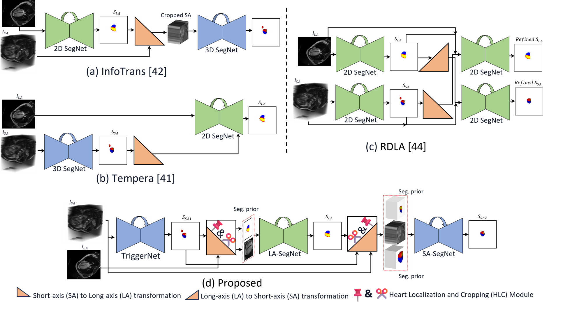
We propose a novel multi-stage trans-dimensional architecture for multi-view cardiac image segmentation. Our method exploits the relationship between long-axis (2D) and short-axis (3D) magnetic resonance (MR) images to perform a sequential 3D-to-2D-to-3D segmentation, segmenting the long-axis and short-axis images. In the first stage, 3D segmentation is performed using the short-axis image, and the prediction is transformed to the long-axis view and used as a segmentation prior in the next stage. In the second step, the heart region is localized and cropped around the segmentation prior using a Heart Localization and Cropping (HLC) module, focusing the subsequent model on the heart region of the image, where a 2D segmentation is performed. Similarly, we transform the long-axis prediction to the short-axis view, localize and crop the heart region and again perform a 3D segmentation to refine the initial short-axis segmentation. We evaluate our proposed method on the Multi-Disease, Multi-View & Multi-Center Right Ventricular Segmentation in Cardiac MRI (M&Ms-2) dataset, where our method outperforms state-of-the-art methods in segmenting cardiac regions of interest in both short-axis and long-axis images. The pre-trained models, source code, and implementation details will be publicly available.
Read more4/26/2024
✨
0
Transforming Heart Chamber Imaging: Self-Supervised Learning for Whole Heart Reconstruction and Segmentation
Abdul Qayyum, Hao Xu, Brian P. Halliday, Cristobal Rodero, Christopher W. Lanyon, Richard D. Wilkinson, Steven Alexander Niederer
Automated segmentation of Cardiac Magnetic Resonance (CMR) plays a pivotal role in efficiently assessing cardiac function, offering rapid clinical evaluations that benefit both healthcare practitioners and patients. While recent research has primarily focused on delineating structures in the short-axis orientation, less attention has been given to long-axis representations, mainly due to the complex nature of structures in this orientation. Performing pixel-wise segmentation of the left ventricular (LV) myocardium and the four cardiac chambers in 2-D steady-state free precession (SSFP) cine sequences is a crucial preprocessing stage for various analyses. However, the challenge lies in the significant variability in contrast, appearance, orientation, and positioning of the heart across different patients, clinical views, scanners, and imaging protocols. Consequently, achieving fully automatic semantic segmentation in this context is notoriously challenging. In recent years, several deep learning models have been proposed to accurately quantify and diagnose cardiac pathologies. These automated tools heavily rely on the accurate segmentation of cardiac structures in magnetic resonance images (MRI). Hence, there is a need for new methods to handle such structures' geometrical and textural complexities. We proposed 2D and 3D two-stage self-supervised deep learning segmentation hybrid transformer and CNN-based architectures for 4CH whole heart segmentation. Accurate segmentation of the ventricles and atria in 4CH views is crucial for analyzing heart health and reconstructing four-chamber meshes, which are essential for estimating various parameters to assess overall heart condition. Our proposed method outperformed state-of-the-art techniques, demonstrating superior performance in this domain.
Read more6/12/2024