Complex-valued neural networks to speed-up MR Thermometry during Hyperthermia using Fourier PD and PDUNet

0
🧠
Sign in to get full access
Overview
- This paper explores the use of deep learning techniques to improve the reconstruction of undersampled magnetic resonance imaging (MRI) data for temperature monitoring during hyperthermia cancer treatments.
- Hyperthermia (HT) involves heating tumor tissue to 39-43°C for 60 minutes, which can enhance the effectiveness of radiation and chemotherapy.
- Temperature monitoring is typically done using non-invasive dynamic MRI, but the slow nature of MRI can lead to motion artifacts.
- Undersampling the MRI data can increase the acquisition speed, but this can result in blurry images with aliasing artifacts.
- The researchers aimed to use deep learning to reconstruct highly undersampled MR thermometry data with better resolution and fewer artifacts compared to conventional methods.
Plain English Explanation
Hyperthermia for Cancer Treatment Hyperthermia is a cancer treatment that involves heating up tumor tissue to a specific temperature range for a period of time. This can make the tumor cells more vulnerable to other cancer treatments like radiation or chemotherapy.
Monitoring Temperature with MRI To monitor the temperature during hyperthermia treatment, doctors often use a technique called magnetic resonance imaging (MRI). MRI can measure the temperature of the tumor tissue non-invasively. However, MRI scans can be quite slow, which can lead to blurry images due to patient movement during the scan.
Improving MRI Scans with Deep Learning The researchers in this study wanted to use a technique called deep learning to improve the quality of the MRI scans used for temperature monitoring during hyperthermia. Deep learning is a type of artificial intelligence that can be trained to solve complex problems.
The researchers tested two different deep learning models to reconstruct the MRI data that had been intentionally undersampled (i.e., only partial data was collected) to speed up the scans. Their deep learning models were able to produce higher-quality images with fewer artifacts compared to conventional reconstruction methods.
This improvement in MRI image quality could help doctors more accurately monitor the temperature of the tumor during hyperthermia treatment, allowing them to optimize the therapy and potentially improve outcomes for cancer patients.
Technical Explanation
The researchers explored the use of two deep learning models, the Fourier Primal-Dual network and the Fourier Primal-Dual UNet, to reconstruct highly undersampled complex MR thermometry data.
Undersampling the MRI data can increase the acquisition speed, but this can result in blurry images with aliasing artifacts due to the violation of the Nyquist criterion. The deep learning models were trained to reconstruct the undersampled data and produce higher-quality images with fewer artifacts.
The Fourier Primal-Dual network and the Fourier Primal-Dual UNet both leveraged the Fourier domain to efficiently process the complex-valued MRI data. The models were trained on pairs of undersampled and fully sampled MRI data to learn the mapping between the two.
Evaluation of the models showed that they were able to reduce the temperature difference between the undersampled MRIs and the fully sampled MRIs from 1.3°C to 0.6°C in the full volume and from 0.49°C to 0.06°C in the tumor region for an acceleration factor of 10.
Critical Analysis
The researchers acknowledged that their deep learning-based approach focused solely on improving the reconstruction of the MRI data and did not address other potential sources of error, such as patient motion or physiological changes during the hyperthermia treatment. Further research would be needed to assess the overall impact of the improved MRI reconstruction on the accuracy and reliability of temperature monitoring in a clinical setting.
Additionally, the study was conducted using simulated undersampled data, and the performance of the deep learning models on real-world, clinically acquired undersampled MRI data remains to be evaluated. The researchers also noted that the training of the deep learning models required a large dataset of paired undersampled and fully sampled MRI data, which may not always be readily available in a clinical context.
Conclusion
This research demonstrates the potential of deep learning techniques to enhance the reconstruction of undersampled MRI data for temperature monitoring during hyperthermia cancer treatments. By improving the quality of the MRI images, the deep learning models could enable more accurate temperature measurements, potentially optimizing the delivery of hyperthermia therapy and ultimately improving outcomes for cancer patients.
This summary was produced with help from an AI and may contain inaccuracies - check out the links to read the original source documents!
Related Papers
🧠
0
Complex-valued neural networks to speed-up MR Thermometry during Hyperthermia using Fourier PD and PDUNet
Rupali Khatun, Soumick Chatterjee, Christoph Bert, Martin Wadepohl, Oliver J. Ott, Rainer Fietkau, Andreas Nurnberger, Udo S. Gaipl, Benjamin Frey
Hyperthermia (HT) in combination with radio- and/or chemotherapy has become an accepted cancer treatment for distinct solid tumour entities. In HT, tumour tissue is exogenously heated to temperatures between 39 and 43 $^circ$C for 60 minutes. Temperature monitoring can be performed non-invasively using dynamic magnetic resonance imaging (MRI). However, the slow nature of MRI leads to motion artefacts in the images due to the movements of patients during image acquisition. By discarding parts of the data, the speed of the acquisition can be increased - known as undersampling. However, due to the invalidation of the Nyquist criterion, the acquired images might be blurry and can also produce aliasing artefacts. The aim of this work was, therefore, to reconstruct highly undersampled MR thermometry acquisitions with better resolution and with fewer artefacts compared to conventional methods. The use of deep learning in the medical field has emerged in recent times, and various studies have shown that deep learning has the potential to solve inverse problems such as MR image reconstruction. However, most of the published work only focuses on the magnitude images, while the phase images are ignored, which are fundamental requirements for MR thermometry. This work, for the first time, presents deep learning-based solutions for reconstructing undersampled MR thermometry data. Two different deep learning models have been employed here, the Fourier Primal-Dual network and the Fourier Primal-Dual UNet, to reconstruct highly undersampled complex images of MR thermometry. The method reduced the temperature difference between the undersampled MRIs and the fully sampled MRIs from 1.3 $^circ$C to 0.6 $^circ$C in full volume and 0.49 $^circ$C to 0.06 $^circ$C in the tumour region for an acceleration factor of 10.
Read more7/24/2024
0
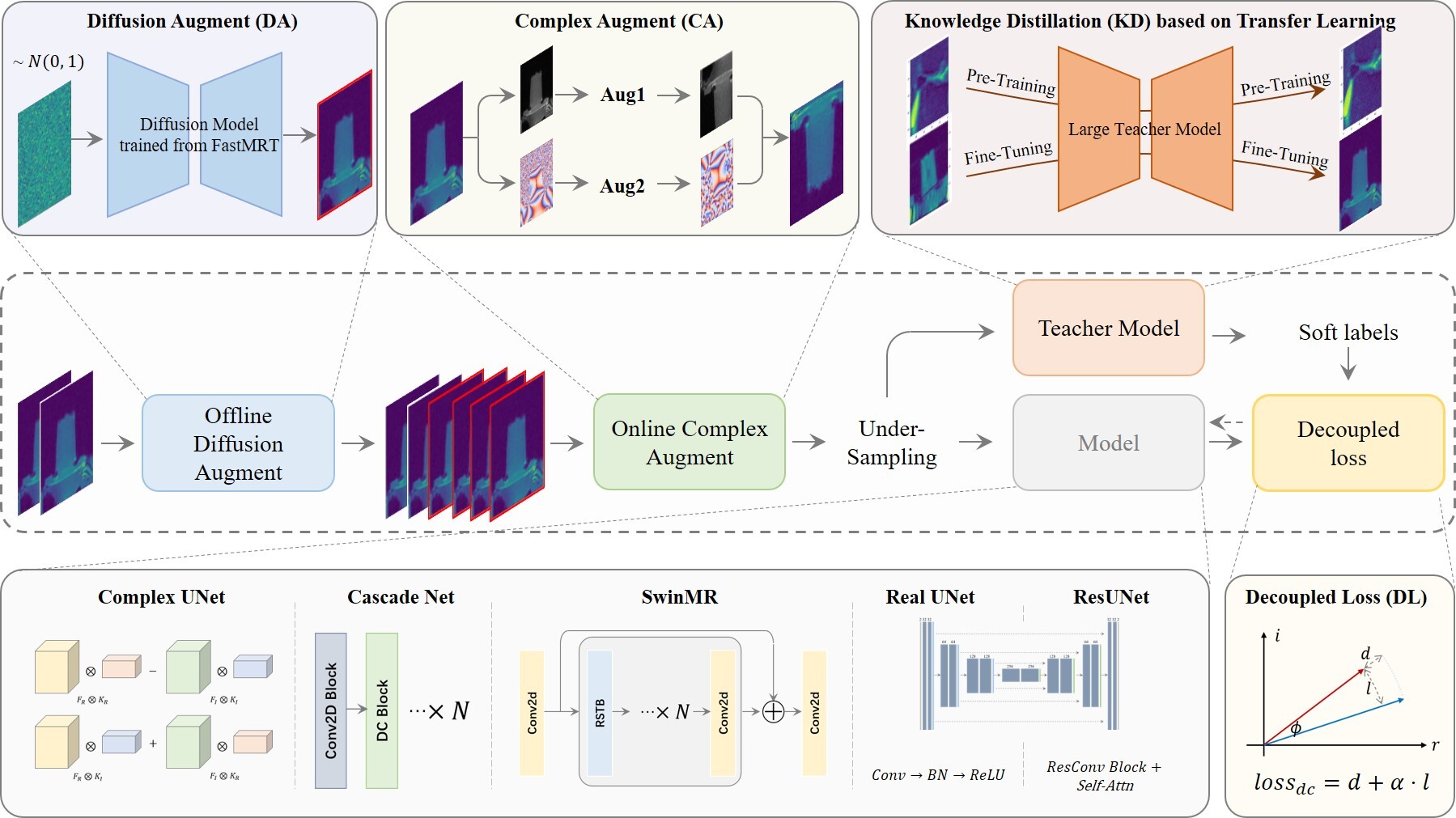
Accelerated Proton Resonance Frequency-based Magnetic Resonance Thermometry by Optimized Deep Learning Method
Sijie Xu, Shenyan Zong, Chang-Sheng Mei, Guofeng Shen, Yueran Zhao, He Wang
Proton resonance frequency (PRF) based MR thermometry is essential for focused ultrasound (FUS) thermal ablation therapies. This work aims to enhance temporal resolution in dynamic MR temperature map reconstruction using an improved deep learning method. The training-optimized methods and five classical neural networks were applied on the 2-fold and 4-fold under-sampling k-space data to reconstruct the temperature maps. The enhanced training modules included offline/online data augmentations, knowledge distillation, and the amplitude-phase decoupling loss function. The heating experiments were performed by a FUS transducer on phantom and ex vivo tissues, respectively. These data were manually under-sampled to imitate acceleration procedures and trained in our method to get the reconstruction model. The additional dozen or so testing datasets were separately obtained for evaluating the real-time performance and temperature accuracy. Acceleration factors of 1.9 and 3.7 were found for 2 times and 4 times k-space under-sampling strategies and the ResUNet-based deep learning reconstruction performed exceptionally well. In 2-fold acceleration scenario, the RMSE of temperature map patches provided the values of 0.888 degree centigrade and 1.145 degree centigrade on phantom and ex vivo testing datasets. The DICE value of temperature areas enclosed by 43 degree centigrade isotherm was 0.809, and the Bland-Altman analysis showed a bias of -0.253 degree centigrade with the apart of plus or minus 2.16 degree centigrade. In 4 times under-sampling case, these evaluating values decreased by approximately 10%. This study demonstrates that deep learning-based reconstruction can significantly enhance the accuracy and efficiency of MR thermometry for clinical FUS thermal therapies.
Read more7/4/2024

0
Fever Detection with Infrared Thermography: Enhancing Accuracy through Machine Learning Techniques
Parsa Razmara, Tina Khezresmaeilzadeh, B. Keith Jenkins
The COVID-19 pandemic has underscored the necessity for advanced diagnostic tools in global health systems. Infrared Thermography (IRT) has proven to be a crucial non-contact method for measuring body temperature, vital for identifying febrile conditions associated with infectious diseases like COVID-19. Traditional non-contact infrared thermometers (NCITs) often exhibit significant variability in readings. To address this, we integrated machine learning algorithms with IRT to enhance the accuracy and reliability of temperature measurements. Our study systematically evaluated various regression models using heuristic feature engineering techniques, focusing on features' physiological relevance and statistical significance. The Convolutional Neural Network (CNN) model, utilizing these techniques, achieved the lowest RMSE of 0.2223, demonstrating superior performance compared to results reported in previous literature. Among non-neural network models, the Binning method achieved the best performance with an RMSE of 0.2296. Our findings highlight the potential of combining advanced feature engineering with machine learning to improve diagnostic tools' effectiveness, with implications extending to other non-contact or remote sensing biomedical applications. This paper offers a comprehensive analysis of these methodologies, providing a foundation for future research in the field of non-invasive medical diagnostics.
Read more8/13/2024
🤿
0
Accelerated MR Cholangiopancreatography with Deep Learning-based Reconstruction
Jinho Kim, Marcel Dominik Nickel, Florian Knoll
This study accelerates MR cholangiopancreatography (MRCP) acquisitions using deep learning-based (DL) reconstruction at 3T and 0.55T. Thirty healthy volunteers underwent conventional two-fold MRCP scans at field strengths of 3T or 0.55T. We trained a variational network (VN) using retrospectively six-fold undersampled data obtained at 3T. We then evaluated our method against standard techniques such as parallel imaging (PI) and compressed sensing (CS), focusing on peak signal-to-noise ratio (PSNR) and structural similarity (SSIM) as metrics. Furthermore, considering acquiring fully-sampled MRCP is impractical, we added a self-supervised DL reconstruction (SSDU) to the evaluating group. We also tested our method in a prospective accelerated scenario to reflect real-world clinical applications and evaluated its adaptability to MRCP at 0.55T. Our method demonstrated a remarkable reduction of average acquisition time from 599/542 to 255/180 seconds for MRCP at 3T/0.55T. In both retrospective and prospective undersampling scenarios, the PSNR and SSIM of VN were higher than those of PI, CS, and SSDU. At the same time, VN preserved the image quality of undersampled data, i.e., sharpness and the visibility of hepatobiliary ducts. In addition, VN also produced high quality reconstructions at 0.55T resulting in the highest PSNR and SSIM. In summary, VN trained for highly accelerated MRCP allows to reduce the acquisition time by a factor of 2.4/3.0 at 3T/0.55T while maintaining the image quality of the conventional acquisition.
Read more5/8/2024