Compressive Electron Backscatter Diffraction Imaging

0
Sign in to get full access
Overview
- This paper introduces a new method called Compressive Electron Backscatter Diffraction (CEBSD) Imaging that can quickly capture high-resolution electron backscatter diffraction (EBSD) data.
- The technique uses a compressive sensing approach to reconstruct full EBSD patterns from a small number of measurements, reducing the time and data required compared to traditional EBSD.
- The authors demonstrate the effectiveness of CEBSD Imaging on experimental data, showing it can capture detailed microstructural information while enabling faster, more efficient EBSD data collection.
Plain English Explanation
Compressive Electron Backscatter Diffraction Imaging is a new technique that can quickly and efficiently capture high-resolution images of a material's microstructure using a method called electron backscatter diffraction (EBSD).
Traditionally, EBSD has required collecting a large amount of data to produce detailed images. This Compressive Electron Backscatter Diffraction Imaging approach uses a clever mathematical technique called "compressive sensing" to reconstruct the full EBSD pattern from just a small number of measurements.
This allows the researchers to collect EBSD data much faster and with less information, without losing important details about the microstructure of the material. The authors show this Compressive Electron Backscatter Diffraction Imaging method works well on experimental data, demonstrating its potential to enable more efficient and practical EBSD imaging.
Technical Explanation
The paper introduces a new technique called Compressive Electron Backscatter Diffraction (CEBSD) Imaging that leverages compressive sensing to reconstruct full EBSD patterns from a small number of measurements.
Traditional EBSD relies on collecting a dense grid of diffraction patterns across the sample surface to build up a detailed microstructural map. In contrast, CEBSD Imaging uses a compressive sensing approach to reconstruct the full EBSD pattern from just a few strategically-chosen measurements.
The authors demonstrate this approach on experimental data, showing CEBSD Imaging can capture the same level of microstructural detail as traditional EBSD but with significantly less data collection. This has the potential to enable faster and more efficient EBSD data acquisition compared to existing methods.
The paper also explores the use of deep learning techniques to further enhance the CEBSD Imaging reconstruction process, improving both accuracy and computational efficiency.
Critical Analysis
The Compressive Electron Backscatter Diffraction Imaging approach presented in this paper offers a promising new direction for EBSD data collection and analysis. The ability to reconstruct detailed microstructural information from a small number of measurements could lead to significant time and cost savings compared to traditional EBSD techniques.
However, the paper does not fully address some potential limitations of the CEBSD Imaging method. For example, the authors do not explore how the technique might perform on more complex or heterogeneous samples, where the underlying assumptions of the compressive sensing approach may be more challenged. Additionally, the computational complexity of the reconstruction process, even with the use of deep learning, could limit the practical deployment of CEBSD Imaging in certain real-world scenarios.
Further research and validation on a wider range of materials and applications would help to better understand the strengths, weaknesses, and appropriate use cases for this Compressive Electron Backscatter Diffraction Imaging approach. Comparisons to other emerging EBSD data acquisition and analysis techniques, such as HD-Snapshot Diffractive Spectral Imaging, would also be valuable for assessing its relative merits and limitations.
Conclusion
The Compressive Electron Backscatter Diffraction Imaging technique presented in this paper offers a compelling new approach to EBSD data collection and analysis. By leveraging compressive sensing and deep learning, the method can capture detailed microstructural information while significantly reducing the time and data required compared to traditional EBSD techniques.
This has the potential to enable faster, more efficient EBSD data acquisition, which could benefit a wide range of materials science and engineering applications. Further research and validation will be important to fully understand the strengths, limitations, and appropriate use cases of this innovative imaging approach.
This summary was produced with help from an AI and may contain inaccuracies - check out the links to read the original source documents!
Related Papers
0
Compressive Electron Backscatter Diffraction Imaging
Zoe Broad, Alex W. Robinson, Jack Wells, Daniel Nicholls, Amirafshar Moshtaghpour, Angus I. Kirkland, Nigel D. Browning
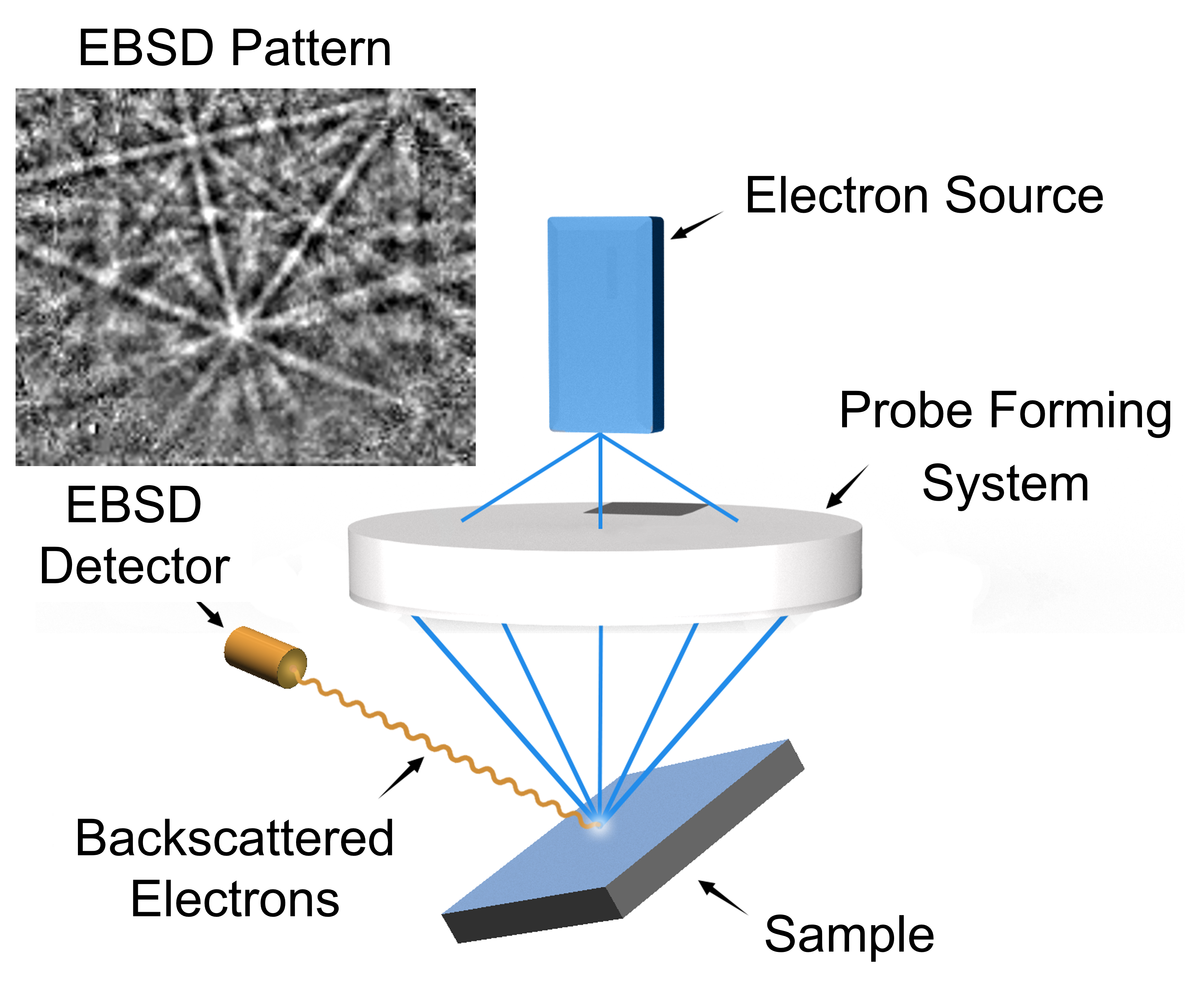
Electron backscatter diffraction (EBSD) has developed over the last few decades into a valuable crystallographic characterisation method for a wide range of sample types. Despite these advances, issues such as the complexity of sample preparation, relatively slow acquisition, and damage in beam-sensitive samples, still limit the quantity and quality of interpretable data that can be obtained. To mitigate these issues, here we propose a method based on the subsampling of probe positions and subsequent reconstruction of an incomplete dataset. The missing probe locations (or pixels in the image) are recovered via an inpainting process using a dictionary-learning based method called beta-process factor analysis (BPFA). To investigate the robustness of both our inpainting method and Hough-based indexing, we simulate subsampled and noisy EBSD datasets from a real fully sampled Ni-superalloy dataset for different subsampling ratios of probe positions using both Gaussian and Poisson noise models. We find that zero solution pixel detection (inpainting un-indexed pixels) enables higher quality reconstructions to be obtained. Numerical tests confirm high quality reconstruction of band contrast and inverse pole figure maps from only 10% of the probe positions, with the potential to reduce this to 5% if only inverse pole figure maps are needed. These results show the potential application of this method in EBSD, allowing for faster analysis and extending the use of this technique to beam sensitive materials.
Read more7/17/2024
🔄
0
Automated high-resolution backscattered-electron imaging at macroscopic scale
Zhiyuan Lang, Zunshuai Zhang, Lei Wang, Yuhan Liu, Weixiong Qian, Shenghua Zhou, Ying Jiang, Tongyi Zhang, Jiong Yang
Scanning electron microscopy (SEM) has been widely utilized in the field of materials science due to its significant advantages, such as large depth of field, wide field of view, and excellent stereoscopic imaging. However, at high magnification, the limited imaging range in SEM cannot cover all the possible inhomogeneous microstructures. In this research, we propose a novel approach for generating high-resolution SEM images across multiple scales, enabling a single image to capture physical dimensions at the centimeter level while preserving submicron-level details. We adopted the SEM imaging on the AlCoCrFeNi2.1 eutectic high entropy alloy (EHEA) as an example. SEM videos and image stitching are combined to fulfill this goal, and the video-extracted low-definition (LD) images are clarified by a well-trained denoising model. Furthermore, we segment the macroscopic image of the EHEA, and area of various microstructures are distinguished. Combining the segmentation results and hardness experiments, we found that the hardness is positively correlated with the content of body-centered cubic (BCC) phase, negatively correlated with the lamella width, and the relationship with the proportion of lamellar structures was not significant. Our work provides a feasible solution to generate macroscopic images based on SEMs for further analysis of the correlations between the microstructures and spatial distribution, and can be widely applied to other types of microscope.
Read more7/16/2024
0
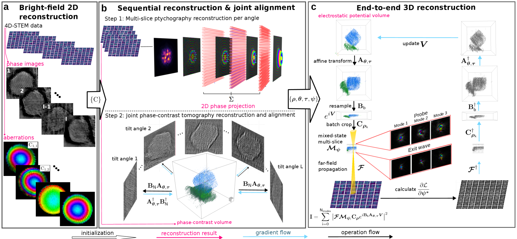
Near-Isotropic Sub-{AA}ngstrom 3D Resolution Phase Contrast Imaging Achieved by End-to-End Ptychographic Electron Tomography
Shengboy You, Andrey Romanov, Philipp Pelz
Three-dimensional atomic resolution imaging using transmission electron microscopes is a unique capability that requires challenging experiments. Linear electron tomography methods are limited by the missing wedge effect, requiring a high tilt range. Multislice ptychography can achieve deep sub-{AA}ngstrom resolution in the transverse direction, but the depth resolution is limited to 2 to 3 nanometers. In this paper, we propose and demonstrate an end-to-end approach to reconstructing the electrostatic potential volume of the sample directly from the 4D-STEM datasets. End-to-end multi-slice ptychographic tomography recovers several slices at each tomography tilt angle and compensates for the missing wedge effect. The algorithm is initially tested in simulation with a Pt@$mathrm{Al_2O_3}$ core-shell nanoparticle, where both heavy and light atoms are recovered in 3D from an unaligned 4D-STEM tilt series with a restricted tilt range of 90 degrees. We also demonstrate the algorithm experimentally, recovering a Te nanoparticle with sub-{AA}ngstrom resolution.
Read more7/30/2024

0
Dual-Domain Deep D-bar Method for Solving Electrical Impedance Tomography
Xiang Cao, Qiaoqiao Ding, Xiaoqun Zhang
The regularized D-bar method is one of the most prominent methods for solving Electrical Impedance Tomography (EIT) problems due to its efficiency and simplicity. It provides a direct approach by applying low-pass filtering to the scattering data in the non-linear Fourier domain, thereby yielding a smoothed conductivity approximation. However, D-bar images often present low contrast and low resolution due to the absence of accurate high-frequency information and ill-posedness of the problem. In this paper, we proposed a dual-domain neural network architecture to retrieve high-contrast D-bar image sequences from low-contrast D-bar images. To further accentuate the spatial features of the conductivity distribution, the widely adopted U-net has been tailored for conductivity image calibration from the predicted D-bar image sequences. We call such a hybrid approach by Dual-Domain Deep D-bar method due to the consideration of both scattering data and image information. Compared to the single-scale structure, our proposed multi-scale structure exhibits superior capabilities in reducing artifacts and refining conductivity approximation. Additionally, solving discrete D-bar systems using the GMRES algorithm entails significant computational complexity, which is extremely time-consuming on CPU-based devices. To remedy this, we designed a surrogate GPU-based Richardson iterative method to accelerate the data enhancement process by D-bar. Numerical results are presented for simulated EIT data from the KIT4 and ACT4 systems to demonstrate notable improvements in absolute EIT imaging quality when compared to existing methodologies.
Read more7/8/2024