Near-Isotropic Sub-{AA}ngstrom 3D Resolution Phase Contrast Imaging Achieved by End-to-End Ptychographic Electron Tomography

0
Sign in to get full access
Overview
- This paper presents a new technique called "end-to-end ptychographic electron tomography" that can achieve near-isotropic sub-Ångstrom 3D resolution phase contrast imaging.
- The technique combines ptychographic imaging, which uses computational methods to reconstruct high-resolution images from diffraction patterns, with electron tomography to create 3D reconstructions.
- The researchers demonstrate the method on a variety of samples, including carbon nanotubes and a zeolite catalyst, achieving resolutions down to 0.7 Ångstrom (Å) in all three dimensions.
Plain English Explanation
The paper describes a new way to take extremely high-resolution 3D images using electron microscopes. Normally, electron microscopes can only provide detailed 2D images of a sample. This new technique, called "end-to-end ptychographic electron tomography," combines two different imaging methods to create 3D images with unprecedented clarity and resolution.
The first part of the technique, ptychography, uses sophisticated computational algorithms to reconstruct high-resolution images from the patterns of scattered electrons. The second part, electron tomography, takes many 2D images of the sample from different angles and combines them to create a 3D reconstruction.
By integrating these two methods, the researchers were able to produce 3D images with a resolution of less than 1 Ångstrom (Å), which is about the size of a single atom. This is an extremely high level of detail, allowing them to clearly see the individual atoms and structures within their samples, which included carbon nanotubes and a type of catalyst material called zeolites.
Technical Explanation
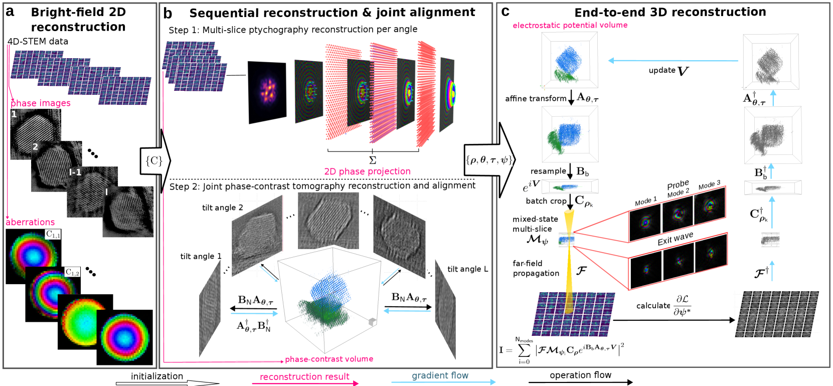
The key innovation presented in this paper is the end-to-end ptychographic electron tomography technique, which combines ptychographic phase contrast imaging with electron tomography to achieve near-isotropic sub-Ångstrom 3D resolution.
In the ptychographic step, the sample is illuminated with a focused electron beam that is scanned across the surface. At each position, the scattered electron waves are recorded as a diffraction pattern. Sophisticated algorithms are then used to computationally reconstruct a high-resolution phase contrast image of the sample from these diffraction patterns.
For the tomography step, the sample is rotated, and a series of these high-resolution 2D ptychographic images are acquired from multiple angles. These 2D projections are then combined using tomographic reconstruction algorithms to create a detailed 3D model of the sample's structure.
The researchers demonstrate this system-sample agnostic isotropic 3D microscopy technique on a variety of samples, including carbon nanotubes and a zeolite catalyst. They achieve resolutions down to 0.7 Å in all three dimensions, allowing them to clearly resolve individual atoms and defects within the samples.
Critical Analysis
The paper provides a compelling demonstration of the capabilities of end-to-end ptychographic electron tomography, showing that it can achieve truly remarkable 3D resolution at the atomic scale. However, the technique does have some limitations:
- The sample preparation and data acquisition process is still relatively complex and time-consuming, which may limit its broader applicability.
- The reconstructions can be sensitive to factors like sample thickness and electron beam coherence, requiring careful optimization of the experimental parameters.
- There are also potential concerns about radiation damage to sensitive samples from the high electron doses required.
Overall, this work represents a significant advance in 3D electron microscopy, pushing the boundaries of what is possible in terms of resolution and sensitivity. With further refinements and improvements, the technique could become a powerful tool for nanoscale materials characterization across a wide range of fields.
Conclusion
This paper presents a new end-to-end ptychographic electron tomography technique that can achieve near-isotropic sub-Ångstrom 3D resolution phase contrast imaging. By combining ptychographic imaging and electron tomography, the researchers demonstrated the ability to resolve individual atoms and defects within a variety of nanostructured materials. While the method has some practical limitations, it represents a major breakthrough in 3D electron microscopy, paving the way for exciting new discoveries in materials science, chemistry, and beyond.
This summary was produced with help from an AI and may contain inaccuracies - check out the links to read the original source documents!
Related Papers
0
Near-Isotropic Sub-{AA}ngstrom 3D Resolution Phase Contrast Imaging Achieved by End-to-End Ptychographic Electron Tomography
Shengboy You, Andrey Romanov, Philipp Pelz
Three-dimensional atomic resolution imaging using transmission electron microscopes is a unique capability that requires challenging experiments. Linear electron tomography methods are limited by the missing wedge effect, requiring a high tilt range. Multislice ptychography can achieve deep sub-{AA}ngstrom resolution in the transverse direction, but the depth resolution is limited to 2 to 3 nanometers. In this paper, we propose and demonstrate an end-to-end approach to reconstructing the electrostatic potential volume of the sample directly from the 4D-STEM datasets. End-to-end multi-slice ptychographic tomography recovers several slices at each tomography tilt angle and compensates for the missing wedge effect. The algorithm is initially tested in simulation with a Pt@$mathrm{Al_2O_3}$ core-shell nanoparticle, where both heavy and light atoms are recovered in 3D from an unaligned 4D-STEM tilt series with a restricted tilt range of 90 degrees. We also demonstrate the algorithm experimentally, recovering a Te nanoparticle with sub-{AA}ngstrom resolution.
Read more7/30/2024

0
X-ray nano-holotomography reconstruction with simultaneous probe retrieval
Viktor Nikitin, Marcus Carlsson, Doga Gursoy, Rajmund Mokso, Peter Cloetens
In conventional tomographic reconstruction, the pre-processing step includes flat-field correction, where each sample projection on the detector is divided by a reference image taken without the sample. When using coherent X-rays as probe, this approach overlooks the phase component of the illumination field (probe), leading to artifacts in phase-retrieved projection images, which are then propagated to the reconstructed 3D sample representation. The problem intensifies in nano-holotomography with focusing optics, that due to various imperfections create high-frequency components in the probe function. Here, we present a new iterative reconstruction scheme for holotomography, simultaneously retrieving the complex-valued probe function. Implemented on GPUs, this algorithm results in 3D reconstruction resolving twice thinner layers in a 3D ALD standard sample measured using nano-holotomography.
Read more9/2/2024
↗️
0
A high-performance reconstruction method for partially coherent ptychography
Wenhui Xu, Shoucong Ning, Pengju Sheng, Huixiang Lin, Angus I Kirkland, Yong Peng, Fucai Zhang
Ptychography is now integrated as a tool in mainstream microscopy allowing quantitative and high-resolution imaging capabilities over a wide field of view. However, its ultimate performance is inevitably limited by the available coherent flux when implemented using electrons or laboratory X-ray sources. We present a universal reconstruction algorithm with high tolerance to low coherence for both far-field and near-field ptychography. The approach is practical for partial temporal and spatial coherence and requires no prior knowledge of the source properties. Our initial visible-light and electron data show that the method can dramatically improve the reconstruction quality and accelerate the convergence rate of the reconstruction. The approach also integrates well into existing ptychographic engines. It can also improve mixed-state and numerical monochromatisation methods, requiring a smaller number of coherent modes or lower dimensionality of Krylov subspace while providing more stable and faster convergence. We propose that this approach could have significant impact on ptychography of weakly scattering samples.
Read more6/12/2024
🔎
0
Improved ACOM pattern matching in 4D STEM through adaptive sub pixel peak detection and image reconstruction
Nicolas Folastre, Junhao Cao, Gozde Oney, Sunkyu Park, Arash Jamali, Christian Masquelier, Laurence Croguennec, Muriel Veron, Edgar F. Rauch, Arnaud Demorti`ere
The technique known as 4D-STEM has recently emerged as a powerful tool for the local characterization of crystalline structures in materials, such as cathode materials for Li-ion batteries or perovskite materials for photovoltaics. However, the use of new detectors optimized for electron diffraction patterns and other advanced techniques requires constant adaptation of methodologies to address the challenges associated with crystalline materials. In this study, we present a novel image processing method to improve pattern matching in the determination of crystalline orientations and phases. Our approach uses sub-pixelar adaptative image processing to register and reconstruct electron diffraction signals in large 4D-STEM datasets. By using adaptive prominence and linear filters such as mean and gaussian blur, we are able to improve the quality of the diffraction pattern registration. The resulting data compression rate of 103 is well-suited for the era of big data and provides a significant enhancement in the performance of the entire ACOM data processing method. Our approach is evaluated using dedicated metrics, which demonstrate a high improvement in phase recognition. Our results demonstrate that this data preparation method not only enhances the quality of the resulting image but also boosts the confidence level in the analysis of the outcomes related to determining crystal orientation and phase. Additionally, it mitigates the impact of user bias that may occur during the application of the method through the manipulation of parameters.
Read more9/10/2024