Three-dimensional Microstructural Image Synthesis from 2D Backscattered Electron Image of Cement Paste

0
🖼️
Sign in to get full access
Overview
- This paper proposes a deep learning-based method for generating high-quality, realistic 3D microstructure images from a single 2D backscattered electron (BSE) image.
- The method, called CEM3DMG, can synthesize 3D images of arbitrary size with a resolution of 0.47 μm per pixel.
- Experimental results show the generated 3D images closely match the real 2D microstructure in terms of features like pore and particle morphology, gray level histogram, phase proportions, and pore size distribution.
Plain English Explanation
The researchers have developed a deep learning-based technique that can take a 2D image of a material's microstructure and generate a realistic 3D version of it. This is useful because 3D images can provide more detailed information about a material's structure than 2D images, but capturing 3D data is often more expensive and time-consuming.
The key idea is to train a machine learning model to learn the relationship between the 2D image and the corresponding 3D structure. Once trained, the model can then generate a 3D image from a new 2D input, without needing to capture the 3D data directly.
The researchers tested their method, called CEM3DMG, on a set of 2D backscattered electron images and found that the generated 3D images were visually very similar to the real 3D structures. Quantitative analysis also showed the 3D images matched the 2D images in terms of important microstructural features like pore size and distribution.
This 2D-to-3D translation approach could be useful in a variety of applications where capturing high-quality 3D data is challenging or expensive, such as in materials science, medical imaging, and geology.
Technical Explanation
The researchers designed a deep learning framework called CEM3DMG (Convolutional Encoder-decoder Microstructure 3D Model Generator) to synthesize 3D microstructure images from a single 2D backscattered electron (BSE) image. The framework consists of an encoder network that learns a latent representation of the 2D microstructure, and a decoder network that generates the corresponding 3D image.
To train the model, the researchers used a dataset of paired 2D BSE images and their corresponding 3D microstructure volumes obtained through X-ray tomography. The encoder network was trained to map the 2D input to a low-dimensional latent representation, while the decoder network was trained to generate a 3D image from this latent code.
Experimental results showed that CEM3DMG could generate realistic 3D images of arbitrary size with a resolution of 0.47 μm per pixel. Visual inspection confirmed that the generated 3D images exhibited similar microstructural features to the 2D input images, including pore and particle morphology. Quantitative analysis further revealed that the generated 3D microstructures closely matched the real 2D microstructure in terms of gray level histogram, phase proportions, and pore size distribution.
Critical Analysis
The researchers acknowledge that their method has some limitations. For example, the model was trained and tested on a specific material (a nickel-based superalloy), and its performance on other types of materials is not yet known. Additionally, the quality of the generated 3D images may be affected by the quality and resolution of the input 2D images.
Another potential concern is that the method relies on having access to a dataset of paired 2D and 3D microstructure data for training. Obtaining such a dataset can be challenging and expensive, especially for some materials or applications.
Despite these limitations, the researchers' approach represents a significant advancement in the field of 3D microstructure generation from 2D images. The ability to generate high-quality 3D images at low cost could have important implications for materials science, medical imaging, and other domains where 3D data is crucial but difficult to acquire.
Future research could explore ways to extend the method to handle a wider range of materials, reduce the reliance on paired 2D-3D training data, and further improve the fidelity of the generated 3D images.
Conclusion
This paper presents a deep learning-based method for generating realistic 3D microstructure images from a single 2D backscattered electron image. The proposed CEM3DMG framework can synthesize 3D images that closely match the real 2D microstructure in terms of key features like pore and particle morphology, gray level histogram, phase proportions, and pore size distribution.
The ability to generate high-quality 3D data from 2D input could have significant implications for a variety of applications, such as materials science, medical imaging, and geology, where capturing 3D data is often challenging or expensive. While the method has some limitations, it represents an important step forward in the field of 3D microstructure generation and could inspire further advancements in this area.
This summary was produced with help from an AI and may contain inaccuracies - check out the links to read the original source documents!
Related Papers
🖼️
0
Three-dimensional Microstructural Image Synthesis from 2D Backscattered Electron Image of Cement Paste
Xin Zhao, Lin Wang, Qinfei Li, Heng Chen, Shuangrong Liu, Pengkun Hou, Xu Wu, Jianfeng Yuan, Haozhong Gao, Bo Yang
This paper proposes a deep learning-based method for generating 3D microstructures from a single two-dimensional (2D) image, capable of producing high-quality, realistic 3D images at low cost. In the method, a framework (CEM3DMG) is designed to synthesize 3D images by learning microstructural information from a 2D backscattered electron (BSE) image. Experimental results show that CEM3DMG can generate realistic 3D images of arbitrary size with a resolution of 0.47 $mu m$ per pixel. Visual observation confirms that the generated 3D images exhibit similar microstructural features to the 2D images, including pores and particles morphology. Furthermore, quantitative analysis reveals that these 3D microstructures closely match the real 2D microstructure in terms of gray level histogram, phase proportions, and pore size distribution.
Read more7/12/2024
🔄
0
Automated high-resolution backscattered-electron imaging at macroscopic scale
Zhiyuan Lang, Zunshuai Zhang, Lei Wang, Yuhan Liu, Weixiong Qian, Shenghua Zhou, Ying Jiang, Tongyi Zhang, Jiong Yang
Scanning electron microscopy (SEM) has been widely utilized in the field of materials science due to its significant advantages, such as large depth of field, wide field of view, and excellent stereoscopic imaging. However, at high magnification, the limited imaging range in SEM cannot cover all the possible inhomogeneous microstructures. In this research, we propose a novel approach for generating high-resolution SEM images across multiple scales, enabling a single image to capture physical dimensions at the centimeter level while preserving submicron-level details. We adopted the SEM imaging on the AlCoCrFeNi2.1 eutectic high entropy alloy (EHEA) as an example. SEM videos and image stitching are combined to fulfill this goal, and the video-extracted low-definition (LD) images are clarified by a well-trained denoising model. Furthermore, we segment the macroscopic image of the EHEA, and area of various microstructures are distinguished. Combining the segmentation results and hardness experiments, we found that the hardness is positively correlated with the content of body-centered cubic (BCC) phase, negatively correlated with the lamella width, and the relationship with the proportion of lamellar structures was not significant. Our work provides a feasible solution to generate macroscopic images based on SEMs for further analysis of the correlations between the microstructures and spatial distribution, and can be widely applied to other types of microscope.
Read more7/16/2024
0
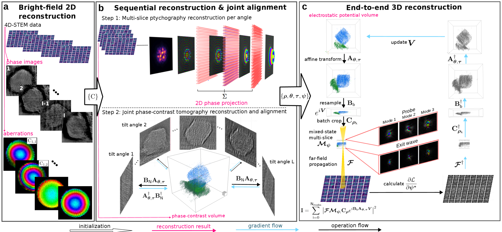
Near-Isotropic Sub-{AA}ngstrom 3D Resolution Phase Contrast Imaging Achieved by End-to-End Ptychographic Electron Tomography
Shengboy You, Andrey Romanov, Philipp Pelz
Three-dimensional atomic resolution imaging using transmission electron microscopes is a unique capability that requires challenging experiments. Linear electron tomography methods are limited by the missing wedge effect, requiring a high tilt range. Multislice ptychography can achieve deep sub-{AA}ngstrom resolution in the transverse direction, but the depth resolution is limited to 2 to 3 nanometers. In this paper, we propose and demonstrate an end-to-end approach to reconstructing the electrostatic potential volume of the sample directly from the 4D-STEM datasets. End-to-end multi-slice ptychographic tomography recovers several slices at each tomography tilt angle and compensates for the missing wedge effect. The algorithm is initially tested in simulation with a Pt@$mathrm{Al_2O_3}$ core-shell nanoparticle, where both heavy and light atoms are recovered in 3D from an unaligned 4D-STEM tilt series with a restricted tilt range of 90 degrees. We also demonstrate the algorithm experimentally, recovering a Te nanoparticle with sub-{AA}ngstrom resolution.
Read more7/30/2024

0
Three-Dimensional, Multimodal Synchrotron Data for Machine Learning Applications
Calum Green, Sharif Ahmed, Shashidhara Marathe, Liam Perera, Alberto Leonardi, Killian Gmyrek, Daniele Dini, James Le Houx
Machine learning techniques are being increasingly applied in medical and physical sciences across a variety of imaging modalities; however, an important issue when developing these tools is the availability of good quality training data. Here we present a unique, multimodal synchrotron dataset of a bespoke zinc-doped Zeolite 13X sample that can be used to develop advanced deep learning and data fusion pipelines. Multi-resolution micro X-ray computed tomography was performed on a zinc-doped Zeolite 13X fragment to characterise its pores and features, before spatially resolved X-ray diffraction computed tomography was carried out to characterise the homogeneous distribution of sodium and zinc phases. Zinc absorption was controlled to create a simple, spatially isolated, two-phase material. Both raw and processed data is available as a series of Zenodo entries. Altogether we present a spatially resolved, three-dimensional, multimodal, multi-resolution dataset that can be used for the development of machine learning techniques. Such techniques include development of super-resolution, multimodal data fusion, and 3D reconstruction algorithm development.
Read more9/12/2024