Computational limits to the legibility of the imaged human brain
2309.07096
0
0
✨
Abstract
Our knowledge of the organisation of the human brain at the population-level is yet to translate into power to predict functional differences at the individual-level, limiting clinical applications, and casting doubt on the generalisability of inferred mechanisms. It remains unknown whether the difficulty arises from the absence of individuating biological patterns within the brain, or from limited power to access them with the models and compute at our disposal. Here we comprehensively investigate the resolvability of such patterns with data and compute at unprecedented scale. Across 23 810 unique participants from UK Biobank, we systematically evaluate the predictability of 25 individual biological characteristics, from all available combinations of structural and functional neuroimaging data. Over 4526 GPU hours of computation, we train, optimize, and evaluate out-of-sample 700 individual predictive models, including fully-connected feed-forward neural networks of demographic, psychological, serological, chronic disease, and functional connectivity characteristics, and both uni- and multi-modal 3D convolutional neural network models of macro- and micro-structural brain imaging. We find a marked discrepancy between the high predictability of sex (balanced accuracy 99.7%), age (mean absolute error 2.048 years, R2 0.859), and weight (mean absolute error 2.609Kg, R2 0.625), for which we set new state-of-the-art performance, and the surprisingly low predictability of other characteristics. Neither structural nor functional imaging predicted psychology better than the coincidence of chronic disease (p<0.05). Serology predicted chronic disease (p<0.05) and was best predicted by it (p<0.001), followed by structural neuroimaging (p<0.05). Our findings suggest either more informative imaging or more powerful models are needed to decipher individual level characteristics from the human brain.
Create account to get full access
Overview
- Researchers investigated the ability to predict individual-level characteristics from brain imaging data in a large population sample.
- They systematically evaluated the predictability of 25 different characteristics using structural and functional brain imaging data.
- The study found a marked discrepancy, where some characteristics like sex, age, and weight could be predicted with high accuracy, but many other characteristics showed surprisingly low predictability.
- The findings suggest that either more informative imaging techniques or more powerful predictive models are needed to decipher individual-level traits from brain data.
Plain English Explanation
Researchers are trying to understand how the human brain works at an individual level. They want to be able to look at someone's brain and predict things about that person, like their personality, health, or behaviors. This could be very useful for medicine and other applications.
However, so far, scientists have had a hard time making accurate predictions about individuals from brain data. The researchers in this study wanted to explore why this is the case. They looked at brain scans from over 23,000 people and tried to use those scans to predict 25 different characteristics about the individuals.
Some things, like a person's sex, age, and weight, could be predicted very accurately from the brain scans. This makes sense - your brain structure is closely tied to your biological sex and age. But other characteristics, like psychological traits and health conditions, could not be predicted well at all, even using sophisticated machine learning models.
The researchers think this discrepancy might mean that the brain scans we currently have are not detailed enough to reveal the complex connections between brain structure/function and individual-level traits. Or it could be that the machine learning methods still need improvement to fully unlock the information hidden in brain data. More work is needed to figure out how to reliably read an individual's unique brain patterns and translate that into meaningful predictions.
Technical Explanation
The researchers took a comprehensive, large-scale approach to investigating the predictability of individual-level characteristics from brain imaging data. They analyzed data from 23,810 participants in the UK Biobank, including structural and functional MRI scans, as well as demographic, psychological, health, and other individual-level information.
Over 4,500 GPU hours, the team trained, optimized, and evaluated 700 different predictive models. These included neural networks that took in demographic, psychological, serological, and chronic disease data, as well as both uni-modal and multi-modal 3D convolutional neural networks that directly processed the brain imaging data.
The key finding was a stark contrast in predictive performance. The models were able to very accurately predict biological traits like sex (99.7% accuracy), age (2 year MAE), and weight (2.6 kg MAE), setting new state-of-the-art benchmarks. However, the models struggled to predict psychological characteristics, chronic diseases, and other individual-level traits based on the neuroimaging data alone. In fact, the chronic disease and serological data were often more predictive of these outcomes than the brain scans.
Critical Analysis
The comprehensive scale and rigorous approach of this study lend strong credibility to the findings. The researchers explored a wide range of predictive models and data modalities, leaving few stones unturned in their quest to maximize the information extracted from the brain imaging data.
That said, the surprisingly low predictability of many individual-level traits raises important questions. The authors note that the limitations may lie in the current brain imaging and modeling techniques, rather than a fundamental absence of individuating patterns in the brain. More advanced neuroimaging methods or more powerful machine learning models could potentially uncover richer insights.
Additionally, the study focuses on group-level patterns and predictive accuracies, but does not delve into the specific brain features or networks that are most informative for each trait. A more granular, interpretable analysis of the predictive models could shed light on the underlying neurobiology.
Overall, this work highlights the significant challenges in translating population-level brain organization into individualized inferences. While we have made remarkable progress in neuroimaging and machine learning, much work remains to fully harness the rich information contained within the human brain.
Conclusion
This large-scale study reveals a striking gap between what we can and cannot predict about individuals from their brain data. While biological traits like sex, age, and weight could be forecast with high accuracy, the predictability of psychological characteristics, health conditions, and other individual-level factors remained surprisingly low.
These findings underscore the limitations of our current tools in deciphering the complex, individualized patterns embedded within the human brain. Developing more advanced neuroimaging techniques and more powerful machine learning models may be necessary to truly unlock the potential of brain-based prediction and pave the way for more personalized, brain-informed applications in medicine and beyond.
This summary was produced with help from an AI and may contain inaccuracies - check out the links to read the original source documents!
Related Papers
Towards Explainable Automated Neuroanatomy
Kui Qian, Litao Qiao, Beth Friedman, Edward O'Donnell, David Kleinfeld, Yoav Freund
0
0
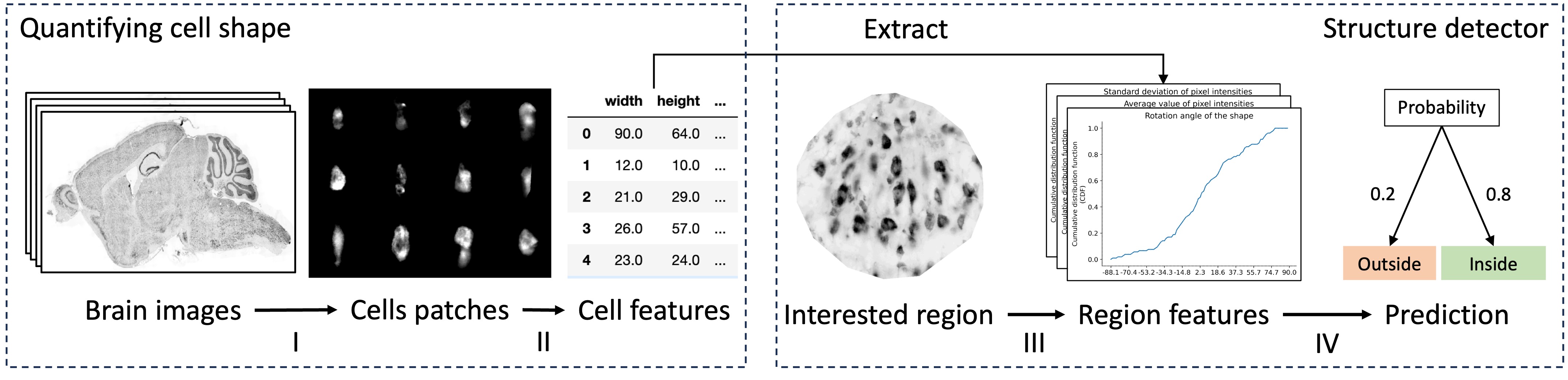
We present a novel method for quantifying the microscopic structure of brain tissue. It is based on the automated recognition of interpretable features obtained by analyzing the shapes of cells. This contrasts with prevailing methods of brain anatomical analysis in two ways. First, contemporary methods use gray-scale values derived from smoothed version of the anatomical images, which dissipated valuable information from the texture of the images. Second, contemporary analysis uses the output of black-box Convolutional Neural Networks, while our system makes decisions based on interpretable features obtained by analyzing the shapes of individual cells. An important benefit of this open-box approach is that the anatomist can understand and correct the decisions made by the computer. Our proposed system can accurately localize and identify existing brain structures. This can be used to align and coregistar brains and will facilitate connectomic studies for reverse engineering of brain circuitry.
4/10/2024
Brainformer: Mimic Human Visual Brain Functions to Machine Vision Models via fMRI
Xuan-Bac Nguyen, Xin Li, Pawan Sinha, Samee U. Khan, Khoa Luu
0
0
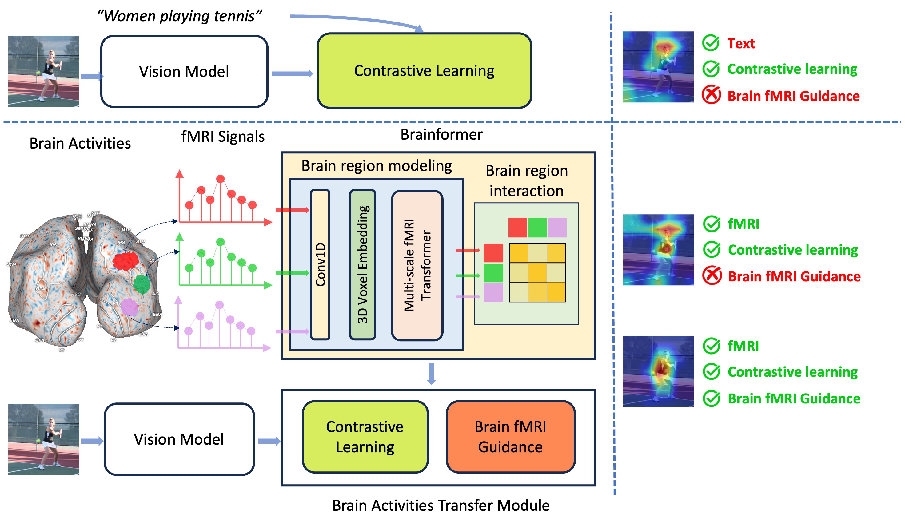
Human perception plays a vital role in forming beliefs and understanding reality. A deeper understanding of brain functionality will lead to the development of novel deep neural networks. In this work, we introduce a novel framework named Brainformer, a straightforward yet effective Transformer-based framework, to analyze Functional Magnetic Resonance Imaging (fMRI) patterns in the human perception system from a machine-learning perspective. Specifically, we present the Multi-scale fMRI Transformer to explore brain activity patterns through fMRI signals. This architecture includes a simple yet efficient module for high-dimensional fMRI signal encoding and incorporates a novel embedding technique called 3D Voxels Embedding. Secondly, drawing inspiration from the functionality of the brain's Region of Interest, we introduce a novel loss function called Brain fMRI Guidance Loss. This loss function mimics brain activity patterns from these regions in the deep neural network using fMRI data. This work introduces a prospective approach to transfer knowledge from human perception to neural networks. Our experiments demonstrate that leveraging fMRI information allows the machine vision model to achieve results comparable to State-of-the-Art methods in various image recognition tasks.
5/30/2024

Automating the Diagnosis of Human Vision Disorders by Cross-modal 3D Generation
Li Zhang, Yuankun Yang, Ziyang Xie, Zhiyuan Yuan, Jianfeng Feng, Xiatian Zhu, Yu-Gang Jiang
0
0
Understanding the hidden mechanisms behind human's visual perception is a fundamental quest in neuroscience, underpins a wide variety of critical applications, e.g. clinical diagnosis. To that end, investigating into the neural responses of human mind activities, such as functional Magnetic Resonance Imaging (fMRI), has been a significant research vehicle. However, analyzing fMRI signals is challenging, costly, daunting, and demanding for professional training. Despite remarkable progress in artificial intelligence (AI) based fMRI analysis, existing solutions are limited and far away from being clinically meaningful. In this context, we leap forward to demonstrate how AI can go beyond the current state of the art by decoding fMRI into visually plausible 3D visuals, enabling automatic clinical analysis of fMRI data, even without healthcare professionals. Innovationally, we reformulate the task of analyzing fMRI data as a conditional 3D scene reconstruction problem. We design a novel cross-modal 3D scene representation learning method, Brain3D, that takes as input the fMRI data of a subject who was presented with a 2D object image, and yields as output the corresponding 3D object visuals. Importantly, we show that in simulated scenarios our AI agent captures the distinct functionalities of each region of human vision system as well as their intricate interplay relationships, aligning remarkably with the established discoveries of neuroscience. Non-expert diagnosis indicate that Brain3D can successfully identify the disordered brain regions, such as V1, V2, V3, V4, and the medial temporal lobe (MTL) within the human visual system. We also present results in cross-modal 3D visual construction setting, showcasing the perception quality of our 3D scene generation.
5/27/2024
BrainFounder: Towards Brain Foundation Models for Neuroimage Analysis
Joseph Cox, Peng Liu, Skylar E. Stolte, Yunchao Yang, Kang Liu, Kyle B. See, Huiwen Ju, Ruogu Fang
0
0
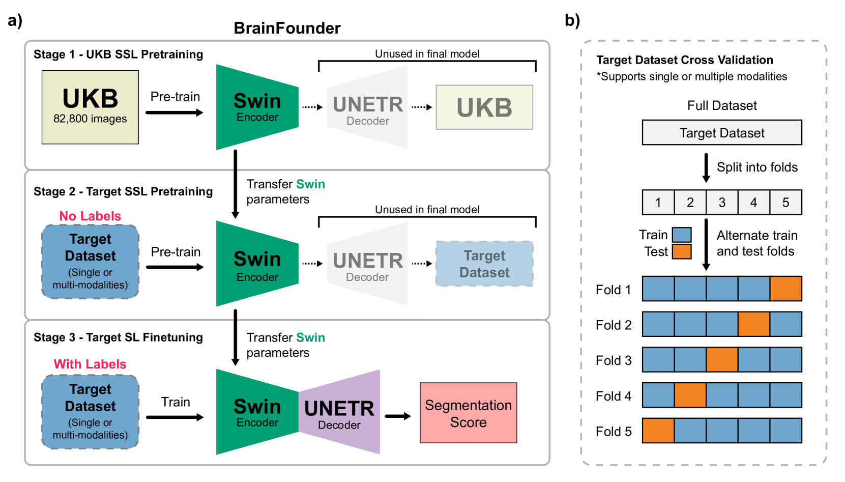
The burgeoning field of brain health research increasingly leverages artificial intelligence (AI) to interpret and analyze neurological data. This study introduces a novel approach towards the creation of medical foundation models by integrating a large-scale multi-modal magnetic resonance imaging (MRI) dataset derived from 41,400 participants in its own. Our method involves a novel two-stage pretraining approach using vision transformers. The first stage is dedicated to encoding anatomical structures in generally healthy brains, identifying key features such as shapes and sizes of different brain regions. The second stage concentrates on spatial information, encompassing aspects like location and the relative positioning of brain structures. We rigorously evaluate our model, BrainFounder, using the Brain Tumor Segmentation (BraTS) challenge and Anatomical Tracings of Lesions After Stroke v2.0 (ATLAS v2.0) datasets. BrainFounder demonstrates a significant performance gain, surpassing the achievements of the previous winning solutions using fully supervised learning. Our findings underscore the impact of scaling up both the complexity of the model and the volume of unlabeled training data derived from generally healthy brains, which enhances the accuracy and predictive capabilities of the model in complex neuroimaging tasks with MRI. The implications of this research provide transformative insights and practical applications in healthcare and make substantial steps towards the creation of foundation models for Medical AI. Our pretrained models and training code can be found at https://github.com/lab-smile/GatorBrain.
6/18/2024