Detection of Peri-Pancreatic Edema using Deep Learning and Radiomics Techniques

0

Sign in to get full access
Overview
- This paper explores the use of deep learning and radiomics techniques for the detection of peri-pancreatic edema, a condition characterized by swelling around the pancreas.
- The researchers developed a deep learning model and a radiomics-based model to analyze computed tomography (CT) images of the abdomen and identify the presence of peri-pancreatic edema.
- The performance of these models was evaluated and compared to assess their effectiveness in detecting this medical condition.
Plain English Explanation
Peri-pancreatic edema is a medical condition where the area around the pancreas becomes swollen. This can be an indication of various underlying health issues, such as pancreatitis or other pancreatic disorders. To detect this condition, the researchers in this study used two different approaches: deep learning and radiomics.
Deep learning is a type of artificial intelligence that can analyze medical images, like CT scans, and identify patterns that may indicate the presence of a disease. The researchers developed a deep learning model specifically trained to detect peri-pancreatic edema in CT images.
Radiomics, on the other hand, involves extracting and analyzing various numerical features, or "radiomics features," from medical images. These features can provide insights into the underlying tissue characteristics and help identify abnormalities. The researchers also created a radiomics-based model to detect peri-pancreatic edema.
By comparing the performance of these two models, the researchers aimed to determine which approach, deep learning or radiomics, is more effective in accurately identifying peri-pancreatic edema from CT scans. This information can help healthcare professionals make more informed decisions when diagnosing and treating patients with this condition.
Technical Explanation
The researchers in this study developed a deep learning model and a radiomics-based model to detect peri-pancreatic edema from computed tomography (CT) images.
For the deep learning approach, the team used a convolutional neural network (CNN) architecture, which is well-suited for analyzing and extracting features from medical images. The CNN model was trained on a dataset of CT scans, with the ground truth labels indicating the presence or absence of peri-pancreatic edema.
In the radiomics-based approach, the researchers extracted a comprehensive set of radiomics features from the CT images, including shape, intensity, and texture-based features. These features were then used to train a machine learning classifier, such as a random forest or a support vector machine, to detect peri-pancreatic edema.
The performance of the deep learning and radiomics-based models was evaluated using various metrics, including accuracy, sensitivity, specificity, and area under the receiver operating characteristic (ROC) curve. The researchers compared the results of the two models to determine the most effective approach for detecting peri-pancreatic edema.
Critical Analysis
The researchers in this study have made a valuable contribution to the field of medical image analysis by exploring the use of deep learning and radiomics techniques for the detection of peri-pancreatic edema. However, it is important to consider some potential limitations and areas for further research.
One potential limitation of the study is the size and diversity of the dataset used for model training and evaluation. The researchers did not provide detailed information about the dataset, such as the number of patients, the distribution of positive and negative cases, or the demographic characteristics of the study population. A larger and more diverse dataset would help to ensure the robustness and generalizability of the models.
Additionally, the study did not compare the performance of the deep learning and radiomics-based models to the current standard of care for peri-pancreatic edema detection, such as manual assessment by radiologists. Evaluating the models against the current clinical practice would provide valuable insights into the potential benefits and limitations of the proposed automated approaches.
Further research could also explore the integration of the deep learning and radiomics-based models, as the combination of these two techniques may potentially lead to improved detection accuracy and more robust diagnostic tools for peri-pancreatic edema.
Conclusion
This study demonstrates the potential of deep learning and radiomics techniques for the detection of peri-pancreatic edema, a condition that can be indicative of various underlying health issues. The researchers developed and evaluated two different models, one based on deep learning and the other on radiomics, to analyze CT images and identify the presence of peri-pancreatic edema.
The findings of this study suggest that both deep learning and radiomics-based approaches can be effective in detecting peri-pancreatic edema, but further research is needed to fully understand the strengths and limitations of each approach. The integration of these techniques or their comparison to current clinical practice may lead to the development of more accurate and reliable diagnostic tools for this medical condition, ultimately improving patient outcomes.
This summary was produced with help from an AI and may contain inaccuracies - check out the links to read the original source documents!
Related Papers
0
Detection of Peri-Pancreatic Edema using Deep Learning and Radiomics Techniques
Ziliang Hong, Debesh Jha, Koushik Biswas, Zheyuan Zhang, Yury Velichko, Cemal Yazici, Temel Tirkes, Amir Borhani, Baris Turkbey, Alpay Medetalibeyoglu, Gorkem Durak, Ulas Bagci
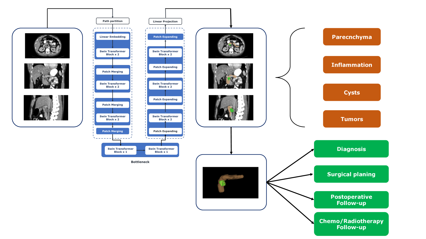
Identifying peri-pancreatic edema is a pivotal indicator for identifying disease progression and prognosis, emphasizing the critical need for accurate detection and assessment in pancreatitis diagnosis and management. This study textit{introduces a novel CT dataset sourced from 255 patients with pancreatic diseases, featuring annotated pancreas segmentation masks and corresponding diagnostic labels for peri-pancreatic edema condition}. With the novel dataset, we first evaluate the efficacy of the textit{LinTransUNet} model, a linear Transformer based segmentation algorithm, to segment the pancreas accurately from CT imaging data. Then, we use segmented pancreas regions with two distinctive machine learning classifiers to identify existence of peri-pancreatic edema: deep learning-based models and a radiomics-based eXtreme Gradient Boosting (XGBoost). The LinTransUNet achieved promising results, with a dice coefficient of 80.85%, and mIoU of 68.73%. Among the nine benchmarked classification models for peri-pancreatic edema detection, textit{Swin-Tiny} transformer model demonstrated the highest recall of $98.85 pm 0.42$ and precision of $98.38pm 0.17$. Comparatively, the radiomics-based XGBoost model achieved an accuracy of $79.61pm4.04$ and recall of $91.05pm3.28$, showcasing its potential as a supplementary diagnostic tool given its rapid processing speed and reduced training time. Our code is available url{https://github.com/NUBagciLab/Peri-Pancreatic-Edema-Detection}.
Read more4/29/2024
🤿
0
Large-Scale Multi-Center CT and MRI Segmentation of Pancreas with Deep Learning
Zheyuan Zhang, Elif Keles, Gorkem Durak, Yavuz Taktak, Onkar Susladkar, Vandan Gorade, Debesh Jha, Asli C. Ormeci, Alpay Medetalibeyoglu, Lanhong Yao, Bin Wang, Ilkin Sevgi Isler, Linkai Peng, Hongyi Pan, Camila Lopes Vendrami, Amir Bourhani, Yury Velichko, Boqing Gong, Concetto Spampinato, Ayis Pyrros, Pallavi Tiwari, Derk C. F. Klatte, Megan Engels, Sanne Hoogenboom, Candice W. Bolan, Emil Agarunov, Nassier Harfouch, Chenchan Huang, Marco J. Bruno, Ivo Schoots, Rajesh N. Keswani, Frank H. Miller, Tamas Gonda, Cemal Yazici, Temel Tirkes, Baris Turkbey, Michael B. Wallace, Ulas Bagci
Automated volumetric segmentation of the pancreas on cross-sectional imaging is needed for diagnosis and follow-up of pancreatic diseases. While CT-based pancreatic segmentation is more established, MRI-based segmentation methods are understudied, largely due to a lack of publicly available datasets, benchmarking research efforts, and domain-specific deep learning methods. In this retrospective study, we collected a large dataset (767 scans from 499 participants) of T1-weighted (T1W) and T2-weighted (T2W) abdominal MRI series from five centers between March 2004 and November 2022. We also collected CT scans of 1,350 patients from publicly available sources for benchmarking purposes. We developed a new pancreas segmentation method, called PanSegNet, combining the strengths of nnUNet and a Transformer network with a new linear attention module enabling volumetric computation. We tested PanSegNet's accuracy in cross-modality (a total of 2,117 scans) and cross-center settings with Dice and Hausdorff distance (HD95) evaluation metrics. We used Cohen's kappa statistics for intra and inter-rater agreement evaluation and paired t-tests for volume and Dice comparisons, respectively. For segmentation accuracy, we achieved Dice coefficients of 88.3% (std: 7.2%, at case level) with CT, 85.0% (std: 7.9%) with T1W MRI, and 86.3% (std: 6.4%) with T2W MRI. There was a high correlation for pancreas volume prediction with R^2 of 0.91, 0.84, and 0.85 for CT, T1W, and T2W, respectively. We found moderate inter-observer (0.624 and 0.638 for T1W and T2W MRI, respectively) and high intra-observer agreement scores. All MRI data is made available at https://osf.io/kysnj/. Our source code is available at https://github.com/NUBagciLab/PaNSegNet.
Read more5/28/2024
0
Deep Learning for Pancreas Segmentation: a Systematic Review
Andrea Moglia, Matteo Cavicchioli, Luca Mainardi, Pietro Cerveri
Pancreas segmentation has been traditionally challenging due to its small size in computed tomography abdominal volumes, high variability of shape and positions among patients, and blurred boundaries due to low contrast between the pancreas and surrounding organs. Many deep learning models for pancreas segmentation have been proposed in the past few years. We present a thorough systematic review based on the Preferred Reporting Items for Systematic Reviews and Meta-analyses (PRISMA) statement. The literature search was conducted on PubMed, Web of Science, Scopus, and IEEE Xplore on original studies published in peer-reviewed journals from 2013 to 2023. Overall, 130 studies were retrieved. We initially provided an overview of the technical background of the most common network architectures and publicly available datasets. Then, the analysis of the studies combining visual presentation in tabular form and text description was reported. The tables grouped the studies specifying the application, dataset size, design (model architecture, learning strategy, and loss function), results, and main contributions. We first analyzed the studies focusing on parenchyma segmentation using coarse-to-fine approaches, multi-organ segmentation, semi-supervised learning, and unsupervised learning, followed by those studies on generalization to other datasets and those concerning the design of new loss functions. Then, we analyzed the studies on segmentation of tumors, cysts, and inflammation reporting multi-stage methods, semi-supervised learning, generalization to other datasets, and design of new loss functions. Finally, we provided a critical discussion on the subject based on the published evidence underlining current issues that need to be addressed before clinical translation.
Read more7/24/2024

0
Pancreatic Tumor Segmentation as Anomaly Detection in CT Images Using Denoising Diffusion Models
Reza Babaei, Samuel Cheng, Theresa Thai, Shangqing Zhao
Despite the advances in medicine, cancer has remained a formidable challenge. Particularly in the case of pancreatic tumors, characterized by their diversity and late diagnosis, early detection poses a significant challenge crucial for effective treatment. The advancement of deep learning techniques, particularly supervised algorithms, has significantly propelled pancreatic tumor detection in the medical field. However, supervised deep learning approaches necessitate extensive labeled medical images for training, yet acquiring such annotations is both limited and costly. Conversely, weakly supervised anomaly detection methods, requiring only image-level annotations, have garnered interest. Existing methodologies predominantly hinge on generative adversarial networks (GANs) or autoencoder models, which can pose complexity in training and, these models may face difficulties in accurately preserving fine image details. This research presents a novel approach to pancreatic tumor detection, employing weak supervision anomaly detection through denoising diffusion algorithms. By incorporating a deterministic iterative process of adding and removing noise along with classifier guidance, the method enables seamless translation of images between diseased and healthy subjects, resulting in detailed anomaly maps without requiring complex training protocols and segmentation masks. This study explores denoising diffusion models as a recent advancement over traditional generative models like GANs, contributing to the field of pancreatic tumor detection. Recognizing the low survival rates of pancreatic cancer, this study emphasizes the need for continued research to leverage diffusion models' efficiency in medical segmentation tasks.
Read more6/6/2024