Large-Scale Multi-Center CT and MRI Segmentation of Pancreas with Deep Learning

0
🤿
Sign in to get full access
Overview
- This study focused on developing a new method for automated volumetric segmentation of the pancreas on MRI scans.
- The researchers collected a large dataset of abdominal MRI and CT scans from multiple centers to train and evaluate their model.
- They developed a new segmentation model called PanSegNet that combines the strengths of nnUNet and a Transformer network with a novel attention module.
- The model was tested on both CT and MRI data, achieving strong performance on segmentation accuracy and volume prediction.
Plain English Explanation
The pancreas is an important organ in the body that plays a key role in digestion and blood sugar regulation. Being able to accurately measure the size and shape of the pancreas on medical scans is important for diagnosing and monitoring pancreatic diseases. While CT scans have been more commonly used for this purpose, the researchers in this study focused on developing methods for pancreas segmentation using MRI data.
The researchers collected a large dataset of MRI and CT scans from multiple hospitals and clinics, which is important because it allows the model to be trained and tested on a diverse set of data. They then developed a new deep learning model called PanSegNet that is able to automatically identify and outline the pancreas in these scans.
PanSegNet combines two powerful machine learning techniques - a convolutional neural network called nnUNet and a Transformer network - in a novel way that allows it to efficiently process 3D volumetric data like MRI scans. The researchers found that PanSegNet was able to accurately segment the pancreas on both CT and MRI scans, achieving Dice scores (a common metric for segmentation accuracy) around 85-88%.
The ability to accurately measure the pancreas volume is also important, and the researchers found a strong correlation between the model's volume predictions and the true pancreas volumes. Additionally, they evaluated the consistency of the model's results by having human experts review the segmentations, finding high levels of agreement.
Overall, this research represents an important step forward in developing automated tools to assist clinicians in diagnosing and monitoring pancreatic diseases using medical imaging data, especially MRI scans which can provide detailed information about the internal structure of the pancreas.
Technical Explanation
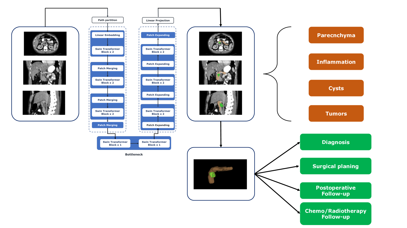
The researchers in this study developed a new deep learning model called PanSegNet for automated volumetric segmentation of the pancreas on cross-sectional imaging data, with a focus on MRI. While CT-based pancreas segmentation methods are more established, MRI-based approaches have been understudied due to a lack of publicly available datasets and specialized deep learning architectures.
To address this gap, the researchers collected a large dataset of 767 abdominal MRI scans (T1-weighted and T2-weighted) from 499 participants across 5 different clinical centers, as well as 1,350 CT scans from public sources for benchmarking. They then developed PanSegNet, which combines the strengths of the nnUNet architecture and a Transformer network with a novel linear attention module to enable efficient volumetric processing.
The model was evaluated on both the CT and MRI data using Dice scores and Hausdorff distance (HD95) as accuracy metrics. On the CT scans, PanSegNet achieved a Dice score of 88.3%, and on the T1-weighted and T2-weighted MRI scans, it achieved Dice scores of 85.0% and 86.3% respectively. The model also showed a strong correlation between its predicted pancreas volumes and the ground truth, with R^2 values of 0.91, 0.84, and 0.85 for CT, T1-weighted, and T2-weighted MRI, respectively.
To further assess the reliability of the segmentations, the researchers conducted intra- and inter-rater agreement studies with human experts, finding moderate to high levels of agreement. All of the MRI data collected for this study has been made publicly available at https://osf.io/kysnj/, and the source code for PanSegNet is available at https://github.com/NUBagciLab/PaNSegNet.
Critical Analysis
The researchers in this study have made a valuable contribution to the field of pancreas segmentation on medical imaging by developing a new deep learning model that achieves strong performance on both CT and MRI data. The large and diverse dataset they collected, combined with the novel architectural elements of PanSegNet, represent important advancements over prior work in this area.
That said, there are a few potential limitations and areas for further research that could be considered. For example, the model's performance on certain types of pancreatic pathologies or challenging imaging conditions is not thoroughly explored. Additionally, while the intra- and inter-rater agreement studies provide insights into the reliability of the segmentations, it would be helpful to understand how the model's performance compares to human experts in a more direct way.
Another area that could benefit from further investigation is the model's generalizability to other imaging modalities or scanners. While the cross-center evaluation suggests the model can handle some degree of variability, a more comprehensive assessment of its robustness would strengthen the conclusions.
Overall, this study represents a significant advancement in the field of automated pancreas segmentation, with the potential to support clinicians in the diagnosis and monitoring of pancreatic diseases. The open-sourcing of the dataset and code will also enable other researchers to build upon this work and continue pushing the boundaries of what is possible with deep learning-based medical image analysis.
Conclusion
This study presents a new deep learning model, called PanSegNet, for automated volumetric segmentation of the pancreas on cross-sectional imaging data, with a focus on MRI scans. The researchers collected a large, multi-center dataset of abdominal MRI and CT scans, which they used to train and evaluate their model.
PanSegNet combines the strengths of nnUNet and a Transformer network with a novel attention module, enabling efficient processing of 3D volumetric data. The model demonstrated strong performance on both CT and MRI scans, achieving Dice scores around 85-88% and a high correlation between predicted and true pancreas volumes.
The availability of the dataset and open-source code will facilitate further research and development in this important area of medical image analysis. While the study has some limitations, it represents a significant step forward in the quest to create robust, automated tools for diagnosing and monitoring pancreatic diseases using advanced imaging techniques.
This summary was produced with help from an AI and may contain inaccuracies - check out the links to read the original source documents!
Related Papers
🤿
0
Large-Scale Multi-Center CT and MRI Segmentation of Pancreas with Deep Learning
Zheyuan Zhang, Elif Keles, Gorkem Durak, Yavuz Taktak, Onkar Susladkar, Vandan Gorade, Debesh Jha, Asli C. Ormeci, Alpay Medetalibeyoglu, Lanhong Yao, Bin Wang, Ilkin Sevgi Isler, Linkai Peng, Hongyi Pan, Camila Lopes Vendrami, Amir Bourhani, Yury Velichko, Boqing Gong, Concetto Spampinato, Ayis Pyrros, Pallavi Tiwari, Derk C. F. Klatte, Megan Engels, Sanne Hoogenboom, Candice W. Bolan, Emil Agarunov, Nassier Harfouch, Chenchan Huang, Marco J. Bruno, Ivo Schoots, Rajesh N. Keswani, Frank H. Miller, Tamas Gonda, Cemal Yazici, Temel Tirkes, Baris Turkbey, Michael B. Wallace, Ulas Bagci
Automated volumetric segmentation of the pancreas on cross-sectional imaging is needed for diagnosis and follow-up of pancreatic diseases. While CT-based pancreatic segmentation is more established, MRI-based segmentation methods are understudied, largely due to a lack of publicly available datasets, benchmarking research efforts, and domain-specific deep learning methods. In this retrospective study, we collected a large dataset (767 scans from 499 participants) of T1-weighted (T1W) and T2-weighted (T2W) abdominal MRI series from five centers between March 2004 and November 2022. We also collected CT scans of 1,350 patients from publicly available sources for benchmarking purposes. We developed a new pancreas segmentation method, called PanSegNet, combining the strengths of nnUNet and a Transformer network with a new linear attention module enabling volumetric computation. We tested PanSegNet's accuracy in cross-modality (a total of 2,117 scans) and cross-center settings with Dice and Hausdorff distance (HD95) evaluation metrics. We used Cohen's kappa statistics for intra and inter-rater agreement evaluation and paired t-tests for volume and Dice comparisons, respectively. For segmentation accuracy, we achieved Dice coefficients of 88.3% (std: 7.2%, at case level) with CT, 85.0% (std: 7.9%) with T1W MRI, and 86.3% (std: 6.4%) with T2W MRI. There was a high correlation for pancreas volume prediction with R^2 of 0.91, 0.84, and 0.85 for CT, T1W, and T2W, respectively. We found moderate inter-observer (0.624 and 0.638 for T1W and T2W MRI, respectively) and high intra-observer agreement scores. All MRI data is made available at https://osf.io/kysnj/. Our source code is available at https://github.com/NUBagciLab/PaNSegNet.
Read more5/28/2024
0
Deep Learning for Pancreas Segmentation: a Systematic Review
Andrea Moglia, Matteo Cavicchioli, Luca Mainardi, Pietro Cerveri
Pancreas segmentation has been traditionally challenging due to its small size in computed tomography abdominal volumes, high variability of shape and positions among patients, and blurred boundaries due to low contrast between the pancreas and surrounding organs. Many deep learning models for pancreas segmentation have been proposed in the past few years. We present a thorough systematic review based on the Preferred Reporting Items for Systematic Reviews and Meta-analyses (PRISMA) statement. The literature search was conducted on PubMed, Web of Science, Scopus, and IEEE Xplore on original studies published in peer-reviewed journals from 2013 to 2023. Overall, 130 studies were retrieved. We initially provided an overview of the technical background of the most common network architectures and publicly available datasets. Then, the analysis of the studies combining visual presentation in tabular form and text description was reported. The tables grouped the studies specifying the application, dataset size, design (model architecture, learning strategy, and loss function), results, and main contributions. We first analyzed the studies focusing on parenchyma segmentation using coarse-to-fine approaches, multi-organ segmentation, semi-supervised learning, and unsupervised learning, followed by those studies on generalization to other datasets and those concerning the design of new loss functions. Then, we analyzed the studies on segmentation of tumors, cysts, and inflammation reporting multi-stage methods, semi-supervised learning, generalization to other datasets, and design of new loss functions. Finally, we provided a critical discussion on the subject based on the published evidence underlining current issues that need to be addressed before clinical translation.
Read more7/24/2024

0
Detection of Peri-Pancreatic Edema using Deep Learning and Radiomics Techniques
Ziliang Hong, Debesh Jha, Koushik Biswas, Zheyuan Zhang, Yury Velichko, Cemal Yazici, Temel Tirkes, Amir Borhani, Baris Turkbey, Alpay Medetalibeyoglu, Gorkem Durak, Ulas Bagci
Identifying peri-pancreatic edema is a pivotal indicator for identifying disease progression and prognosis, emphasizing the critical need for accurate detection and assessment in pancreatitis diagnosis and management. This study textit{introduces a novel CT dataset sourced from 255 patients with pancreatic diseases, featuring annotated pancreas segmentation masks and corresponding diagnostic labels for peri-pancreatic edema condition}. With the novel dataset, we first evaluate the efficacy of the textit{LinTransUNet} model, a linear Transformer based segmentation algorithm, to segment the pancreas accurately from CT imaging data. Then, we use segmented pancreas regions with two distinctive machine learning classifiers to identify existence of peri-pancreatic edema: deep learning-based models and a radiomics-based eXtreme Gradient Boosting (XGBoost). The LinTransUNet achieved promising results, with a dice coefficient of 80.85%, and mIoU of 68.73%. Among the nine benchmarked classification models for peri-pancreatic edema detection, textit{Swin-Tiny} transformer model demonstrated the highest recall of $98.85 pm 0.42$ and precision of $98.38pm 0.17$. Comparatively, the radiomics-based XGBoost model achieved an accuracy of $79.61pm4.04$ and recall of $91.05pm3.28$, showcasing its potential as a supplementary diagnostic tool given its rapid processing speed and reduced training time. Our code is available url{https://github.com/NUBagciLab/Peri-Pancreatic-Edema-Detection}.
Read more4/29/2024

0
Automatic Organ and Pan-cancer Segmentation in Abdomen CT: the FLARE 2023 Challenge
Jun Ma, Yao Zhang, Song Gu, Cheng Ge, Ershuai Wang, Qin Zhou, Ziyan Huang, Pengju Lyu, Jian He, Bo Wang
Organ and cancer segmentation in abdomen Computed Tomography (CT) scans is the prerequisite for precise cancer diagnosis and treatment. Most existing benchmarks and algorithms are tailored to specific cancer types, limiting their ability to provide comprehensive cancer analysis. This work presents the first international competition on abdominal organ and pan-cancer segmentation by providing a large-scale and diverse dataset, including 4650 CT scans with various cancer types from over 40 medical centers. The winning team established a new state-of-the-art with a deep learning-based cascaded framework, achieving average Dice Similarity Coefficient scores of 92.3% for organs and 64.9% for lesions on the hidden multi-national testing set. The dataset and code of top teams are publicly available, offering a benchmark platform to drive further innovations https://codalab.lisn.upsaclay.fr/competitions/12239.
Read more8/23/2024