A Diffusion Model for Simulation Ready Coronary Anatomy with Morpho-skeletal Control

0

Sign in to get full access
Overview
- This paper presents a diffusion model for generating realistic and anatomically accurate 3D models of coronary arteries.
- The model allows for fine-grained control over the morphological and skeletal features of the generated coronary anatomy.
- The generated models can be used for medical simulations, virtual interventions, and the creation of digital twins.
Plain English Explanation
The research paper describes a new technique for creating detailed 3D models of the coronary arteries - the blood vessels that supply the heart with oxygen-rich blood. These models can be used for a variety of medical applications, such as simulating surgical procedures or developing personalized treatments.
The key innovation is the use of a diffusion model, which is a type of machine learning algorithm that can generate new data that resembles a training dataset. In this case, the model is trained on a database of real coronary artery scans, allowing it to produce synthetic but realistic 3D models.
What makes this approach unique is that the researchers added the ability to fine-tune the generated models by specifying certain desired morphological and skeletal features. For example, you could tell the model to generate a model with a longer or more twisted coronary artery. This gives medical researchers and clinicians more control over the virtual coronary anatomy for their specific needs.
Technical Explanation
The core of the system is a conditional diffusion model that learns to generate 3D coronary artery geometries from a training dataset of real scans. The model takes in a latent code that encodes high-level morphological and skeletal features of the target anatomy, and then uses a diffusion process to iteratively refine a random noise input into a final 3D model.
The key innovations include:
- A novel diffusion guidance mechanism that allows the model to respect the specified morpho-skeletal constraints during generation.
- A progressive growing strategy that gradually increases the resolution of the generated models.
- A post-processing pipeline that ensures the final models are simulation-ready with consistent topology and no self-intersections.
Extensive experiments demonstrate that the proposed method can generate high-fidelity coronary artery models with user-specified morphological and skeletal characteristics. The generated models are shown to be useful for applications like virtual surgical planning and digital twin creation.
Critical Analysis
The paper presents a compelling approach for generating controllable 3D models of coronary arteries. The use of a diffusion model enables the generation of realistic anatomical structures, while the ability to specify morphological and skeletal features provides a valuable level of control for medical applications.
That said, the paper does not address some potential limitations and areas for further research:
- The training dataset size and diversity are not discussed, which could impact the generalizability of the generated models.
- The computational efficiency and generation speed of the system are not evaluated, which would be an important consideration for real-time applications.
- The validation of the generated models' suitability for downstream tasks like surgical simulation is limited, and more thorough testing would be needed.
Overall, the proposed diffusion-based approach is a promising step forward in the field of anatomically controllable medical image generation, but further research and development would be needed to fully realize its potential.
Conclusion
This research presents a novel diffusion model for generating high-fidelity 3D models of coronary arteries with user-specified morphological and skeletal characteristics. The ability to fine-tune the generated models opens up new possibilities for medical simulations, virtual interventions, and the creation of personalized digital twins. While the approach shows promise, further work is needed to address potential limitations and expand its real-world applications.
This summary was produced with help from an AI and may contain inaccuracies - check out the links to read the original source documents!
Related Papers
0
A Diffusion Model for Simulation Ready Coronary Anatomy with Morpho-skeletal Control
Karim Kadry, Shreya Gupta, Jonas Sogbadji, Michiel Schaap, Kersten Petersen, Takuya Mizukami, Carlos Collet, Farhad R. Nezami, Elazer R. Edelman
Virtual interventions enable the physics-based simulation of device deployment within coronary arteries. This framework allows for counterfactual reasoning by deploying the same device in different arterial anatomies. However, current methods to create such counterfactual arteries face a trade-off between controllability and realism. In this study, we investigate how Latent Diffusion Models (LDMs) can custom synthesize coronary anatomy for virtual intervention studies based on mid-level anatomic constraints such as topological validity, local morphological shape, and global skeletal structure. We also extend diffusion model guidance strategies to the context of morpho-skeletal conditioning and propose a novel guidance method for continuous attributes that adaptively updates the negative guiding condition throughout sampling. Our framework enables the generation and editing of coronary anatomy in a controllable manner, allowing device designers to derive mechanistic insights regarding anatomic variation and simulated device deployment.
Read more7/24/2024
0
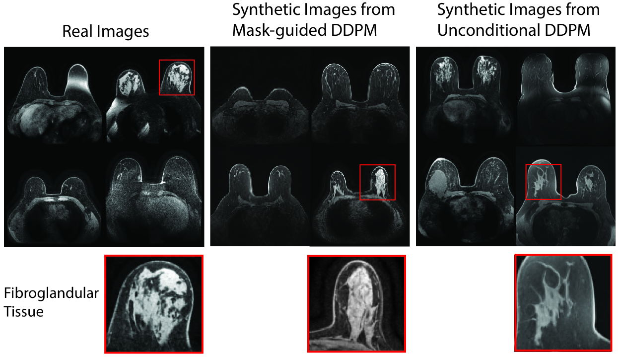
Anatomically-Controllable Medical Image Generation with Segmentation-Guided Diffusion Models
Nicholas Konz, Yuwen Chen, Haoyu Dong, Maciej A. Mazurowski
Diffusion models have enabled remarkably high-quality medical image generation, yet it is challenging to enforce anatomical constraints in generated images. To this end, we propose a diffusion model-based method that supports anatomically-controllable medical image generation, by following a multi-class anatomical segmentation mask at each sampling step. We additionally introduce a random mask ablation training algorithm to enable conditioning on a selected combination of anatomical constraints while allowing flexibility in other anatomical areas. We compare our method (SegGuidedDiff) to existing methods on breast MRI and abdominal/neck-to-pelvis CT datasets with a wide range of anatomical objects. Results show that our method reaches a new state-of-the-art in the faithfulness of generated images to input anatomical masks on both datasets, and is on par for general anatomical realism. Finally, our model also enjoys the extra benefit of being able to adjust the anatomical similarity of generated images to real images of choice through interpolation in its latent space. SegGuidedDiff has many applications, including cross-modality translation, and the generation of paired or counterfactual data. Our code is available at https://github.com/mazurowski-lab/segmentation-guided-diffusion.
Read more6/21/2024
0
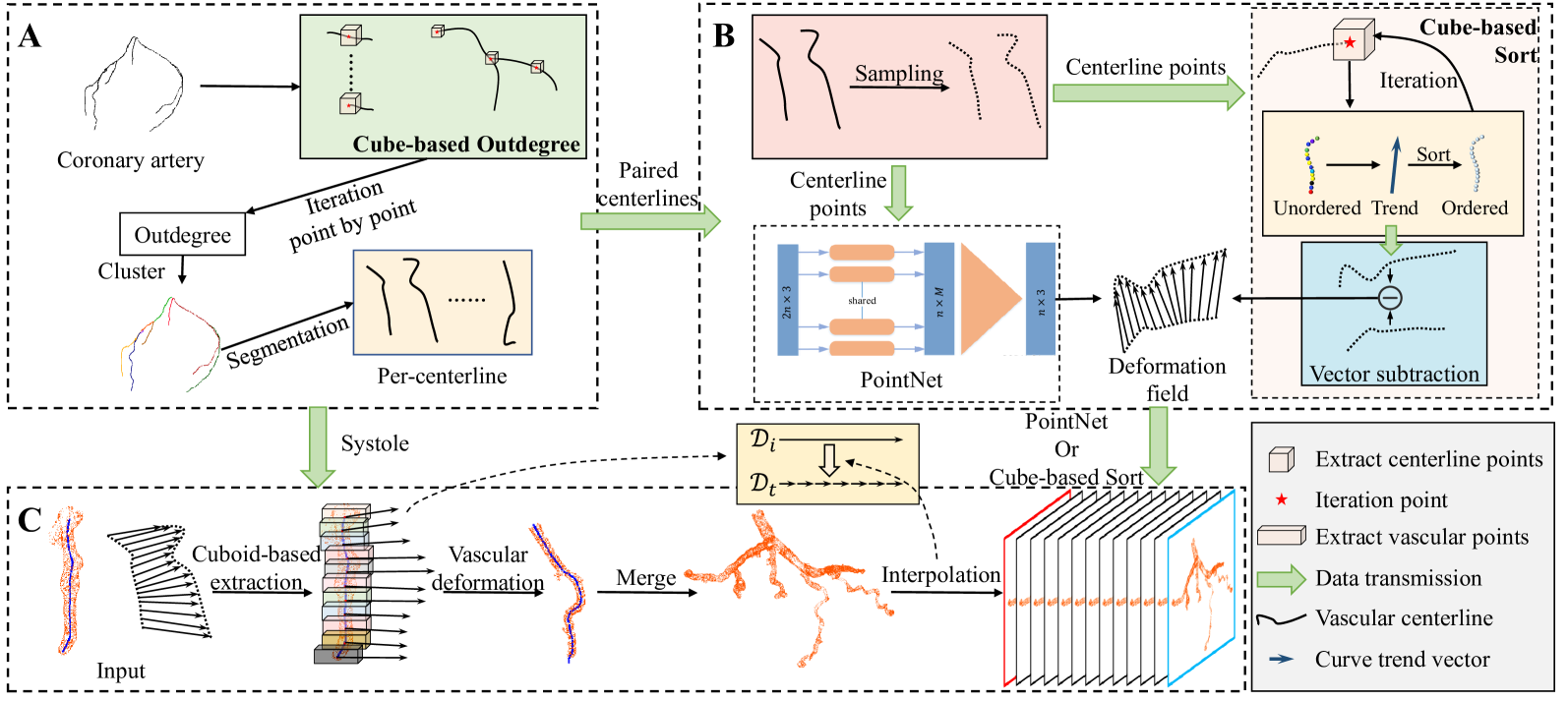
4D-CAT: Synthesis of 4D Coronary Artery Trees from Systole and Diastole
Daosong Hu, Ruomeng Wang, Liang Zhao, Mingyue Cui, Song Ding, Kai Huang
The three-dimensional vascular model reconstructed from CT images is widely used in medical diagnosis. At different phases, the beating of the heart can cause deformation of vessels, resulting in different vascular imaging states and false positive diagnostic results. The 4D model can simulate a complete cardiac cycle. Due to the dose limitation of contrast agent injection in patients, it is valuable to synthesize a 4D coronary artery trees through finite phases imaging. In this paper, we propose a method for generating a 4D coronary artery trees, which maps the systole to the diastole through deformation field prediction, interpolates on the timeline, and the motion trajectory of points are obtained. Specifically, the centerline is used to represent vessels and to infer deformation fields using cube-based sorting and neural networks. Adjacent vessel points are aggregated and interpolated based on the deformation field of the centerline point to obtain displacement vectors of different phases. Finally, the proposed method is validated through experiments to achieve the registration of non-rigid vascular points and the generation of 4D coronary trees.
Read more9/4/2024
📊
0
Post-processing of coronary and myocardial spatial data
Jay Aodh Mackenzie, Megan Jeanne Miller, Nicholas Hill, Mette Olufsen
Numerical simulations of real-world phenomenon are implemented with at least two parts: the computational scheme and the computational domain. In the context of hemodynamics, the computational domain of a simulation represents the blood vessel network through which blood flows. Such blood vessel networks can contain millions of individual vessels that are joined together to form a in series and parallel to form the network. It is computationally unfeasible to explicitly simulate blood flow in all blood vessels. Here, from imaged data of a single porcine left coronary arterial tree, we develop a data-pipeline to obtain computational domains for hemodynmaic simulations from a graph representing the coronary vascular tree. Further, we develop a method to ascertain which subregions of the left ventricle are most likely to be perfused via a given artery using a comparison with the American Heart Association division of the left ventricle as a sense check.
Read more4/16/2024