A Disease Labeler for Chinese Chest X-Ray Report Generation

0
🛸
Sign in to get full access
Overview
- The scarcity of Chinese chest X-ray report datasets has hindered the development of medical image analysis technology for generating Chinese chest X-ray reports.
- Constructing a Chinese chest X-ray report dataset is limited by the time-consuming and costly process of accurate expert disease annotation.
- Evaluating the similarity between generated and ground-truth reports using a single natural language generation metric does not guarantee the clinical accuracy and effectiveness of the generated reports.
- To address these issues, the study proposes a disease labeler tailored for the generation of Chinese chest X-ray reports.
Plain English Explanation
Analyzing medical images, such as chest X-rays, is crucial for diagnosing and treating various health conditions. However, the development of technology to automatically generate detailed reports on Chinese chest X-ray images has been hindered by the lack of available datasets.
One of the main challenges is the difficulty in creating a comprehensive dataset of Chinese chest X-ray reports. Accurately annotating the diseases and abnormalities shown in the X-rays requires significant time and effort from medical experts, making the process costly and time-consuming.
Additionally, the common practice of evaluating the similarity between generated reports and ground-truth (expert-written) reports using a single natural language generation metric may not be sufficient. This approach does not necessarily ensure the clinical accuracy and effectiveness of the generated reports in aiding medical professionals.
To address these issues, the researchers in this study have developed a specialized "disease labeler" - a machine learning model that can accurately identify and classify the various medical conditions visible in Chinese chest X-ray images. This labeler uses a unique dual-BERT architecture to handle the diagnostic reports and clinical information separately, and a hierarchical label learning algorithm to enhance the text classification performance.
By using this disease labeler, the researchers were able to establish a new dataset of 51,262 Chinese chest X-ray report samples. This dataset can be used to train and evaluate models for automatically generating Chinese chest X-ray reports, which can ultimately support medical professionals in their diagnosis and treatment of patients.
Technical Explanation
The study proposes a disease labeler tailored for the generation of Chinese chest X-ray reports. This labeler leverages a dual BERT (Bidirectional Encoder Representations from Transformers) architecture to handle diagnostic reports and clinical information separately. It also constructs a hierarchical label learning algorithm based on the affiliation between diseases and body parts to enhance text classification performance.
The dual BERT architecture consists of two BERT models: one for processing the diagnostic reports and another for handling the clinical information. This approach allows the model to capture the nuanced relationships between the textual data and the underlying medical conditions.
The hierarchical label learning algorithm is designed to improve the model's ability to accurately classify the various diseases and abnormalities present in the chest X-ray images. By incorporating the affiliation between diseases and body parts, the algorithm can leverage the inherent structure and relationships within the medical domain, leading to more precise disease labeling.
Using the proposed disease labeler, the researchers were able to establish a Chinese chest X-ray report dataset comprising 51,262 report samples. This dataset can be used to train and evaluate models for automatically generating Chinese chest X-ray reports, addressing the initial scarcity of such datasets.
Experiments and analyses were conducted on a subset of expert-annotated Chinese chest X-ray reports, validating the effectiveness of the proposed disease labeler. The results demonstrate the model's ability to accurately identify and classify the medical conditions depicted in the chest X-ray images, laying the foundation for the development of more robust and clinically-relevant medical image analysis systems.
Critical Analysis
The researchers have addressed an important challenge in the field of medical image analysis - the lack of Chinese chest X-ray report datasets. By developing a specialized disease labeler, they have been able to create a more comprehensive dataset, which can be used to train and evaluate models for generating Chinese chest X-ray reports.
However, the study does not provide a detailed discussion of the potential limitations or caveats of the proposed disease labeler. For example, it would be helpful to understand the model's performance on rare or uncommon medical conditions, as well as its ability to handle the nuances and uncertainties inherent in medical diagnoses.
Additionally, the researchers mention that the clinical accuracy and effectiveness of the generated reports rely on an accurate disease labeler (classifier). It would be valuable to see a more thorough evaluation of the model's clinical relevance and its impact on medical decision-making, rather than just focusing on natural language generation metrics.
Further research could explore the integration of the disease labeler with advanced natural language generation models to produce high-quality, clinically-relevant Chinese chest X-ray reports. Investigating the transferability of the proposed approach to other medical imaging modalities or languages could also expand the impact of this work.
Overall, the study presents a promising step towards addressing the data scarcity challenge in Chinese chest X-ray report generation, but additional research and validation may be needed to fully realize the clinical benefits of the proposed disease labeler.
Conclusion
This study addresses the critical issue of the scarcity of Chinese chest X-ray report datasets, which has hindered the development of medical image analysis technology for generating Chinese chest X-ray reports. By proposing a specialized disease labeler that leverages a dual BERT architecture and a hierarchical label learning algorithm, the researchers were able to establish a new dataset of 51,262 Chinese chest X-ray report samples.
The establishment of this dataset lays the foundation for the development of more robust and clinically-relevant models for automatically generating Chinese chest X-ray reports. This technology can ultimately support medical professionals in their diagnosis and treatment of patients, improving healthcare outcomes. While the study presents a promising approach, further research and validation are needed to fully realize the clinical benefits of the proposed disease labeler.
This summary was produced with help from an AI and may contain inaccuracies - check out the links to read the original source documents!
Related Papers
🛸
0
A Disease Labeler for Chinese Chest X-Ray Report Generation
Mengwei Wang, Ruixin Yan, Zeyi Hou, Ning Lang, Xiuzhuang Zhou
In the field of medical image analysis, the scarcity of Chinese chest X-ray report datasets has hindered the development of technology for generating Chinese chest X-ray reports. On one hand, the construction of a Chinese chest X-ray report dataset is limited by the time-consuming and costly process of accurate expert disease annotation. On the other hand, a single natural language generation metric is commonly used to evaluate the similarity between generated and ground-truth reports, while the clinical accuracy and effectiveness of the generated reports rely on an accurate disease labeler (classifier). To address the issues, this study proposes a disease labeler tailored for the generation of Chinese chest X-ray reports. This labeler leverages a dual BERT architecture to handle diagnostic reports and clinical information separately and constructs a hierarchical label learning algorithm based on the affiliation between diseases and body parts to enhance text classification performance. Utilizing this disease labeler, a Chinese chest X-ray report dataset comprising 51,262 report samples was established. Finally, experiments and analyses were conducted on a subset of expert-annotated Chinese chest X-ray reports, validating the effectiveness of the proposed disease labeler.
Read more4/29/2024

0
Enhancing chest X-ray datasets with privacy-preserving large language models and multi-type annotations: a data-driven approach for improved classification
Ricardo Bigolin Lanfredi, Pritam Mukherjee, Ronald Summers
In chest X-ray (CXR) image analysis, rule-based systems are usually employed to extract labels from reports for dataset releases. However, there is still room for improvement in label quality. These labelers typically output only presence labels, sometimes with binary uncertainty indicators, which limits their usefulness. Supervised deep learning models have also been developed for report labeling but lack adaptability, similar to rule-based systems. In this work, we present MAPLEZ (Medical report Annotations with Privacy-preserving Large language model using Expeditious Zero shot answers), a novel approach leveraging a locally executable Large Language Model (LLM) to extract and enhance findings labels on CXR reports. MAPLEZ extracts not only binary labels indicating the presence or absence of a finding but also the location, severity, and radiologists' uncertainty about the finding. Over eight abnormalities from five test sets, we show that our method can extract these annotations with an increase of 3.6 percentage points (pp) in macro F1 score for categorical presence annotations and more than 20 pp increase in F1 score for the location annotations over competing labelers. Additionally, using the combination of improved annotations and multi-type annotations in classification supervision, we demonstrate substantial advancements in model quality, with an increase of 1.1 pp in AUROC over models trained with annotations from the best alternative approach. We share code and annotations.
Read more8/16/2024
🛸
0
Expert Insight-Enhanced Follow-up Chest X-Ray Summary Generation
Zhichuan Wang, Kinhei Lee, Qiao Deng, Tiffany Y. So, Wan Hang Chiu, Yeung Yu Hui, Bingjing Zhou, Edward S. Hui
A chest X-ray radiology report describes abnormal findings not only from X-ray obtained at current examination, but also findings on disease progression or change in device placement with reference to the X-ray from previous examination. Majority of the efforts on automatic generation of radiology report pertain to reporting the former, but not the latter, type of findings. To the best of the authors' knowledge, there is only one work dedicated to generating summary of the latter findings, i.e., follow-up summary. In this study, we therefore propose a transformer-based framework to tackle this task. Motivated by our observations on the significance of medical lexicon on the fidelity of summary generation, we introduce two mechanisms to bestow expert insight to our model, namely expert soft guidance and masked entity modeling loss. The former mechanism employs a pretrained expert disease classifier to guide the presence level of specific abnormalities, while the latter directs the model's attention toward medical lexicon. Extensive experiments were conducted to demonstrate that the performance of our model is competitive with or exceeds the state-of-the-art.
Read more5/7/2024
0
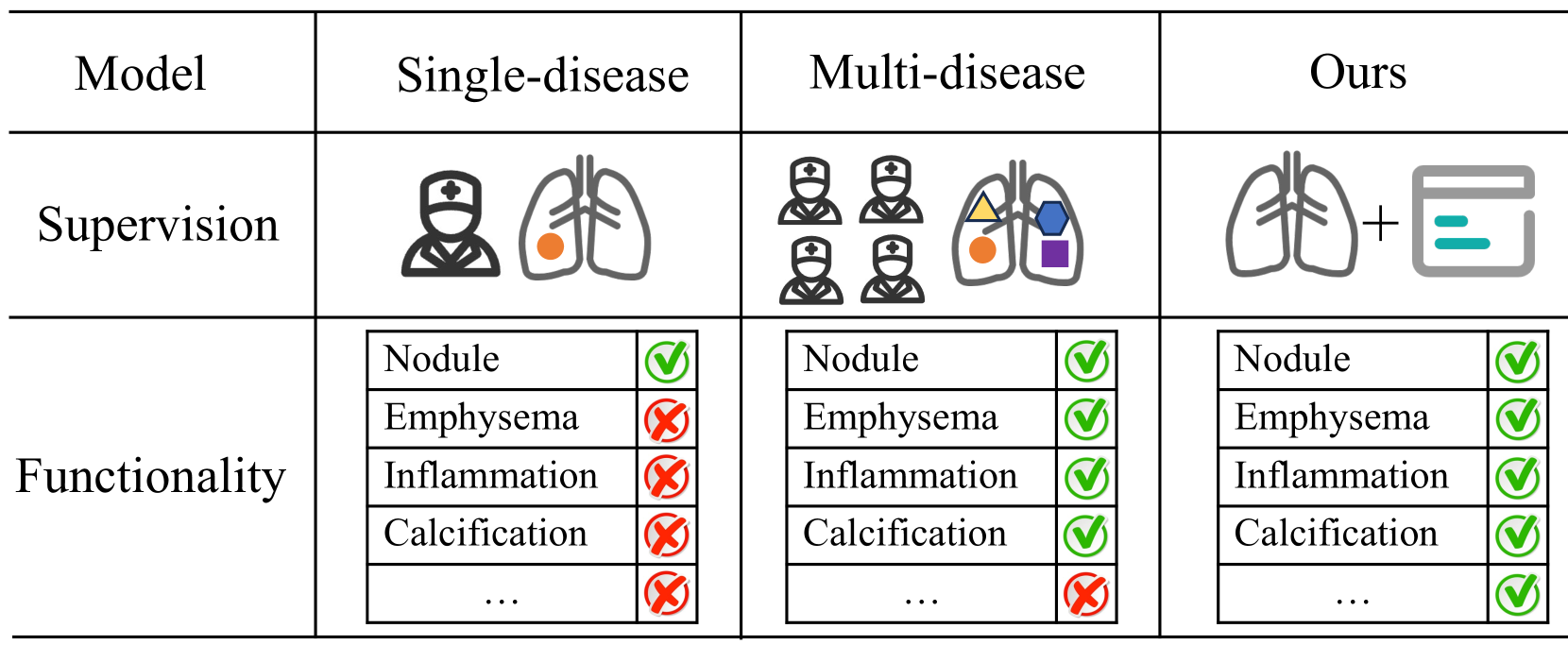
Bootstrapping Chest CT Image Understanding by Distilling Knowledge from X-ray Expert Models
Weiwei Cao, Jianpeng Zhang, Yingda Xia, Tony C. W. Mok, Zi Li, Xianghua Ye, Le Lu, Jian Zheng, Yuxing Tang, Ling Zhang
Radiologists highly desire fully automated versatile AI for medical imaging interpretation. However, the lack of extensively annotated large-scale multi-disease datasets has hindered the achievement of this goal. In this paper, we explore the feasibility of leveraging language as a naturally high-quality supervision for chest CT imaging. In light of the limited availability of image-report pairs, we bootstrap the understanding of 3D chest CT images by distilling chest-related diagnostic knowledge from an extensively pre-trained 2D X-ray expert model. Specifically, we propose a language-guided retrieval method to match each 3D CT image with its semantically closest 2D X-ray image, and perform pair-wise and semantic relation knowledge distillation. Subsequently, we use contrastive learning to align images and reports within the same patient while distinguishing them from the other patients. However, the challenge arises when patients have similar semantic diagnoses, such as healthy patients, potentially confusing if treated as negatives. We introduce a robust contrastive learning that identifies and corrects these false negatives. We train our model with over 12,000 pairs of chest CT images and radiology reports. Extensive experiments across multiple scenarios, including zero-shot learning, report generation, and fine-tuning processes, demonstrate the model's feasibility in interpreting chest CT images.
Read more4/9/2024