Enhancing chest X-ray datasets with privacy-preserving large language models and multi-type annotations: a data-driven approach for improved classification

0

Sign in to get full access
Overview
- This paper explores a data-driven approach for enhancing chest X-ray datasets using privacy-preserving large language models and multi-type annotations to improve classification performance.
- The researchers developed a method to generate synthetic chest X-ray images and corresponding annotations using large language models, while preserving patient privacy.
- They demonstrated that incorporating this synthetic data into existing chest X-ray datasets can lead to improved classification performance on downstream tasks.
Plain English Explanation
Chest X-rays are commonly used in medical imaging to diagnose various lung and heart conditions. However, the availability of high-quality, annotated chest X-ray datasets can be limited, which can hinder the development of advanced AI models for medical image analysis.
To address this challenge, the researchers in this paper proposed a novel approach that leverages large language models to generate synthetic chest X-ray images and corresponding annotations, while preserving patient privacy. This synthetic data can then be combined with existing chest X-ray datasets to improve the performance of AI models trained for chest X-ray classification tasks.
The key idea is to use a large language model that has been trained on a vast amount of text data to generate realistic-looking descriptions of chest X-ray images, including information about the various medical conditions visible in the images. These descriptions can then be used to create synthetic chest X-ray images that are closely aligned with the text, without the need to access or share actual patient data.
By incorporating this synthetic data into existing chest X-ray datasets, the researchers were able to demonstrate improved classification performance on a range of chest X-ray analysis tasks. This approach can help address the data scarcity challenge in medical imaging and enable the development of more accurate and reliable AI-based diagnostic tools.
Technical Explanation
The researchers in this paper developed a method to enhance chest X-ray datasets using synthetic data generated by privacy-preserving large language models and multi-type annotations.
The key steps of their approach are:
-
Large Language Model Training: The researchers trained a large language model on a vast corpus of text data, including medical literature and clinical reports, to enable the generation of realistic-looking descriptions of chest X-ray images.
-
Synthetic Data Generation: Using the trained language model, the researchers generated synthetic text descriptions of chest X-ray images, including information about various medical conditions visible in the images. These textual descriptions were then used to create corresponding synthetic chest X-ray images.
-
Multi-type Annotation: In addition to generating synthetic images, the researchers also created multi-type annotations for the synthetic data, including labels for different medical conditions, segmentation masks, and other relevant metadata.
-
Dataset Augmentation: The researchers combined the synthetic chest X-ray images and annotations with existing chest X-ray datasets to create a more comprehensive and diverse training dataset.
-
Model Training and Evaluation: The researchers trained various AI models for chest X-ray classification tasks using the augmented dataset and evaluated their performance, demonstrating improvements over models trained on the original datasets alone.
The key innovation of this work is the use of privacy-preserving large language models to generate synthetic chest X-ray data, which can help address the data scarcity challenge in medical imaging without compromising patient privacy. The multi-type annotations further enhance the utility of the synthetic data for a range of medical imaging tasks.
Critical Analysis
The researchers in this paper have presented a promising approach for enhancing chest X-ray datasets using synthetic data generated by privacy-preserving large language models. However, there are a few potential limitations and areas for further research that could be considered:
-
Realism of Synthetic Data: While the researchers claim that the synthetic chest X-ray images and annotations are realistic, it's important to evaluate the extent to which they accurately reflect the characteristics and variability of real-world medical imaging data. Further validation and comparison with human-annotated data may be needed to ensure the synthetic data is truly representative.
-
Generalization Across Datasets: The researchers primarily evaluated their approach on a single chest X-ray dataset. It would be valuable to assess the generalizability of their method by applying it to a wider range of chest X-ray datasets, potentially from different healthcare systems or geographic regions, to ensure the synthetic data can benefit a diverse set of medical imaging applications.
-
Clinical Validation: Ultimately, the true test of this approach's effectiveness will be its ability to improve the performance of AI-based medical imaging tools in real-world clinical settings. Further research is needed to evaluate the impact of the synthetic data-augmented models on clinician decision-making and patient outcomes.
-
Ethical Considerations: While the researchers emphasize the privacy-preserving nature of their approach, there may be additional ethical and regulatory considerations around the use of synthetic medical data, particularly in sensitive healthcare contexts. Careful evaluation of these issues is essential to ensure the responsible deployment of this technology.
Despite these potential areas for further exploration, the research presented in this paper represents a significant step forward in addressing the data scarcity challenge in medical imaging and holds promise for improving the development and deployment of AI-powered diagnostic tools.
Conclusion
This paper introduces a novel data-driven approach for enhancing chest X-ray datasets using privacy-preserving large language models and multi-type annotations. By generating synthetic chest X-ray images and corresponding annotations, the researchers were able to demonstrate improved classification performance on downstream tasks compared to models trained on existing datasets alone.
The key innovation of this work lies in the use of large language models to generate realistic-looking synthetic data while preserving patient privacy, addressing a critical challenge in medical imaging research. The incorporation of multi-type annotations further enhances the utility of the synthetic data for a range of medical imaging applications.
While the researchers have identified some potential limitations and areas for further exploration, this work represents an important step forward in the field of medical image analysis and the development of more accurate and reliable AI-based diagnostic tools. As the healthcare industry continues to grapple with data scarcity and privacy concerns, approaches like the one presented in this paper could play a crucial role in enabling the widespread adoption of advanced AI technologies in clinical practice.
This summary was produced with help from an AI and may contain inaccuracies - check out the links to read the original source documents!
Related Papers
0
Enhancing chest X-ray datasets with privacy-preserving large language models and multi-type annotations: a data-driven approach for improved classification
Ricardo Bigolin Lanfredi, Pritam Mukherjee, Ronald Summers
In chest X-ray (CXR) image analysis, rule-based systems are usually employed to extract labels from reports for dataset releases. However, there is still room for improvement in label quality. These labelers typically output only presence labels, sometimes with binary uncertainty indicators, which limits their usefulness. Supervised deep learning models have also been developed for report labeling but lack adaptability, similar to rule-based systems. In this work, we present MAPLEZ (Medical report Annotations with Privacy-preserving Large language model using Expeditious Zero shot answers), a novel approach leveraging a locally executable Large Language Model (LLM) to extract and enhance findings labels on CXR reports. MAPLEZ extracts not only binary labels indicating the presence or absence of a finding but also the location, severity, and radiologists' uncertainty about the finding. Over eight abnormalities from five test sets, we show that our method can extract these annotations with an increase of 3.6 percentage points (pp) in macro F1 score for categorical presence annotations and more than 20 pp increase in F1 score for the location annotations over competing labelers. Additionally, using the combination of improved annotations and multi-type annotations in classification supervision, we demonstrate substantial advancements in model quality, with an increase of 1.1 pp in AUROC over models trained with annotations from the best alternative approach. We share code and annotations.
Read more8/16/2024
0
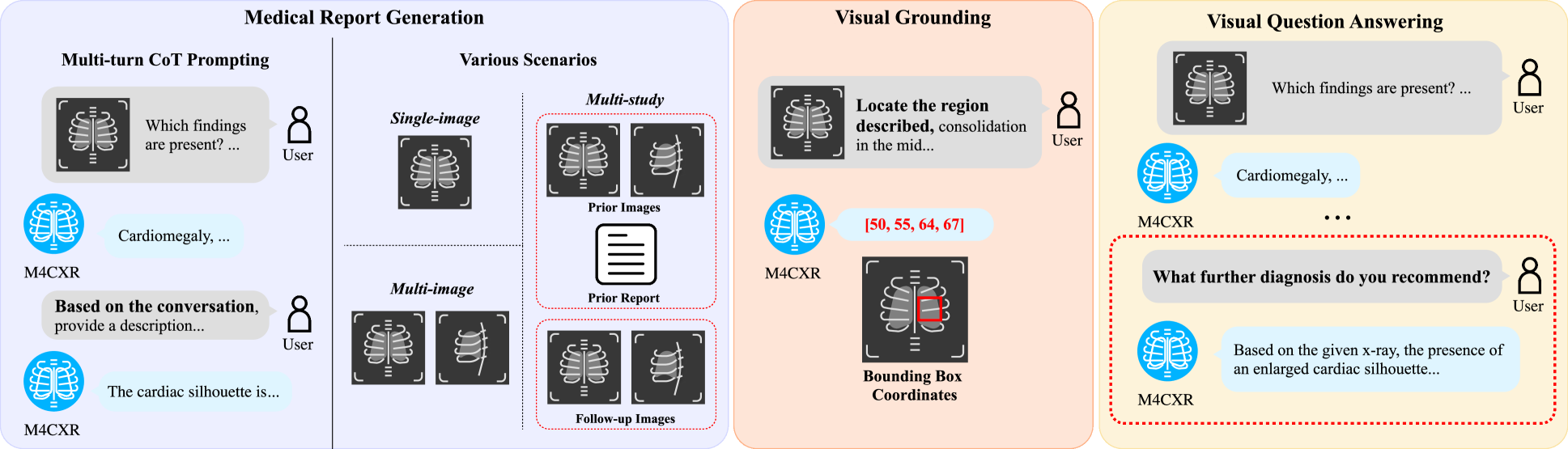
M4CXR: Exploring Multi-task Potentials of Multi-modal Large Language Models for Chest X-ray Interpretation
Jonggwon Park, Soobum Kim, Byungmu Yoon, Jihun Hyun, Kyoyun Choi
The rapid evolution of artificial intelligence, especially in large language models (LLMs), has significantly impacted various domains, including healthcare. In chest X-ray (CXR) analysis, previous studies have employed LLMs, but with limitations: either underutilizing the multi-tasking capabilities of LLMs or lacking clinical accuracy. This paper presents M4CXR, a multi-modal LLM designed to enhance CXR interpretation. The model is trained on a visual instruction-following dataset that integrates various task-specific datasets in a conversational format. As a result, the model supports multiple tasks such as medical report generation (MRG), visual grounding, and visual question answering (VQA). M4CXR achieves state-of-the-art clinical accuracy in MRG by employing a chain-of-thought prompting strategy, in which it identifies findings in CXR images and subsequently generates corresponding reports. The model is adaptable to various MRG scenarios depending on the available inputs, such as single-image, multi-image, and multi-study contexts. In addition to MRG, M4CXR performs visual grounding at a level comparable to specialized models and also demonstrates outstanding performance in VQA. Both quantitative and qualitative assessments reveal M4CXR's versatility in MRG, visual grounding, and VQA, while consistently maintaining clinical accuracy.
Read more8/30/2024

0
DALL-M: Context-Aware Clinical Data Augmentation with LLMs
Chihcheng Hsieh, Catarina Moreira, Isabel Blanco Nobre, Sandra Costa Sousa, Chun Ouyang, Margot Brereton, Joaquim Jorge, Jacinto C. Nascimento
X-ray images are vital in medical diagnostics, but their effectiveness is limited without clinical context. Radiologists often find chest X-rays insufficient for diagnosing underlying diseases, necessitating comprehensive clinical features and data integration. We present a novel technique to enhance the clinical context through augmentation techniques with clinical tabular data, thereby improving its applicability and reliability in AI medical diagnostics. To address this, we introduce a pioneering approach to clinical data augmentation that employs large language models (LLMs) to generate patient contextual synthetic data. This methodology is crucial for training more robust deep learning models in healthcare. It preserves the integrity of real patient data while enriching the dataset with contextually relevant synthetic features, significantly enhancing model performance. DALL-M uses a three-phase feature generation process: (i) clinical context storage, (ii) expert query generation, and (iii) context-aware feature augmentation. DALL-M generates new, clinically relevant features by synthesizing chest X-ray images and reports. Applied to 799 cases using nine features from the MIMIC-IV dataset, it created an augmented set of 91 features. This is the first work to generate contextual values for existing and new features based on patients' X-ray reports, gender, and age and to produce new contextual knowledge during data augmentation. Empirical validation with machine learning models, including Decision Trees, Random Forests, XGBoost, and TabNET, showed significant performance improvements. Incorporating augmented features increased the F1 score by 16.5% and Precision and Recall by approximately 25%. DALL-M addresses a critical gap in clinical data augmentation, offering a robust framework for generating contextually enriched datasets.
Read more7/12/2024
🛸
0
A Disease Labeler for Chinese Chest X-Ray Report Generation
Mengwei Wang, Ruixin Yan, Zeyi Hou, Ning Lang, Xiuzhuang Zhou
In the field of medical image analysis, the scarcity of Chinese chest X-ray report datasets has hindered the development of technology for generating Chinese chest X-ray reports. On one hand, the construction of a Chinese chest X-ray report dataset is limited by the time-consuming and costly process of accurate expert disease annotation. On the other hand, a single natural language generation metric is commonly used to evaluate the similarity between generated and ground-truth reports, while the clinical accuracy and effectiveness of the generated reports rely on an accurate disease labeler (classifier). To address the issues, this study proposes a disease labeler tailored for the generation of Chinese chest X-ray reports. This labeler leverages a dual BERT architecture to handle diagnostic reports and clinical information separately and constructs a hierarchical label learning algorithm based on the affiliation between diseases and body parts to enhance text classification performance. Utilizing this disease labeler, a Chinese chest X-ray report dataset comprising 51,262 report samples was established. Finally, experiments and analyses were conducted on a subset of expert-annotated Chinese chest X-ray reports, validating the effectiveness of the proposed disease labeler.
Read more4/29/2024