Enhanced Muscle and Fat Segmentation for CT-Based Body Composition Analysis: A Comparative Study

0

Sign in to get full access
Overview
- This paper presents a comparative study on enhancing muscle and fat segmentation in CT-based body composition analysis.
- The researchers developed improved algorithms for accurately separating muscle and fat tissues in CT scans, which is crucial for assessing body composition and health.
- The study compares the performance of their new methods against existing techniques, demonstrating significant improvements in segmentation accuracy.
Plain English Explanation
Computed tomography (CT) scans can provide detailed information about the body's internal structures, including the muscle and fat tissues. Accurately separating and measuring these tissues is important for assessing a person's overall health and risk of conditions like obesity or muscle wasting. However, existing methods for doing this can be imprecise.
In this study, the researchers developed new algorithms that can more accurately identify and differentiate between muscle and fat in CT scans. They tested these new methods against existing techniques and found that their approaches resulted in significantly better segmentation of the tissues. This means the new algorithms were better able to correctly separate the muscle and fat regions in the CT images.
The improved ability to measure muscle and fat composition from CT scans could lead to better monitoring and management of conditions related to body composition, such as obesity or muscle wasting. It could also enable more detailed analysis of body composition changes over time or in response to interventions. Overall, the research represents an important advance in the field of body composition assessment using CT imaging.
Technical Explanation
The paper describes the development and evaluation of enhanced algorithms for segmenting muscle and fat tissues in CT scans. The researchers used a deep learning-based approach to train models that could accurately identify and differentiate between muscle and fat regions in the CT images.
The study included a patient population of 100 individuals who underwent full-body CT scans. The researchers compared the performance of their new muscle and fat segmentation methods against a manual segmentation by radiologists, as well as several existing automated techniques.
Evaluation metrics showed that the researchers' approaches significantly outperformed the existing methods, achieving higher Dice similarity coefficients and lower Hausdorff distances in separating muscle and fat. This indicates their algorithms were better able to match the precise boundaries and volumes of the manually segmented tissues.
The paper also discusses potential limitations, such as the need to further validate the methods on larger and more diverse patient populations. The researchers note that continued refinement of annotator simulations could help improve the realism and reliability of the evaluations.
Critical Analysis
The researchers present a compelling case for the value of their enhanced muscle and fat segmentation algorithms. The comparative evaluation against manual segmentation and existing techniques demonstrates clear performance improvements, which could have meaningful clinical implications.
However, the study is limited by its relatively small patient cohort. While the results are promising, further validation on larger and more diverse datasets would be needed to fully establish the generalizability and robustness of the new methods.
Additionally, the paper does not provide much insight into the specific network architectures or training approaches used for the deep learning models. More technical details about the model design and optimization could help other researchers build upon this work.
It would also be interesting to see how the segmentation accuracy translates to downstream analysis and clinical decision-making. The paper focuses on the segmentation metrics, but exploring the impact on body composition assessment and health risk evaluation could further showcase the practical benefits of the enhanced algorithms.
Conclusion
This research presents a significant advance in the field of CT-based body composition analysis through the development of improved muscle and fat segmentation techniques. The new deep learning-based algorithms demonstrate marked improvements over existing methods, suggesting they could enable more accurate and reliable assessment of body composition.
While further validation is needed, this work represents an important step forward in leveraging advanced image analysis to support better monitoring and management of health conditions related to body composition, such as obesity and muscle wasting. Continued refinement and deployment of these techniques could have significant implications for clinical practice and population health.
This summary was produced with help from an AI and may contain inaccuracies - check out the links to read the original source documents!
Related Papers
0
Enhanced Muscle and Fat Segmentation for CT-Based Body Composition Analysis: A Comparative Study
Benjamin Hou, Tejas Sudharshan Mathai, Jianfei Liu, Christopher Parnell, Ronald M. Summers
Purpose: Body composition measurements from routine abdominal CT can yield personalized risk assessments for asymptomatic and diseased patients. In particular, attenuation and volume measures of muscle and fat are associated with important clinical outcomes, such as cardiovascular events, fractures, and death. This study evaluates the reliability of an Internal tool for the segmentation of muscle and fat (subcutaneous and visceral) as compared to the well-established public TotalSegmentator tool. Methods: We assessed the tools across 900 CT series from the publicly available SAROS dataset, focusing on muscle, subcutaneous fat, and visceral fat. The Dice score was employed to assess accuracy in subcutaneous fat and muscle segmentation. Due to the lack of ground truth segmentations for visceral fat, Cohen's Kappa was utilized to assess segmentation agreement between the tools. Results: Our Internal tool achieved a 3% higher Dice (83.8 vs. 80.8) for subcutaneous fat and a 5% improvement (87.6 vs. 83.2) for muscle segmentation respectively. A Wilcoxon signed-rank test revealed that our results were statistically different with p<0.01. For visceral fat, the Cohen's kappa score of 0.856 indicated near-perfect agreement between the two tools. Our internal tool also showed very strong correlations for muscle volume (R^2=0.99), muscle attenuation (R^2=0.93), and subcutaneous fat volume (R^2=0.99) with a moderate correlation for subcutaneous fat attenuation (R^2=0.45). Conclusion: Our findings indicated that our Internal tool outperformed TotalSegmentator in measuring subcutaneous fat and muscle. The high Cohen's Kappa score for visceral fat suggests a reliable level of agreement between the two tools. These results demonstrate the potential of our tool in advancing the accuracy of body composition analysis.
Read more4/16/2024
✅
0
Validation of musculoskeletal segmentation model with uncertainty estimation for bone and muscle assessment in hip-to-knee clinical CT images
Mazen Soufi, Yoshito Otake, Makoto Iwasa, Keisuke Uemura, Tomoki Hakotani, Masahiro Hashimoto, Yoshitake Yamada, Minoru Yamada, Yoichi Yokoyama, Masahiro Jinzaki, Suzushi Kusano, Masaki Takao, Seiji Okada, Nobuhiko Sugano, Yoshinobu Sato
Deep learning-based image segmentation has allowed for the fully automated, accurate, and rapid analysis of musculoskeletal (MSK) structures from medical images. However, current approaches were either applied only to 2D cross-sectional images, addressed few structures, or were validated on small datasets, which limit the application in large-scale databases. This study aimed to validate an improved deep learning model for volumetric MSK segmentation of the hip and thigh with uncertainty estimation from clinical computed tomography (CT) images. Databases of CT images from multiple manufacturers/scanners, disease status, and patient positioning were used. The segmentation accuracy, and accuracy in estimating the structures volume and density, i.e., mean HU, were evaluated. An approach for segmentation failure detection based on predictive uncertainty was also investigated. The model has shown an overall improvement with respect to all segmentation accuracy and structure volume/density evaluation metrics. The predictive uncertainty yielded large areas under the receiver operating characteristic (AUROC) curves (AUROCs>=.95) in detecting inaccurate and failed segmentations. The high segmentation and muscle volume/density estimation accuracy, along with the high accuracy in failure detection based on the predictive uncertainty, exhibited the model's reliability for analyzing individual MSK structures in large-scale CT databases.
Read more9/5/2024
✅
0
Automated Body Composition Analysis Using DAFS Express on 2D MRI Slices at L3 Vertebral Level
Varun Akella, Razeyeh Bagherinasab, Jia Ming Li, Long Nguyen, Vincent Tze Yang Chow, Hyunwoo Lee, Karteek Popuri, Mirza Faisal Beg
Body composition analysis is vital in assessing health conditions such as obesity, sarcopenia, and metabolic syndromes. MRI provides detailed images of skeletal muscle (SKM), visceral adipose tissue (VAT), and subcutaneous adipose tissue (SAT), but their manual segmentation is labor-intensive and limits clinical applicability. This study validates an automated tool for MRI-based 2D body composition analysis- (Data Analysis Facilitation Suite (DAFS) Express), comparing its automated measurements with expert manual segmentations using UK Biobank data. A cohort of 399 participants from the UK Biobank dataset was selected, yielding 423 single L3 slices for analysis. DAFS Express performed automated segmentations of SKM, VAT, and SAT, which were then manually corrected by expert raters for validation. Evaluation metrics included Jaccard coefficients, Dice scores, Intraclass Correlation Coefficients (ICCs), and Bland-Altman Plots to assess segmentation agreement and reliability. High agreements were observed between automated and manual segmentations with mean Jaccard scores: SKM 99.03%, VAT 95.25%, and SAT 99.57%; and mean Dice scores: SKM 99.51%, VAT 97.41%, and SAT 99.78%. Cross-sectional area comparisons showed consistent measurements with automated methods closely matching manual measurements for SKM and SAT, and slightly higher values for VAT (SKM: Auto 132.51 cm^2, Manual 132.36 cm^2; VAT: Auto 137.07 cm^2, Manual 134.46 cm^2; SAT: Auto 203.39 cm^2, Manual 202.85 cm^2). ICCs confirmed strong reliability (SKM: 0.998, VAT: 0.994, SAT: 0.994). Bland-Altman plots revealed minimal biases, and boxplots illustrated distribution similarities across SKM, VAT, and SAT areas. On average DAFS Express took 18 seconds per DICOM. This underscores its potential to streamline image analysis processes in research and clinical settings, enhancing diagnostic accuracy and efficiency.
Read more9/12/2024
0
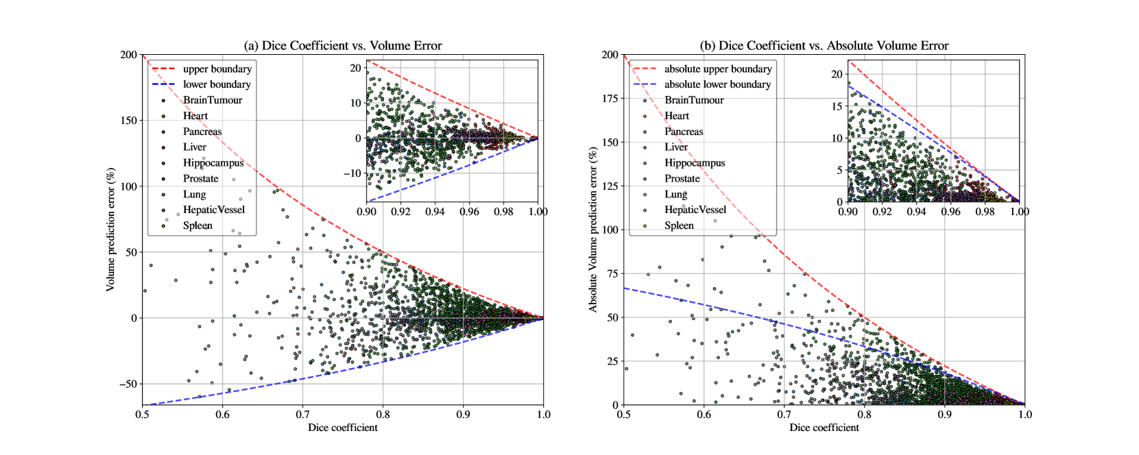
Segmentation Quality and Volumetric Accuracy in Medical Imaging
Zheyuan Zhang, Ulas Bagci
Current medical image segmentation relies on the region-based (Dice, F1-score) and boundary-based (Hausdorff distance, surface distance) metrics as the de-facto standard. While these metrics are widely used, they lack a unified interpretation, particularly regarding volume agreement. Clinicians often lack clear benchmarks to gauge the goodness of segmentation results based on these metrics. Recognizing the clinical relevance of volumetry, we utilize relative volume prediction error (vpe) to directly assess the accuracy of volume predictions derived from segmentation tasks. Our work integrates theoretical analysis and empirical validation across diverse datasets. We delve into the often-ambiguous relationship between segmentation quality (measured by Dice) and volumetric accuracy in clinical practice. Our findings highlight the critical role of incorporating volumetric prediction accuracy into segmentation evaluation. This approach empowers clinicians with a more nuanced understanding of segmentation performance, ultimately improving the interpretation and utility of these metrics in real-world healthcare settings.
Read more5/15/2024