Explicit Differentiable Slicing and Global Deformation for Cardiac Mesh Reconstruction

0
Sign in to get full access
Overview
- This paper presents a novel method for cardiac mesh reconstruction using explicit differentiable slicing and global deformation.
- The proposed approach enables accurate and efficient reconstruction of 3D cardiac meshes from 2D medical images.
- The method involves explicit differentiable slicing to extract 2D cardiac boundaries and global deformation to reconstruct the 3D cardiac mesh.
Plain English Explanation
The human heart is a complex 3D structure, and accurately reconstructing its shape from 2D medical images can be challenging. This paper introduces a new technique that makes this process more efficient and precise.
The key idea is to first extract the 2D outlines or "boundaries" of the heart from the 2D images using an explicit and differentiable slicing process. This means the method can automatically learn how to identify the heart's edges in the images, without requiring manual labeling.
Next, the method uses a "global deformation" approach to reconstruct the full 3D shape of the heart by bending and stretching a generic 3D heart model to match the extracted 2D boundaries. This global deformation allows the method to capture the complex 3D structure of the heart.
By combining these two key innovations - explicit differentiable slicing and global deformation - the paper presents an effective way to reconstruct accurate 3D cardiac meshes from standard 2D medical scans. This could have important applications in fields like cardiac disease diagnosis and surgical planning.
Technical Explanation
The proposed method consists of two main components:
-
Explicit Differentiable Slicing: This module takes 2D medical images as input and extracts the 2D cardiac boundaries using an explicit and differentiable slicing process. This allows the method to automatically learn to identify the heart's edges without requiring manual annotations.
-
Global Deformation: Given the extracted 2D cardiac boundaries, this component uses a global deformation approach to reconstruct the full 3D cardiac mesh. It does this by deforming a generic 3D heart model to match the 2D boundaries, capturing the complex 3D structure of the heart.
The explicit differentiable slicing is implemented using a neural network that predicts the cardiac boundaries in a differentiable manner, enabling end-to-end training. The global deformation module uses a deformation field to warp the generic 3D heart mesh to fit the extracted 2D boundaries.
Experiments on public cardiac imaging datasets show that the proposed method achieves state-of-the-art performance in 3D cardiac mesh reconstruction, demonstrating the effectiveness of the explicit differentiable slicing and global deformation approach.
Critical Analysis
The paper presents a compelling approach for cardiac mesh reconstruction that addresses key challenges in this domain. The explicit differentiable slicing and global deformation techniques are innovative and well-designed.
One potential limitation is that the method relies on a generic 3D heart model as the starting point for reconstruction. This model may not capture the full diversity of heart shapes and sizes seen in the real world. Exploring ways to make the method more adaptable to patient-specific anatomies could be an area for future research.
Additionally, the paper does not provide extensive analysis of failure cases or potential sources of error in the reconstructed meshes. Further investigation into the method's robustness and failure modes would help users better understand its strengths and weaknesses.
Overall, this is a high-quality piece of research that makes valuable contributions to the field of cardiac imaging and modeling. The authors have demonstrated the effectiveness of their approach and provided a solid foundation for future work in this important area.
Conclusion
This paper presents a novel method for 3D cardiac mesh reconstruction that combines explicit differentiable slicing and global deformation. By extracting 2D cardiac boundaries and deforming a generic 3D heart model, the approach can efficiently and accurately reconstruct the complex 3D structure of the human heart from standard 2D medical images.
The explicit differentiable slicing and global deformation techniques are key innovations that enable this method to outperform previous state-of-the-art approaches. This work has significant potential for applications in cardiac disease diagnosis, surgical planning, and other areas of healthcare that rely on accurate 3D modeling of the heart.
Overall, this research makes an important contribution to the field of cardiac imaging and modeling, and lays the groundwork for further advancements in this critical domain.
This summary was produced with help from an AI and may contain inaccuracies - check out the links to read the original source documents!
Related Papers
0
Explicit Differentiable Slicing and Global Deformation for Cardiac Mesh Reconstruction
Yihao Luo, Dario Sesia, Fanwen Wang, Yinzhe Wu, Wenhao Ding, Jiahao Huang, Fadong Shi Anoop Shah, Amit Kaural, Jamil Mayet, Guang Yang, ChoonHwai Yap
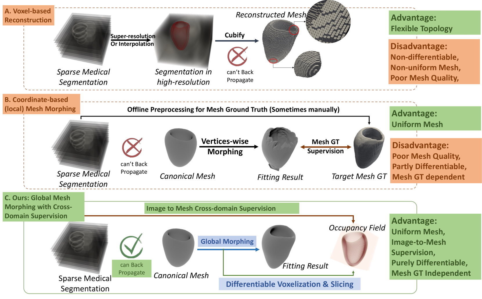
Mesh reconstruction of the cardiac anatomy from medical images is useful for shape and motion measurements and biophysics simulations to facilitate the assessment of cardiac function and health. However, 3D medical images are often acquired as 2D slices that are sparsely sampled and noisy, and mesh reconstruction on such data is a challenging task. Traditional voxel-based approaches rely on pre- and post-processing that compromises image fidelity, while mesh-level deep learning approaches require mesh annotations that are difficult to get. Therefore, direct cross-domain supervision from 2D images to meshes is a key technique for advancing 3D learning in medical imaging, but it has not been well-developed. While there have been attempts to approximate the optimized meshes' slicing, few existing methods directly use 2D slices to supervise mesh reconstruction in a differentiable manner. Here, we propose a novel explicit differentiable voxelization and slicing (DVS) algorithm that allows gradient backpropagation to a mesh from its slices, facilitating refined mesh optimization directly supervised by the losses defined on 2D images. Further, we propose an innovative framework for extracting patient-specific left ventricle (LV) meshes from medical images by coupling DVS with a graph harmonic deformation (GHD) mesh morphing descriptor of cardiac shape that naturally preserves mesh quality and smoothness during optimization. Experimental results demonstrate that our method achieves state-of-the-art performance in cardiac mesh reconstruction tasks from CT and MRI, with an overall Dice score of 90% on multi-datasets, outperforming existing approaches. The proposed method can further quantify clinically useful parameters such as ejection fraction and global myocardial strains, closely matching the ground truth and surpassing the traditional voxel-based approach in sparse images.
Read more9/4/2024

0
Multi-view Hybrid Graph Convolutional Network for Volume-to-mesh Reconstruction in Cardiovascular MRI
Nicol'as Gaggion, Benjamin A. Matheson, Yan Xia, Rodrigo Bonazzola, Nishant Ravikumar, Zeike A. Taylor, Diego H. Milone, Alejandro F. Frangi, Enzo Ferrante
Cardiovascular magnetic resonance imaging is emerging as a crucial tool to examine cardiac morphology and function. Essential to this endeavour are anatomical 3D surface and volumetric meshes derived from CMR images, which facilitate computational anatomy studies, biomarker discovery, and in-silico simulations. However, conventional surface mesh generation methods, such as active shape models and multi-atlas segmentation, are highly time-consuming and require complex processing pipelines to generate simulation-ready 3D meshes. In response, we introduce HybridVNet, a novel architecture for direct image-to-mesh extraction seamlessly integrating standard convolutional neural networks with graph convolutions, which we prove can efficiently handle surface and volumetric meshes by encoding them as graph structures. To further enhance accuracy, we propose a multiview HybridVNet architecture which processes both long axis and short axis CMR, showing that it can increase the performance of cardiac MR mesh generation. Our model combines traditional convolutional networks with variational graph generative models, deep supervision and mesh-specific regularisation. Experiments on a comprehensive dataset from the UK Biobank confirm the potential of HybridVNet to significantly advance cardiac imaging and computational cardiology by efficiently generating high-fidelity and simulation ready meshes from CMR images.
Read more8/15/2024

0
Synthetic Data Generation for 3D Myocardium Deformation Analysis
Shahar Zuler, Dan Raviv
Accurate analysis of 3D myocardium deformation using high-resolution computerized tomography (CT) datasets with ground truth (GT) annotations is crucial for advancing cardiovascular imaging research. However, the scarcity of such datasets poses a significant challenge for developing robust myocardium deformation analysis models. To address this, we propose a novel approach to synthetic data generation for enriching cardiovascular imaging datasets. We introduce a synthetic data generation method, enriched with crucial GT 3D optical flow annotations. We outline the data preparation from a cardiac four-dimensional (4D) CT scan, selection of parameters, and the subsequent creation of synthetic data from the same or other sources of 3D cardiac CT data for training. Our work contributes to overcoming the limitations imposed by the scarcity of high-resolution CT datasets with precise annotations, thereby facilitating the development of accurate and reliable myocardium deformation analysis algorithms for clinical applications and diagnostics. Our code is available at: http://www.github.com/shaharzuler/cardio_volume_skewer
Read more6/4/2024
0
3D MRI Synthesis with Slice-Based Latent Diffusion Models: Improving Tumor Segmentation Tasks in Data-Scarce Regimes
Aghiles Kebaili, J'er^ome Lapuyade-Lahorgue, Pierre Vera, Su Ruan
Despite the increasing use of deep learning in medical image segmentation, the limited availability of annotated training data remains a major challenge due to the time-consuming data acquisition and privacy regulations. In the context of segmentation tasks, providing both medical images and their corresponding target masks is essential. However, conventional data augmentation approaches mainly focus on image synthesis. In this study, we propose a novel slice-based latent diffusion architecture designed to address the complexities of volumetric data generation in a slice-by-slice fashion. This approach extends the joint distribution modeling of medical images and their associated masks, allowing a simultaneous generation of both under data-scarce regimes. Our approach mitigates the computational complexity and memory expensiveness typically associated with diffusion models. Furthermore, our architecture can be conditioned by tumor characteristics, including size, shape, and relative position, thereby providing a diverse range of tumor variations. Experiments on a segmentation task using the BRATS2022 confirm the effectiveness of the synthesized volumes and masks for data augmentation.
Read more6/11/2024