Multi-view Hybrid Graph Convolutional Network for Volume-to-mesh Reconstruction in Cardiovascular MRI

0

Sign in to get full access
Overview
- The paper proposes a novel method called Multi-view Hybrid Graph Convolutional Network (MHGCN) for volume-to-mesh reconstruction in cardiovascular MRI.
- The method leverages multiple 2D views of the 3D volume to learn a graph representation for accurate mesh reconstruction.
- It combines graph convolutional networks with multi-view fusion to capture both local and global shape features.
Plain English Explanation
The paper presents a technique to Reconstructing 3D Meshes from 3D Volumes in Cardiac MRI. Traditionally, this task has been challenging because 3D volumes lack the detailed shape information needed for accurate mesh reconstruction.
The researchers' innovation is to use Multiple 2D Views of the 3D Volume to learn a graph-based representation of the 3D shape. By combining Graph Convolutional Networks with Multi-view Fusion, their method can capture both local and global shape features to reconstruct detailed 3D meshes from the 3D volume data.
The key innovation is using multiple 2D views to learn a more comprehensive 3D shape representation, rather than relying on the 3D volume alone. This allows the model to reconstruct 3D meshes that better match the true anatomical structures compared to prior approaches.
Technical Explanation
The Multi-view Hybrid Graph Convolutional Network (MHGCN) takes a 3D cardiovascular MRI volume as input and outputs a 3D mesh representation of the cardiac anatomy.
The architecture consists of three main components:
-
Multi-view Encoder: This module extracts 2D feature maps from multiple 2D views of the 3D volume using a convolutional neural network.
-
Graph Convolutional Network: The 2D feature maps are then used to construct a graph representation of the 3D shape. A graph convolutional network operates on this graph to learn local shape features.
-
Multi-view Fusion: The local features from the graph ConvNet are combined with global features from the multi-view encoder using an attention-based fusion module. This allows the network to capture both local and global shape information for accurate mesh reconstruction.
The key innovation is the use of multiple 2D views to learn a more comprehensive 3D shape representation compared to using the 3D volume directly. This multi-view approach enables the model to reconstruct detailed 3D meshes that closely match the true cardiac anatomy.
Critical Analysis
The paper provides a thorough evaluation of the MHGCN method on a large cardiac MRI dataset, demonstrating significant improvements over prior state-of-the-art approaches. However, a few potential limitations are worth noting:
-
Computational Complexity: The multi-view architecture and graph ConvNet components may increase the computational cost and memory requirements compared to simpler 3D volume-to-mesh methods. The authors do not provide detailed runtime or memory usage analysis.
-
Generalization to Other Organs: The paper focuses on cardiac MRI, but it's unclear how well the MHGCN method would generalize to volume-to-mesh reconstruction of other anatomical structures beyond the heart. Further validation on diverse medical imaging datasets would be helpful.
-
Clinical Validation: While the paper demonstrates improved mesh reconstruction accuracy, the clinical relevance and impact of these improvements are not fully explored. Deeper collaboration with medical experts would be valuable to assess the practical utility of the method in real-world clinical settings.
Conclusion
The Multi-view Hybrid Graph Convolutional Network (MHGCN) presented in this paper offers a novel approach to volume-to-mesh reconstruction in cardiovascular MRI. By leveraging multiple 2D views of the 3D volume and combining graph convolutional networks with multi-view fusion, the method can capture both local and global shape features to produce detailed 3D mesh reconstructions.
This work represents an important step forward in medical image analysis, as accurate 3D mesh representations of anatomical structures are crucial for various clinical applications, such as surgical planning, disease diagnosis, and patient-specific modeling. While the method shows promise, further research is needed to address potential limitations and ensure its widespread clinical adoption.
This summary was produced with help from an AI and may contain inaccuracies - check out the links to read the original source documents!
Related Papers
0
Multi-view Hybrid Graph Convolutional Network for Volume-to-mesh Reconstruction in Cardiovascular MRI
Nicol'as Gaggion, Benjamin A. Matheson, Yan Xia, Rodrigo Bonazzola, Nishant Ravikumar, Zeike A. Taylor, Diego H. Milone, Alejandro F. Frangi, Enzo Ferrante
Cardiovascular magnetic resonance imaging is emerging as a crucial tool to examine cardiac morphology and function. Essential to this endeavour are anatomical 3D surface and volumetric meshes derived from CMR images, which facilitate computational anatomy studies, biomarker discovery, and in-silico simulations. However, conventional surface mesh generation methods, such as active shape models and multi-atlas segmentation, are highly time-consuming and require complex processing pipelines to generate simulation-ready 3D meshes. In response, we introduce HybridVNet, a novel architecture for direct image-to-mesh extraction seamlessly integrating standard convolutional neural networks with graph convolutions, which we prove can efficiently handle surface and volumetric meshes by encoding them as graph structures. To further enhance accuracy, we propose a multiview HybridVNet architecture which processes both long axis and short axis CMR, showing that it can increase the performance of cardiac MR mesh generation. Our model combines traditional convolutional networks with variational graph generative models, deep supervision and mesh-specific regularisation. Experiments on a comprehensive dataset from the UK Biobank confirm the potential of HybridVNet to significantly advance cardiac imaging and computational cardiology by efficiently generating high-fidelity and simulation ready meshes from CMR images.
Read more8/15/2024

0
Deep-Motion-Net: GNN-based volumetric organ shape reconstruction from single-view 2D projections
Isuru Wijesinghe, Michael Nix, Arezoo Zakeri, Alireza Hokmabadi, Bashar Al-Qaisieh, Ali Gooya, Zeike A. Taylor
We propose Deep-Motion-Net: an end-to-end graph neural network (GNN) architecture that enables 3D (volumetric) organ shape reconstruction from a single in-treatment kV planar X-ray image acquired at any arbitrary projection angle. Estimating and compensating for true anatomical motion during radiotherapy is essential for improving the delivery of planned radiation dose to target volumes while sparing organs-at-risk, and thereby improving the therapeutic ratio. Achieving this using only limited imaging available during irradiation and without the use of surrogate signals or invasive fiducial markers is attractive. The proposed model learns the mesh regression from a patient-specific template and deep features extracted from kV images at arbitrary projection angles. A 2D-CNN encoder extracts image features, and four feature pooling networks fuse these features to the 3D template organ mesh. A ResNet-based graph attention network then deforms the feature-encoded mesh. The model is trained using synthetically generated organ motion instances and corresponding kV images. The latter is generated by deforming a reference CT volume aligned with the template mesh, creating digitally reconstructed radiographs (DRRs) at required projection angles, and DRR-to-kV style transferring with a conditional CycleGAN model. The overall framework was tested quantitatively on synthetic respiratory motion scenarios and qualitatively on in-treatment images acquired over full scan series for liver cancer patients. Overall mean prediction errors for synthetic motion test datasets were 0.16$pm$0.13 mm, 0.18$pm$0.19 mm, 0.22$pm$0.34 mm, and 0.12$pm$0.11 mm. Mean peak prediction errors were 1.39 mm, 1.99 mm, 3.29 mm, and 1.16 mm.
Read more7/10/2024
0
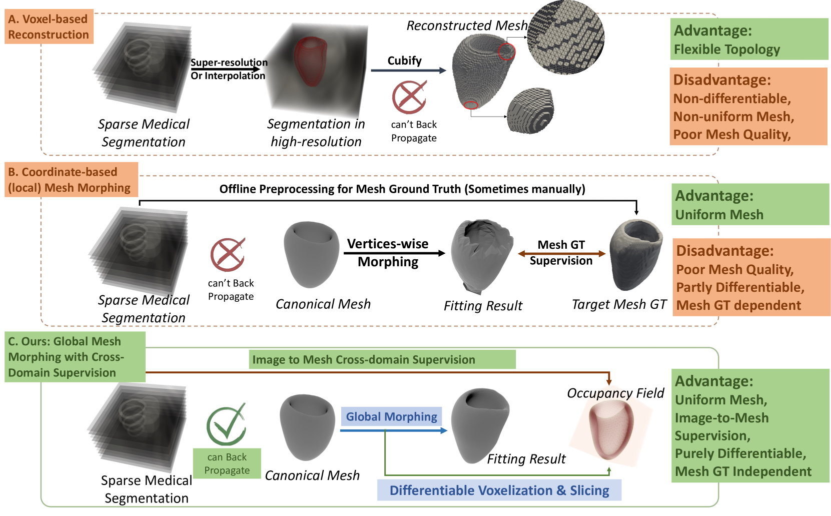
Explicit Differentiable Slicing and Global Deformation for Cardiac Mesh Reconstruction
Yihao Luo, Dario Sesia, Fanwen Wang, Yinzhe Wu, Wenhao Ding, Jiahao Huang, Fadong Shi Anoop Shah, Amit Kaural, Jamil Mayet, Guang Yang, ChoonHwai Yap
Mesh reconstruction of the cardiac anatomy from medical images is useful for shape and motion measurements and biophysics simulations to facilitate the assessment of cardiac function and health. However, 3D medical images are often acquired as 2D slices that are sparsely sampled and noisy, and mesh reconstruction on such data is a challenging task. Traditional voxel-based approaches rely on pre- and post-processing that compromises image fidelity, while mesh-level deep learning approaches require mesh annotations that are difficult to get. Therefore, direct cross-domain supervision from 2D images to meshes is a key technique for advancing 3D learning in medical imaging, but it has not been well-developed. While there have been attempts to approximate the optimized meshes' slicing, few existing methods directly use 2D slices to supervise mesh reconstruction in a differentiable manner. Here, we propose a novel explicit differentiable voxelization and slicing (DVS) algorithm that allows gradient backpropagation to a mesh from its slices, facilitating refined mesh optimization directly supervised by the losses defined on 2D images. Further, we propose an innovative framework for extracting patient-specific left ventricle (LV) meshes from medical images by coupling DVS with a graph harmonic deformation (GHD) mesh morphing descriptor of cardiac shape that naturally preserves mesh quality and smoothness during optimization. Experimental results demonstrate that our method achieves state-of-the-art performance in cardiac mesh reconstruction tasks from CT and MRI, with an overall Dice score of 90% on multi-datasets, outperforming existing approaches. The proposed method can further quantify clinically useful parameters such as ejection fraction and global myocardial strains, closely matching the ground truth and surpassing the traditional voxel-based approach in sparse images.
Read more9/4/2024
0
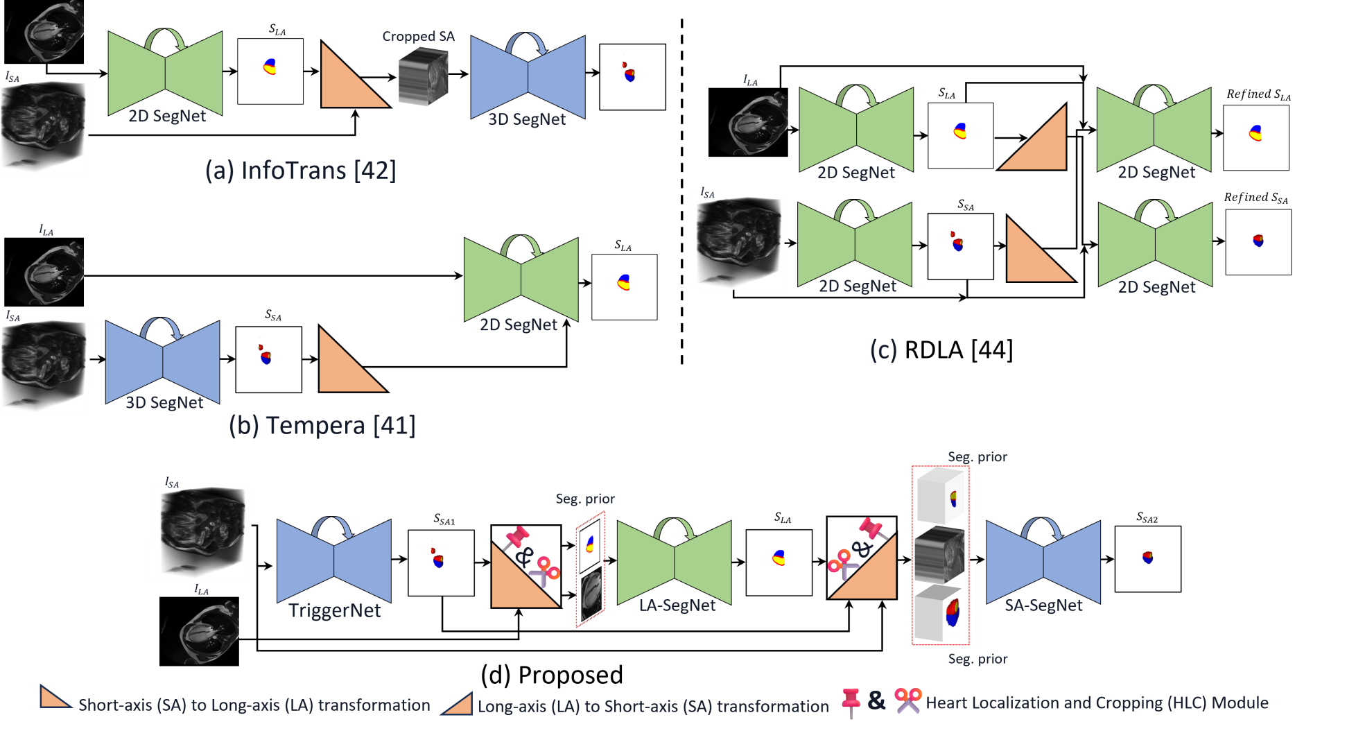
Multi-view Cardiac Image Segmentation via Trans-Dimensional Priors
Abbas Khan, Muhammad Asad, Martin Benning, Caroline Roney, Gregory Slabaugh
We propose a novel multi-stage trans-dimensional architecture for multi-view cardiac image segmentation. Our method exploits the relationship between long-axis (2D) and short-axis (3D) magnetic resonance (MR) images to perform a sequential 3D-to-2D-to-3D segmentation, segmenting the long-axis and short-axis images. In the first stage, 3D segmentation is performed using the short-axis image, and the prediction is transformed to the long-axis view and used as a segmentation prior in the next stage. In the second step, the heart region is localized and cropped around the segmentation prior using a Heart Localization and Cropping (HLC) module, focusing the subsequent model on the heart region of the image, where a 2D segmentation is performed. Similarly, we transform the long-axis prediction to the short-axis view, localize and crop the heart region and again perform a 3D segmentation to refine the initial short-axis segmentation. We evaluate our proposed method on the Multi-Disease, Multi-View & Multi-Center Right Ventricular Segmentation in Cardiac MRI (M&Ms-2) dataset, where our method outperforms state-of-the-art methods in segmenting cardiac regions of interest in both short-axis and long-axis images. The pre-trained models, source code, and implementation details will be publicly available.
Read more4/26/2024