Framework to generate perfusion map from CT and CTA images in patients with acute ischemic stroke: A longitudinal and cross-sectional study

0
Sign in to get full access
Overview
- Presents a framework to generate perfusion maps from CT and CTA images in patients with acute ischemic stroke
- Longitudinal and cross-sectional study design to assess the framework's performance
- Potential to aid in the diagnosis and treatment of acute ischemic stroke
Plain English Explanation
This research paper introduces a framework to generate perfusion maps from CT and CTA images in patients with acute ischemic stroke. Perfusion maps are visual representations of blood flow in the brain, which can help doctors identify areas of the brain that are not receiving enough blood flow during a stroke.
The researchers used a longitudinal and cross-sectional study design to assess the performance of their framework. This means they followed patients over time (longitudinal) and also looked at a group of patients at one point in time (cross-sectional). By doing this, they could evaluate how well the framework works in different scenarios.
Diagnosing and treating acute ischemic stroke, which is a type of stroke caused by a blockage in a blood vessel, is critical. The ability to generate accurate perfusion maps from CT and CTA images could potentially help doctors make better decisions about how to treat patients and improve their outcomes.
Technical Explanation
The researchers developed a framework to generate perfusion maps from CT and CTA images in patients with acute ischemic stroke. This framework involves several steps:
- Image preprocessing: The CT and CTA images are preprocessed to remove noise and enhance features.
- Perfusion parameter estimation: Algorithms are used to estimate perfusion parameters, such as cerebral blood flow and cerebral blood volume, from the preprocessed images.
- Perfusion map generation: The estimated perfusion parameters are used to generate a visual representation of blood flow in the brain, known as a perfusion map.
To assess the performance of their framework, the researchers conducted a longitudinal and cross-sectional study. In the longitudinal study, they followed a group of patients over time, collecting CT and CTA images at multiple time points. In the cross-sectional study, they looked at a separate group of patients at a single time point.
The researchers compared the perfusion maps generated by their framework to reference perfusion maps obtained using other methods, such as MRI. They evaluated the accuracy, reliability, and consistency of their framework in both the longitudinal and cross-sectional studies.
Critical Analysis
The researchers acknowledge several limitations of their study. First, the sample sizes for both the longitudinal and cross-sectional studies were relatively small, which may limit the generalizability of the findings. Additionally, the researchers did not directly compare their framework to other existing methods for generating perfusion maps, which makes it difficult to assess its relative performance.
Furthermore, the researchers do not discuss the potential sources of error or bias in their framework, such as variations in image acquisition or preprocessing techniques. It would be helpful to understand the robustness of the framework to these types of factors.
Despite these limitations, the framework presented in this paper has the potential to significantly improve the diagnosis and treatment of acute ischemic stroke. By providing accurate and reliable perfusion maps from readily available CT and CTA images, the framework could help clinicians make more informed decisions about patient management and intervention.
Conclusion
This research paper introduces a novel framework for generating perfusion maps from CT and CTA images in patients with acute ischemic stroke. The longitudinal and cross-sectional study design suggests that the framework can produce accurate and reliable perfusion maps, which could potentially aid in the diagnosis and treatment of this critical condition.
While the study has some limitations, the researchers' work represents an important step forward in the field of stroke imaging and management. Further research is needed to validate the framework in larger and more diverse patient populations, as well as to compare its performance to other established methods. Nonetheless, this framework has the potential to significantly improve outcomes for patients with acute ischemic stroke.
This summary was produced with help from an AI and may contain inaccuracies - check out the links to read the original source documents!
Related Papers
0
Framework to generate perfusion map from CT and CTA images in patients with acute ischemic stroke: A longitudinal and cross-sectional study
Chayanin Tangwiriyasakul, Pedro Borges, Stefano Moriconi, Paul Wright, Yee-Haur Mah, James Teo, Parashkev Nachev, Sebastien Ourselin, M. Jorge Cardoso
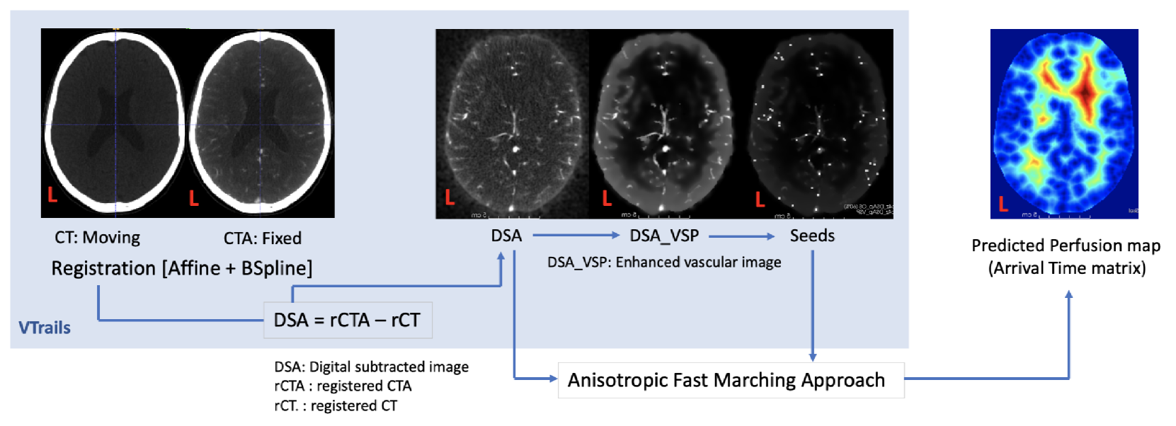
Stroke is a leading cause of disability and death. Effective treatment decisions require early and informative vascular imaging. 4D perfusion imaging is ideal but rarely available within the first hour after stroke, whereas plain CT and CTA usually are. Hence, we propose a framework to extract a predicted perfusion map (PPM) derived from CT and CTA images. In all eighteen patients, we found significantly high spatial similarity (with average Spearman's correlation = 0.7893) between our predicted perfusion map (PPM) and the T-max map derived from 4D-CTP. Voxelwise correlations between the PPM and National Institutes of Health Stroke Scale (NIHSS) subscores for L/R hand motor, gaze, and language on a large cohort of 2,110 subjects reliably mapped symptoms to expected infarct locations. Therefore our PPM could serve as an alternative for 4D perfusion imaging, if the latter is unavailable, to investigate blood perfusion in the first hours after hospital admission.
Read more4/8/2024
🏅
0
A dual-task mutual learning framework for predicting post-thrombectomy cerebral hemorrhage
Caiwen Jiang, Tianyu Wang, Xiaodan Xing, Mianxin Liu, Guang Yang, Zhongxiang Ding, Dinggang Shen
Ischemic stroke is a severe condition caused by the blockage of brain blood vessels, and can lead to the death of brain tissue due to oxygen deprivation. Thrombectomy has become a common treatment choice for ischemic stroke due to its immediate effectiveness. But, it carries the risk of postoperative cerebral hemorrhage. Clinically, multiple CT scans within 0-72 hours post-surgery are used to monitor for hemorrhage. However, this approach exposes radiation dose to patients, and may delay the detection of cerebral hemorrhage. To address this dilemma, we propose a novel prediction framework for measuring postoperative cerebral hemorrhage using only the patient's initial CT scan. Specifically, we introduce a dual-task mutual learning framework to takes the initial CT scan as input and simultaneously estimates both the follow-up CT scan and prognostic label to predict the occurrence of postoperative cerebral hemorrhage. Our proposed framework incorporates two attention mechanisms, i.e., self-attention and interactive attention. Specifically, the self-attention mechanism allows the model to focus more on high-density areas in the image, which are critical for diagnosis (i.e., potential hemorrhage areas). The interactive attention mechanism further models the dependencies between the interrelated generation and classification tasks, enabling both tasks to perform better than the case when conducted individually. Validated on clinical data, our method can generate follow-up CT scans better than state-of-the-art methods, and achieves an accuracy of 86.37% in predicting follow-up prognostic labels. Thus, our work thus contributes to the timely screening of post-thrombectomy cerebral hemorrhage, and could significantly reform the clinical process of thrombectomy and other similar operations related to stroke.
Read more8/6/2024
0
ISLES'24: Improving final infarct prediction in ischemic stroke using multimodal imaging and clinical data
Ezequiel de la Rosa, Ruisheng Su, Mauricio Reyes, Roland Wiest, Evamaria O. Riedel, Florian Kofler, Kaiyuan Yang, Hakim Baazaoui, David Robben, Susanne Wegener, Jan S. Kirschke, Benedikt Wiestler, Bjoern Menze
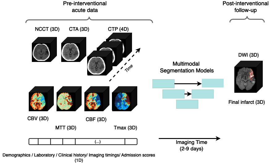
Accurate estimation of core (irreversibly damaged tissue) and penumbra (salvageable tissue) volumes is essential for ischemic stroke treatment decisions. Perfusion CT, the clinical standard, estimates these volumes but is affected by variations in deconvolution algorithms, implementations, and thresholds. Core tissue expands over time, with growth rates influenced by thrombus location, collateral circulation, and inherent patient-specific factors. Understanding this tissue growth is crucial for determining the need to transfer patients to comprehensive stroke centers, predicting the benefits of additional reperfusion attempts during mechanical thrombectomy, and forecasting final clinical outcomes. This work presents the ISLES'24 challenge, which addresses final post-treatment stroke infarct prediction from pre-interventional acute stroke imaging and clinical data. ISLES'24 establishes a unique 360-degree setting where all feasibly accessible clinical data are available for participants, including full CT acute stroke imaging, sub-acute follow-up MRI, and clinical tabular data. The contributions of this work are two-fold: first, we introduce a standardized benchmarking of final stroke infarct segmentation algorithms through the ISLES'24 challenge; second, we provide insights into infarct segmentation using multimodal imaging and clinical data strategies by identifying outperforming methods on a finely curated dataset. The outputs of this challenge are anticipated to enhance clinical decision-making and improve patient outcome predictions. All ISLES'24 materials, including data, performance evaluation scripts, and leading algorithmic strategies, are available to the research community following url{https://isles-24.grand-challenge.org/}.
Read more8/21/2024
⛏️
0
CPAISD: Core-penumbra acute ischemic stroke dataset
D. Umerenkov, S. Kudin, M. Peksheva, D. Pavlov
We introduce the CPAISD: Core-Penumbra Acute Ischemic Stroke Dataset, aimed at enhancing the early detection and segmentation of ischemic stroke using Non-Contrast Computed Tomography (NCCT) scans. Addressing the challenges in diagnosing acute ischemic stroke during its early stages due to often non-revealing native CT findings, the dataset provides a collection of segmented NCCT images. These include annotations of ischemic core and penumbra regions, critical for developing machine learning models for rapid stroke identification and assessment. By offering a carefully collected and annotated dataset, we aim to facilitate the development of advanced diagnostic tools, contributing to improved patient care and outcomes in stroke management. Our dataset's uniqueness lies in its focus on the acute phase of ischemic stroke, with non-informative native CT scans, and includes a baseline model to demonstrate the dataset's application, encouraging further research and innovation in the field of medical imaging and stroke diagnosis.
Read more4/4/2024