ISLES'24: Improving final infarct prediction in ischemic stroke using multimodal imaging and clinical data

0
Sign in to get full access
Overview
- Predicting the final extent of brain tissue damage after an ischemic stroke is crucial for treatment planning.
- This paper explores using multimodal imaging and clinical data to improve the accuracy of final infarct prediction.
- The researchers developed a machine learning model to integrate various data sources and provide more reliable predictions.
Plain English Explanation
The research paper focuses on improving the ability to predict the final size of brain damage, or infarct, after a person has an ischemic stroke. This is important because it can help doctors decide on the best treatment plan for the patient.
The researchers used a combination of different types of medical imaging data, such as CT scans and MRI scans, along with clinical information about the patient, like their medical history. They developed a machine learning model that could take all of this information and make more accurate predictions about the final size of the stroke damage.
By using multiple data sources, the model was able to provide more reliable and detailed predictions compared to using just one type of data. This could help doctors make better-informed decisions about the most appropriate treatments for each individual patient.
Technical Explanation
The paper presents a novel approach to predicting the final infarct size in ischemic stroke using a multimodal machine learning model. The model integrates multimodal imaging data, such as CT perfusion and diffusion-weighted MRI, along with clinical data to provide more accurate and reliable predictions.
The researchers developed a framework to generate perfusion maps from CT imaging and used a dual-task mutual learning approach to jointly predict the final infarct size and other stroke-related outcomes. The model was trained and evaluated on a large dataset of ischemic stroke patients.
The results show that the multimodal approach significantly outperformed models using single data modalities, demonstrating the value of integrating diverse sources of information for more accurate prediction of thrombectomy recanalization from CT imaging.
Critical Analysis
The paper presents a well-designed and thorough study, with a comprehensive evaluation of the proposed multimodal model. However, the authors acknowledge several limitations and areas for further research:
- The model was trained and evaluated on a single dataset, and its performance on other populations or healthcare settings may vary.
- The study did not explore the interpretability of the model's predictions, which could be important for clinical decision-making.
- The researchers did not investigate the impact of the model's predictions on patient outcomes or treatment decisions, which would be a crucial next step.
Additionally, while the multimodal approach showed improved performance, the extent of the improvement and the trade-offs in terms of model complexity and computational requirements could be further explored.
Conclusion
This research demonstrates the potential of integrating multimodal imaging and clinical data to enhance the prediction of final infarct size in ischemic stroke. By leveraging diverse data sources, the proposed machine learning model can provide more accurate and reliable predictions to support clinical decision-making and treatment planning.
The findings highlight the value of interdisciplinary collaboration and the incorporation of advanced analytics in stroke care. Further studies are needed to validate the model's performance in real-world settings and explore its impact on patient outcomes. Nonetheless, this work represents an important step towards improving the management of ischemic stroke and enhancing patient care.
This summary was produced with help from an AI and may contain inaccuracies - check out the links to read the original source documents!
Related Papers
0
ISLES'24: Improving final infarct prediction in ischemic stroke using multimodal imaging and clinical data
Ezequiel de la Rosa, Ruisheng Su, Mauricio Reyes, Roland Wiest, Evamaria O. Riedel, Florian Kofler, Kaiyuan Yang, Hakim Baazaoui, David Robben, Susanne Wegener, Jan S. Kirschke, Benedikt Wiestler, Bjoern Menze
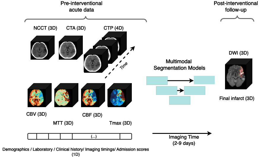
Accurate estimation of core (irreversibly damaged tissue) and penumbra (salvageable tissue) volumes is essential for ischemic stroke treatment decisions. Perfusion CT, the clinical standard, estimates these volumes but is affected by variations in deconvolution algorithms, implementations, and thresholds. Core tissue expands over time, with growth rates influenced by thrombus location, collateral circulation, and inherent patient-specific factors. Understanding this tissue growth is crucial for determining the need to transfer patients to comprehensive stroke centers, predicting the benefits of additional reperfusion attempts during mechanical thrombectomy, and forecasting final clinical outcomes. This work presents the ISLES'24 challenge, which addresses final post-treatment stroke infarct prediction from pre-interventional acute stroke imaging and clinical data. ISLES'24 establishes a unique 360-degree setting where all feasibly accessible clinical data are available for participants, including full CT acute stroke imaging, sub-acute follow-up MRI, and clinical tabular data. The contributions of this work are two-fold: first, we introduce a standardized benchmarking of final stroke infarct segmentation algorithms through the ISLES'24 challenge; second, we provide insights into infarct segmentation using multimodal imaging and clinical data strategies by identifying outperforming methods on a finely curated dataset. The outputs of this challenge are anticipated to enhance clinical decision-making and improve patient outcome predictions. All ISLES'24 materials, including data, performance evaluation scripts, and leading algorithmic strategies, are available to the research community following url{https://isles-24.grand-challenge.org/}.
Read more8/21/2024
🎲
0
ISLES 2024: The first longitudinal multimodal multi-center real-world dataset in (sub-)acute stroke
Evamaria O. Riedel, Ezequiel de la Rosa, The Anh Baran, Moritz Hernandez Petzsche, Hakim Baazaoui, Kaiyuan Yang, David Robben, Joaquin Oscar Seia, Roland Wiest, Mauricio Reyes, Ruisheng Su, Claus Zimmer, Tobias Boeckh-Behrens, Maria Berndt, Bjoern Menze, Benedikt Wiestler, Susanne Wegener, Jan S. Kirschke
Stroke remains a leading cause of global morbidity and mortality, placing a heavy socioeconomic burden. Over the past decade, advances in endovascular reperfusion therapy and the use of CT and MRI imaging for treatment guidance have significantly improved patient outcomes and are now standard in clinical practice. To develop machine learning algorithms that can extract meaningful and reproducible models of brain function for both clinical and research purposes from stroke images - particularly for lesion identification, brain health quantification, and prognosis - large, diverse, and well-annotated public datasets are essential. While only a few datasets with (sub-)acute stroke data were previously available, several large, high-quality datasets have recently been made publicly accessible. However, these existing datasets include only MRI data. In contrast, our dataset is the first to offer comprehensive longitudinal stroke data, including acute CT imaging with angiography and perfusion, follow-up MRI at 2-9 days, as well as acute and longitudinal clinical data up to a three-month outcome. The dataset includes a training dataset of n = 150 and a test dataset of n = 100 scans. Training data is publicly available, while test data will be used exclusively for model validation. We are making this dataset available as part of the 2024 edition of the Ischemic Stroke Lesion Segmentation (ISLES) challenge (https://www.isles-challenge.org/), which continuously aims to establish benchmark methods for acute and sub-acute ischemic stroke lesion segmentation, aiding in creating open stroke imaging datasets and evaluating cutting-edge image processing algorithms.
Read more8/22/2024

0
A Robust Ensemble Algorithm for Ischemic Stroke Lesion Segmentation: Generalizability and Clinical Utility Beyond the ISLES Challenge
Ezequiel de la Rosa, Mauricio Reyes, Sook-Lei Liew, Alexandre Hutton, Roland Wiest, Johannes Kaesmacher, Uta Hanning, Arsany Hakim, Richard Zubal, Waldo Valenzuela, David Robben, Diana M. Sima, Vincenzo Anania, Arne Brys, James A. Meakin, Anne Mickan, Gabriel Broocks, Christian Heitkamp, Shengbo Gao, Kongming Liang, Ziji Zhang, Md Mahfuzur Rahman Siddiquee, Andriy Myronenko, Pooya Ashtari, Sabine Van Huffel, Hyun-su Jeong, Chi-ho Yoon, Chulhong Kim, Jiayu Huo, Sebastien Ourselin, Rachel Sparks, Albert Cl`erigues, Arnau Oliver, Xavier Llad'o, Liam Chalcroft, Ioannis Pappas, Jeroen Bertels, Ewout Heylen, Juliette Moreau, Nima Hatami, Carole Frindel, Abdul Qayyum, Moona Mazher, Domenec Puig, Shao-Chieh Lin, Chun-Jung Juan, Tianxi Hu, Lyndon Boone, Maged Goubran, Yi-Jui Liu, Susanne Wegener, Florian Kofler, Ivan Ezhov, Suprosanna Shit, Moritz R. Hernandez Petzsche, Bjoern Menze, Jan S. Kirschke, Benedikt Wiestler
Diffusion-weighted MRI (DWI) is essential for stroke diagnosis, treatment decisions, and prognosis. However, image and disease variability hinder the development of generalizable AI algorithms with clinical value. We address this gap by presenting a novel ensemble algorithm derived from the 2022 Ischemic Stroke Lesion Segmentation (ISLES) challenge. ISLES'22 provided 400 patient scans with ischemic stroke from various medical centers, facilitating the development of a wide range of cutting-edge segmentation algorithms by the research community. Through collaboration with leading teams, we combined top-performing algorithms into an ensemble model that overcomes the limitations of individual solutions. Our ensemble model achieved superior ischemic lesion detection and segmentation accuracy on our internal test set compared to individual algorithms. This accuracy generalized well across diverse image and disease variables. Furthermore, the model excelled in extracting clinical biomarkers. Notably, in a Turing-like test, neuroradiologists consistently preferred the algorithm's segmentations over manual expert efforts, highlighting increased comprehensiveness and precision. Validation using a real-world external dataset (N=1686) confirmed the model's generalizability. The algorithm's outputs also demonstrated strong correlations with clinical scores (admission NIHSS and 90-day mRS) on par with or exceeding expert-derived results, underlining its clinical relevance. This study offers two key findings. First, we present an ensemble algorithm (https://github.com/Tabrisrei/ISLES22_Ensemble) that detects and segments ischemic stroke lesions on DWI across diverse scenarios on par with expert (neuro)radiologists. Second, we show the potential for biomedical challenge outputs to extend beyond the challenge's initial objectives, demonstrating their real-world clinical applicability.
Read more4/4/2024
⛏️
0
CPAISD: Core-penumbra acute ischemic stroke dataset
D. Umerenkov, S. Kudin, M. Peksheva, D. Pavlov
We introduce the CPAISD: Core-Penumbra Acute Ischemic Stroke Dataset, aimed at enhancing the early detection and segmentation of ischemic stroke using Non-Contrast Computed Tomography (NCCT) scans. Addressing the challenges in diagnosing acute ischemic stroke during its early stages due to often non-revealing native CT findings, the dataset provides a collection of segmented NCCT images. These include annotations of ischemic core and penumbra regions, critical for developing machine learning models for rapid stroke identification and assessment. By offering a carefully collected and annotated dataset, we aim to facilitate the development of advanced diagnostic tools, contributing to improved patient care and outcomes in stroke management. Our dataset's uniqueness lies in its focus on the acute phase of ischemic stroke, with non-informative native CT scans, and includes a baseline model to demonstrate the dataset's application, encouraging further research and innovation in the field of medical imaging and stroke diagnosis.
Read more4/4/2024