GPU-Accelerated RSF Level Set Evolution for Large-Scale Microvascular Segmentation

0
🧪
Sign in to get full access
Overview
- Modeling microvascular networks is challenging due to their small size and complex structure.
- Traditional imaging techniques struggle to clearly capture individual components of these networks.
- The researchers propose a new parallel processing approach to a level set model that can efficiently segment large, high-resolution microvascular data.
- The method was tested on multiple datasets from advanced imaging techniques like micro-CT and light sheet microscopy.
Plain English Explanation
Microvascular networks are the tiny blood vessels that form complex structures throughout the body. Modeling and understanding these networks is important for medical research and applications, but it's incredibly difficult. The vessels are so small that current imaging technologies can barely see them clearly. This makes it hard to accurately identify and map out the individual components of the networks using computer algorithms.
The researchers developed a new approach to solve this problem. They took an existing mathematical model called a "level set" and adapted it to run much faster on powerful parallel processing hardware like graphics cards. This allows the model to efficiently analyze massive, high-resolution datasets of microvascular networks captured through advanced 3D imaging techniques.
By speeding up this level set model, the researchers enable practical, large-scale segmentation and modeling of intricate microvascular structures. This could lead to major advances in our understanding of these tiny blood vessel networks and how they function in the body.
Technical Explanation
The core challenge in modeling microvascular networks is that the vessels are near the diffraction limit of current 3D imaging technologies. This makes it very difficult for computer algorithms to reliably identify and separate the individual components of these dense, tangled networks.
The researchers propose using a "region-scalable fitting" (RSF) level set model to address this problem. Level set methods are well-suited because they can incorporate surface and topological constraints to guide the segmentation. However, traditional level set approaches are extremely computationally intensive, making them impractical for analyzing large, high-resolution microvascular datasets.
To overcome this, the researchers developed a parallel processing implementation of the RSF level set model. This allows the level set equation to be evaluated independently on different regions of the dataset using graphics processing units (GPUs). This parallel approach dramatically speeds up the computation, making large-scale microvascular segmentation feasible.
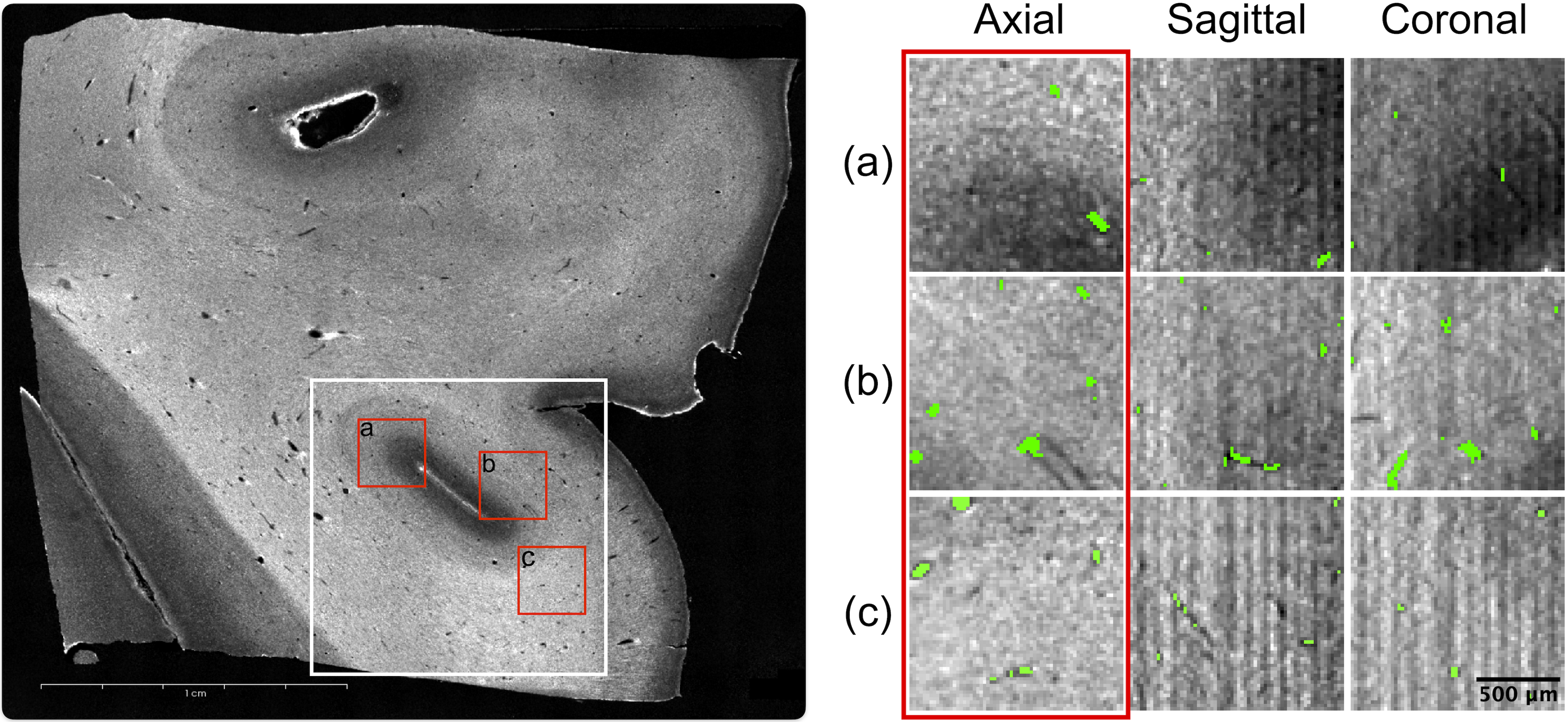
The researchers tested this parallel RSF method on diverse microvascular datasets captured through advanced 3D imaging techniques like micro-CT, light sheet fluorescence microscopy, and milling microscopy. They validated the performance and accuracy of the RSF model through rigorous statistical analysis and comparisons to other segmentation approaches.
Critical Analysis
The researchers acknowledge several important limitations and caveats of their work. First, while the parallel RSF approach significantly improves processing speed, segmenting terabyte-scale microvascular datasets still requires substantial computational resources. The method may not be practical for all research labs or applications.
Additionally, the accuracy validation focused on statistical comparisons to other segmentation algorithms, but did not include direct ground truth validation against manual expert segmentation. This introduces some uncertainty about the true fidelity of the RSF model's results.
The paper also does not explore the potential impact of imaging artifacts or noise on the segmentation performance. Microvascular imaging often suffers from various optical distortions and interference, which could degrade the model's reliability in real-world conditions.
Overall, the researchers have made an important contribution in developing a scalable, parallel processing approach to level set modeling for microvascular segmentation. However, further validation and refinement will be needed to fully assess the practical utility and limitations of this method.
Conclusion
This research showcases an innovative technique to enable large-scale modeling and analysis of intricate microvascular network structures. By adapting a powerful level set segmentation model to run efficiently on parallel hardware, the researchers have overcome a major computational bottleneck in this challenging domain.
The successful application of this parallel RSF method to diverse microvascular imaging datasets demonstrates its practical value and potential for advancing medical research and applications. With further validation and optimization, this approach could become an important tool for building high-fidelity models of the body's complex circulatory systems at unprecedented scales.
This summary was produced with help from an AI and may contain inaccuracies - check out the links to read the original source documents!
Related Papers
🧪
0
GPU-Accelerated RSF Level Set Evolution for Large-Scale Microvascular Segmentation
Meher Niger, Helya Goharbavang, Taeyong Ahn, Emily K. Alley, Joshua D. Wythe, Guoning Chen, David Mayerich
Microvascular networks are challenging to model because these structures are currently near the diffraction limit for most advanced three-dimensional imaging modalities, including confocal and light sheet microscopy. This makes semantic segmentation difficult, because individual components of these networks fluctuate within the confines of individual pixels. Level set methods are ideally suited to solve this problem by providing surface and topological constraints on the resulting model, however these active contour techniques are extremely time intensive and impractical for terabyte-scale images. We propose a reformulation and implementation of the region-scalable fitting (RSF) level set model that makes it amenable to three-dimensional evaluation using both single-instruction multiple data (SIMD) and single-program multiple-data (SPMD) parallel processing. This enables evaluation of the level set equation on independent regions of the data set using graphics processing units (GPUs), making large-scale segmentation of high-resolution networks practical and inexpensive. We tested this 3D parallel RSF approach on multiple data sets acquired using state-of-the-art imaging techniques to acquire microvascular data, including micro-CT, light sheet fluorescence microscopy (LSFM) and milling microscopy. To assess the performance and accuracy of the RSF model, we conducted a Monte-Carlo-based validation technique to compare results to other segmentation methods. We also provide a rigorous profiling to show the gains in processing speed leveraging parallel hardware. This study showcases the practical application of the RSF model, emphasizing its utility in the challenging domain of segmenting large-scale high-topology network structures with a particular focus on building microvascular models.
Read more4/4/2024
🏅
0
RSF-Conv: Rotation-and-Scale Equivariant Fourier Parameterized Convolution for Retinal Vessel Segmentation
Zihong Sun, Hong Wang, Qi Xie, Yefeng Zheng, Deyu Meng
Retinal vessel segmentation is of great clinical significance for the diagnosis of many eye-related diseases, but it is still a formidable challenge due to the intricate vascular morphology. With the skillful characterization of the translation symmetry existing in retinal vessels, convolutional neural networks (CNNs) have achieved great success in retinal vessel segmentation. However, the rotation-and-scale symmetry, as a more widespread image prior in retinal vessels, fails to be characterized by CNNs. Therefore, we propose a rotation-and-scale equivariant Fourier parameterized convolution (RSF-Conv) specifically for retinal vessel segmentation, and provide the corresponding equivariance analysis. As a general module, RSF-Conv can be integrated into existing networks in a plug-and-play manner while significantly reducing the number of parameters. For instance, we replace the traditional convolution filters in U-Net and Iter-Net with RSF-Convs, and faithfully conduct comprehensive experiments. RSF-Conv+U-Net and RSF-Conv+Iter-Net not only have slight advantages under in-domain evaluation, but more importantly, outperform all comparison methods by a significant margin under out-of-domain evaluation. It indicates the remarkable generalization of RSF-Conv, which holds greater practical clinical significance for the prevalent cross-device and cross-hospital challenges in clinical practice. To comprehensively demonstrate the effectiveness of RSF-Conv, we also apply RSF-Conv+U-Net and RSF-Conv+Iter-Net to retinal artery/vein classification and achieve promising performance as well, indicating its clinical application potential.
Read more9/9/2024
🖼️
0
Re-initialization-free Level Set Method via Molecular Beam Epitaxy Equation Regularization for Image Segmentation
Fanghui Song, Jiebao Sun, Shengzhu Shi, Zhichang Guo, Dazhi Zhang
Variational level set method has become a powerful tool in image segmentation due to its ability to handle complex topological changes and maintain continuity and smoothness in the process of evolution. However its evolution process can be unstable, which results in over flatted or over sharpened contours and segmentation failure. To improve the accuracy and stability of evolution, we propose a high-order level set variational segmentation method integrated with molecular beam epitaxy (MBE) equation regularization. This method uses the crystal growth in the MBE process to limit the evolution of the level set function, and thus can avoid the re-initialization in the evolution process and regulate the smoothness of the segmented curve. It also works for noisy images with intensity inhomogeneity, which is a challenge in image segmentation. To solve the variational model, we derive the gradient flow and design scalar auxiliary variable (SAV) scheme coupled with fast Fourier transform (FFT), which can significantly improve the computational efficiency compared with the traditional semi-implicit and semi-explicit scheme. Numerical experiments show that the proposed method can generate smooth segmentation curves, retain fine segmentation targets and obtain robust segmentation results of small objects. Compared to existing level set methods, this model is state-of-the-art in both accuracy and efficiency.
Read more6/27/2024
0
Neurovascular Segmentation in sOCT with Deep Learning and Synthetic Training Data
Etienne Chollet, Yael Balbastre, Chiara Mauri, Caroline Magnain, Bruce Fischl, Hui Wang
Microvascular anatomy is known to be involved in various neurological disorders. However, understanding these disorders is hindered by the lack of imaging modalities capable of capturing the comprehensive three-dimensional vascular network structure at microscopic resolution. With a lateral resolution of $<=$20 {textmu}m and ability to reconstruct large tissue blocks up to tens of cubic centimeters, serial-section optical coherence tomography (sOCT) is well suited for this task. This method uses intrinsic optical properties to visualize the vessels and therefore does not possess a specific contrast, which complicates the extraction of accurate vascular models. The performance of traditional vessel segmentation methods is heavily degraded in the presence of substantial noise and imaging artifacts and is sensitive to domain shifts, while convolutional neural networks (CNNs) require extensive labeled data and are also sensitive the precise intensity characteristics of the data that they are trained on. Building on the emerging field of synthesis-based training, this study demonstrates a synthesis engine for neurovascular segmentation in sOCT images. Characterized by minimal priors and high variance sampling, our highly generalizable method tested on five distinct sOCT acquisitions eliminates the need for manual annotations while attaining human-level precision. Our approach comprises two phases: label synthesis and label-to-image transformation. We demonstrate the efficacy of the former by comparing it to several more realistic sets of training labels, and the latter by an ablation study of synthetic noise and artifact models.
Read more7/2/2024