Hibou: A Family of Foundational Vision Transformers for Pathology

0
Sign in to get full access
Overview
- Presents a family of foundational vision transformers called Hibou for pathology applications
- Explores the use of vision transformers for medical image analysis, a relatively new and promising approach
- Introduces several Hibou model variants with different architectural choices and pretraining strategies
- Evaluates the Hibou models on various pathology datasets, demonstrating strong performance
Plain English Explanation
The paper introduces a new family of models called Hibou, which are a type of vision transformer designed for medical image analysis tasks in pathology. Vision transformers are a relatively new approach in machine learning that has shown promising results for various computer vision problems.
The key idea behind the Hibou models is to leverage the powerful feature representation capabilities of vision transformers and adapt them specifically for pathology applications. The researchers experiment with different architectural choices and pretraining strategies to create a set of Hibou model variants, each with its own strengths and tradeoffs.
The Hibou models are evaluated on several pathology datasets, including tasks such as classifying colon histopathology images and detecting ovarian cancer. The results demonstrate that the Hibou models can achieve state-of-the-art performance, highlighting the potential of vision transformers for medical image analysis.
The paper also discusses the importance of self-supervised pretraining and hierarchical image representations in the context of pathology tasks, which are key aspects of the Hibou models.
Technical Explanation
The Hibou models are a family of vision transformers designed for pathology applications. The researchers explore different architectural choices and pretraining strategies to create several Hibou model variants.
The core Hibou architecture is based on the vision transformer (ViT) model, which has shown promising results for various computer vision tasks. The researchers experiment with different patch sizes, embedding dimensions, and the number of transformer layers to create Hibou-Small, Hibou-Base, and Hibou-Large models.
Additionally, the researchers investigate the impact of pretraining the Hibou models using self-supervised learning on large-scale medical image datasets, such as CheXpert and PatchCamelyon. This pretraining approach aims to learn rich, generalizable features that can be effectively fine-tuned for specific pathology tasks.
The Hibou models are evaluated on several pathology datasets, including histopathology image classification, tumor detection, and cancer diagnosis. The results demonstrate that the Hibou models can achieve state-of-the-art performance, outperforming conventional convolutional neural networks and other vision transformer variants.
Critical Analysis
The paper presents a thorough and well-designed study on the application of vision transformers for pathology tasks. The researchers have carefully explored different architectural choices and pretraining strategies to create a family of Hibou models tailored for medical image analysis.
One potential limitation mentioned in the paper is the need for large-scale, high-quality pathology datasets to fully leverage the capabilities of the Hibou models. The researchers acknowledge that the availability and diversity of such datasets remains a challenge in the field.
Additionally, the paper does not delve deeply into the interpretability and explainability of the Hibou models, which is an important consideration for medical applications. Further research could explore methods to enhance the transparency and understanding of the Hibou models' decision-making processes.
Overall, the Hibou models demonstrate the potential of vision transformers for pathology tasks and provide a solid foundation for future research in this area. The insights and findings presented in the paper contribute to the ongoing advancements in medical image analysis using deep learning techniques.
Conclusion
The paper introduces the Hibou family of foundational vision transformers for pathology applications. The Hibou models leverage the powerful feature representation capabilities of vision transformers and adapt them specifically for medical image analysis tasks.
The researchers explore different architectural choices and pretraining strategies to create several Hibou model variants, each with its own strengths and tradeoffs. The evaluation on various pathology datasets showcases the strong performance of the Hibou models, outperforming conventional approaches and highlighting the potential of vision transformers for medical image analysis.
The insights and findings presented in this paper contribute to the growing body of research on the application of deep learning, and specifically vision transformers, in the field of pathology. The Hibou models provide a solid foundation for further advancements in medical image analysis and have the potential to significantly impact the development of computer-aided diagnostic tools in healthcare.
This summary was produced with help from an AI and may contain inaccuracies - check out the links to read the original source documents!
Related Papers
0
Hibou: A Family of Foundational Vision Transformers for Pathology
Dmitry Nechaev, Alexey Pchelnikov, Ekaterina Ivanova
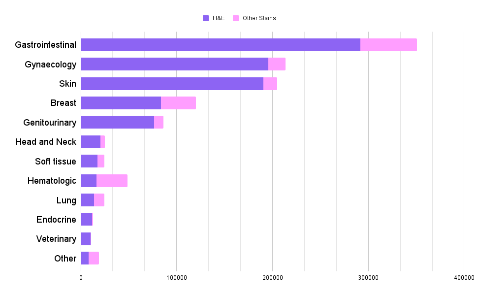
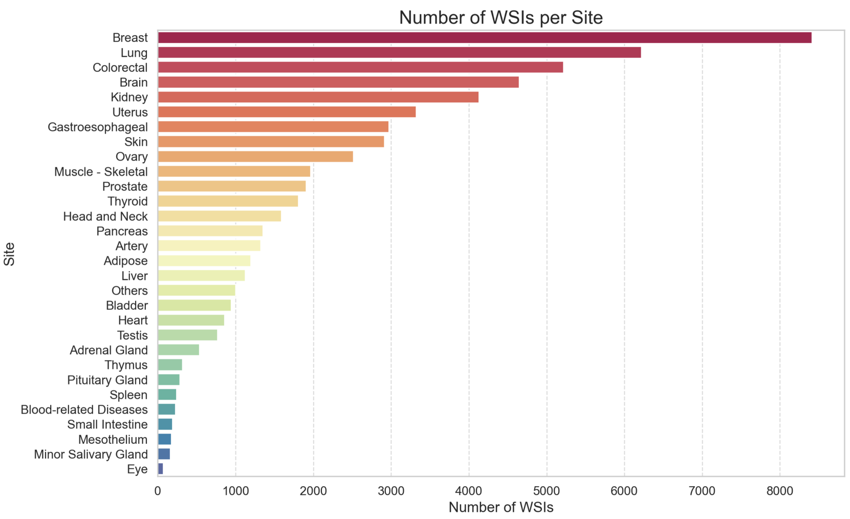
Pathology, the microscopic examination of diseased tissue, is critical for diagnosing various medical conditions, particularly cancers. Traditional methods are labor-intensive and prone to human error. Digital pathology, which converts glass slides into high-resolution digital images for analysis by computer algorithms, revolutionizes the field by enhancing diagnostic accuracy, consistency, and efficiency through automated image analysis and large-scale data processing. Foundational transformer pretraining is crucial for developing robust, generalizable models as it enables learning from vast amounts of unannotated data. This paper introduces the Hibou family of foundational vision transformers for pathology, leveraging the DINOv2 framework to pretrain two model variants, Hibou-B and Hibou-L, on a proprietary dataset of over 1 million whole slide images (WSIs) representing diverse tissue types and staining techniques. Our pretrained models demonstrate superior performance on both patch-level and slide-level benchmarks, surpassing existing state-of-the-art methods. Notably, Hibou-L achieves the highest average accuracy across multiple benchmark datasets. To support further research and application in the field, we have open-sourced the Hibou models, which can be accessed at https://github.com/HistAI/hibou.
Read more8/21/2024

0
PathoDuet: Foundation Models for Pathological Slide Analysis of H&E and IHC Stains
Shengyi Hua, Fang Yan, Tianle Shen, Lei Ma, Xiaofan Zhang
Large amounts of digitized histopathological data display a promising future for developing pathological foundation models via self-supervised learning methods. Foundation models pretrained with these methods serve as a good basis for downstream tasks. However, the gap between natural and histopathological images hinders the direct application of existing methods. In this work, we present PathoDuet, a series of pretrained models on histopathological images, and a new self-supervised learning framework in histopathology. The framework is featured by a newly-introduced pretext token and later task raisers to explicitly utilize certain relations between images, like multiple magnifications and multiple stains. Based on this, two pretext tasks, cross-scale positioning and cross-stain transferring, are designed to pretrain the model on Hematoxylin and Eosin (H&E) images and transfer the model to immunohistochemistry (IHC) images, respectively. To validate the efficacy of our models, we evaluate the performance over a wide variety of downstream tasks, including patch-level colorectal cancer subtyping and whole slide image (WSI)-level classification in H&E field, together with expression level prediction of IHC marker, tumor identification and slide-level qualitative analysis in IHC field. The experimental results show the superiority of our models over most tasks and the efficacy of proposed pretext tasks. The codes and models are available at https://github.com/openmedlab/PathoDuet.
Read more8/7/2024
🔄
0
PLUTO: Pathology-Universal Transformer
Dinkar Juyal, Harshith Padigela, Chintan Shah, Daniel Shenker, Natalia Harguindeguy, Yi Liu, Blake Martin, Yibo Zhang, Michael Nercessian, Miles Markey, Isaac Finberg, Kelsey Luu, Daniel Borders, Syed Ashar Javed, Emma Krause, Raymond Biju, Aashish Sood, Allen Ma, Jackson Nyman, John Shamshoian, Guillaume Chhor, Darpan Sanghavi, Marc Thibault, Limin Yu, Fedaa Najdawi, Jennifer A. Hipp, Darren Fahy, Benjamin Glass, Eric Walk, John Abel, Harsha Pokkalla, Andrew H. Beck, Sean Grullon
Pathology is the study of microscopic inspection of tissue, and a pathology diagnosis is often the medical gold standard to diagnose disease. Pathology images provide a unique challenge for computer-vision-based analysis: a single pathology Whole Slide Image (WSI) is gigapixel-sized and often contains hundreds of thousands to millions of objects of interest across multiple resolutions. In this work, we propose PathoLogy Universal TransfOrmer (PLUTO): a light-weight pathology FM that is pre-trained on a diverse dataset of 195 million image tiles collected from multiple sites and extracts meaningful representations across multiple WSI scales that enable a large variety of downstream pathology tasks. In particular, we design task-specific adaptation heads that utilize PLUTO's output embeddings for tasks which span pathology scales ranging from subcellular to slide-scale, including instance segmentation, tile classification, and slide-level prediction. We compare PLUTO's performance to other state-of-the-art methods on a diverse set of external and internal benchmarks covering multiple biologically relevant tasks, tissue types, resolutions, stains, and scanners. We find that PLUTO matches or outperforms existing task-specific baselines and pathology-specific foundation models, some of which use orders-of-magnitude larger datasets and model sizes when compared to PLUTO. Our findings present a path towards a universal embedding to power pathology image analysis, and motivate further exploration around pathology foundation models in terms of data diversity, architectural improvements, sample efficiency, and practical deployability in real-world applications.
Read more5/14/2024
0
New!Phikon-v2, A large and public feature extractor for biomarker prediction
Alexandre Filiot, Paul Jacob, Alice Mac Kain, Charlie Saillard
Gathering histopathology slides from over 100 publicly available cohorts, we compile a diverse dataset of 460 million pathology tiles covering more than 30 cancer sites. Using this dataset, we train a large self-supervised vision transformer using DINOv2 and publicly release one iteration of this model for further experimentation, coined Phikon-v2. While trained on publicly available histology slides, Phikon-v2 surpasses our previously released model (Phikon) and performs on par with other histopathology foundation models (FM) trained on proprietary data. Our benchmarks include eight slide-level tasks with results reported on external validation cohorts avoiding any data contamination between pre-training and evaluation datasets. Our downstream training procedure follows a simple yet robust ensembling strategy yielding a +1.75 AUC increase across tasks and models compared to one-shot retraining (p<0.001). We compare Phikon (ViT-B) and Phikon-v2 (ViT-L) against 14 different histology feature extractors, making our evaluation the most comprehensive to date. Our result support evidences that DINOv2 handles joint model and data scaling better than iBOT. Also, we show that recent scaling efforts are overall beneficial to downstream performance in the context of biomarker prediction with GigaPath and H-Optimus-0 (two ViT-g with 1.1B parameters each) standing out. However, the statistical margins between the latest top-performing FMs remain mostly non-significant; some even underperform on specific indications or tasks such as MSI prediction - deposed by a 13x smaller model developed internally. While latest foundation models may exhibit limitations for clinical deployment, they nonetheless offer excellent grounds for the development of more specialized and cost-efficient histology encoders fueling AI-guided diagnostic tools.
Read more9/17/2024