Predicting Thrombectomy Recanalization from CT Imaging Using Deep Learning Models

0
🤿
Sign in to get full access
Overview
- Acute ischemic stroke (AIS) patients with large vessel occlusions must be evaluated for mechanical thrombectomy (MTB) treatment
- Pre-treatment computed tomography (CT) and angiography (CTA) imaging are used to characterize the brain's blood vessel blockages
- If eligible, a modified treatment in cerebral ischemia (mTICI) score is used to assess how well blood flow is restored after the MTB procedure
- This study proposes a fully automated system to predict a patient's recanalization score using the pre-treatment CT and CTA images
Plain English Explanation
When someone has a serious stroke caused by a major blood vessel blockage in the brain, doctors must decide if a specialized procedure called mechanical thrombectomy (MTB) is the best treatment option. Before the procedure, the doctors use CT and CTA scans to get a detailed look at the blockage in the brain's blood vessels. If the patient is approved for MTB, a score called mTICI is used to assess how well blood flow is restored after the procedure.
This research aimed to create an AI system that can automatically predict a patient's mTICI score based only on the pre-treatment CT and CTA scans. The researchers developed a spatial cross attention network (SCANet) that uses vision transformers to identify the most important areas in the brain scans. Their top model was able to predict the mTICI score with 77% accuracy on average.
This is an encouraging result that could help doctors better identify which stroke patients are most likely to benefit from the MTB procedure. By automating this prediction, it may be possible to streamline the decision-making process and ensure more eligible patients receive this potentially life-saving treatment.
Technical Explanation
The researchers designed a spatial cross attention network (SCANet) to automatically predict a patient's recanalization score from pre-treatment CT and CTA imaging. SCANet utilizes vision transformers to identify the most relevant slices and regions of the brain scans for making this prediction.
The team trained and evaluated their model using a dataset of AIS patients who had undergone MTB treatment. They assessed the model's performance using the receiver operating characteristic area under the curve (ROC-AUC) metric, which measures how well the model can distinguish between different recanalization outcomes.
The top SCANet model achieved an average cross-validated ROC-AUC of 77.33 ± 3.9%. This is a promising result that suggests deep learning on CT and CTA imaging could be a valuable tool for automating the identification of eligible AIS patients for MTB.
Critical Analysis
The study provides an encouraging proof-of-concept for using deep learning to predict MTB treatment outcomes from pre-procedure imaging. However, the authors acknowledge several limitations that should be addressed in future research.
First, the dataset used for training and evaluating the model was relatively small, consisting of only 150 patients. Larger and more diverse datasets would be needed to ensure the model's robustness and generalizability to broader patient populations.
Additionally, the study focused solely on predicting the mTICI score as the outcome measure. While this is a widely used metric, it may not fully capture all the important factors clinicians consider when deciding on MTB treatment. Incorporating other relevant clinical outcomes could provide a more comprehensive assessment of the model's utility.
Finally, the authors note that their model's performance, while promising, still falls short of the level of accuracy that would be required for it to be reliably deployed in clinical practice. Further refinements to the model architecture and training process may be needed to improve its predictive power.
Overall, this study represents an important step forward in the application of deep learning to support decision-making for acute ischemic stroke treatment. However, additional research and validation will be necessary before such systems can be confidently integrated into real-world clinical workflows.
Conclusion
This study proposes a deep learning-based approach to automatically predict the likelihood of successful recanalization for acute ischemic stroke patients considered for mechanical thrombectomy (MTB) treatment. The researchers developed a spatial cross attention network (SCANet) that can leverage pre-treatment CT and CTA imaging to make this prediction, achieving an average cross-validated ROC-AUC of 77.33%.
If further developed and validated, this type of automated prediction system could help streamline the decision-making process for clinicians evaluating MTB eligibility. By providing a quantitative assessment of a patient's likely treatment outcome, it may ensure more eligible individuals receive this potentially life-saving intervention. However, additional research with larger and more diverse datasets will be needed to improve the model's accuracy and readiness for real-world clinical deployment.
This summary was produced with help from an AI and may contain inaccuracies - check out the links to read the original source documents!
Related Papers
🤿
0
Predicting Thrombectomy Recanalization from CT Imaging Using Deep Learning Models
Haoyue Zhang, Jennifer S. Polson, Eric J. Yang, Kambiz Nael, William Speier, Corey W. Arnold
For acute ischemic stroke (AIS) patients with large vessel occlusions, clinicians must decide if the benefit of mechanical thrombectomy (MTB) outweighs the risks and potential complications following an invasive procedure. Pre-treatment computed tomography (CT) and angiography (CTA) are widely used to characterize occlusions in the brain vasculature. If a patient is deemed eligible, a modified treatment in cerebral ischemia (mTICI) score will be used to grade how well blood flow is reestablished throughout and following the MTB procedure. An estimation of the likelihood of successful recanalization can support treatment decision-making. In this study, we proposed a fully automated prediction of a patient's recanalization score using pre-treatment CT and CTA imaging. We designed a spatial cross attention network (SCANet) that utilizes vision transformers to localize to pertinent slices and brain regions. Our top model achieved an average cross-validated ROC-AUC of 77.33 $pm$ 3.9%. This is a promising result that supports future applications of deep learning on CT and CTA for the identification of eligible AIS patients for MTB.
Read more4/19/2024
🏅
0
A dual-task mutual learning framework for predicting post-thrombectomy cerebral hemorrhage
Caiwen Jiang, Tianyu Wang, Xiaodan Xing, Mianxin Liu, Guang Yang, Zhongxiang Ding, Dinggang Shen
Ischemic stroke is a severe condition caused by the blockage of brain blood vessels, and can lead to the death of brain tissue due to oxygen deprivation. Thrombectomy has become a common treatment choice for ischemic stroke due to its immediate effectiveness. But, it carries the risk of postoperative cerebral hemorrhage. Clinically, multiple CT scans within 0-72 hours post-surgery are used to monitor for hemorrhage. However, this approach exposes radiation dose to patients, and may delay the detection of cerebral hemorrhage. To address this dilemma, we propose a novel prediction framework for measuring postoperative cerebral hemorrhage using only the patient's initial CT scan. Specifically, we introduce a dual-task mutual learning framework to takes the initial CT scan as input and simultaneously estimates both the follow-up CT scan and prognostic label to predict the occurrence of postoperative cerebral hemorrhage. Our proposed framework incorporates two attention mechanisms, i.e., self-attention and interactive attention. Specifically, the self-attention mechanism allows the model to focus more on high-density areas in the image, which are critical for diagnosis (i.e., potential hemorrhage areas). The interactive attention mechanism further models the dependencies between the interrelated generation and classification tasks, enabling both tasks to perform better than the case when conducted individually. Validated on clinical data, our method can generate follow-up CT scans better than state-of-the-art methods, and achieves an accuracy of 86.37% in predicting follow-up prognostic labels. Thus, our work thus contributes to the timely screening of post-thrombectomy cerebral hemorrhage, and could significantly reform the clinical process of thrombectomy and other similar operations related to stroke.
Read more8/6/2024
0
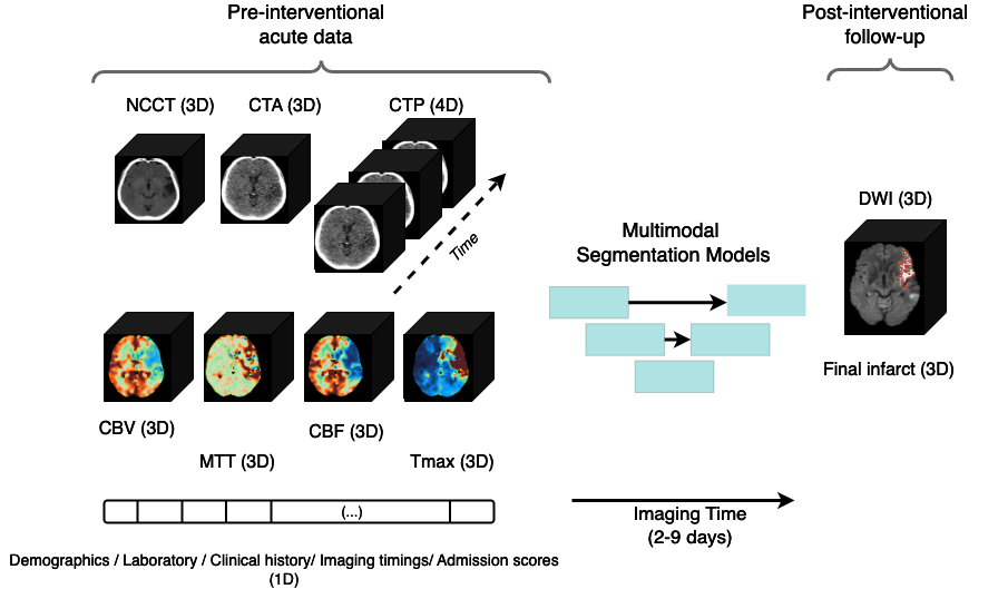
ISLES'24: Improving final infarct prediction in ischemic stroke using multimodal imaging and clinical data
Ezequiel de la Rosa, Ruisheng Su, Mauricio Reyes, Roland Wiest, Evamaria O. Riedel, Florian Kofler, Kaiyuan Yang, Hakim Baazaoui, David Robben, Susanne Wegener, Jan S. Kirschke, Benedikt Wiestler, Bjoern Menze
Accurate estimation of core (irreversibly damaged tissue) and penumbra (salvageable tissue) volumes is essential for ischemic stroke treatment decisions. Perfusion CT, the clinical standard, estimates these volumes but is affected by variations in deconvolution algorithms, implementations, and thresholds. Core tissue expands over time, with growth rates influenced by thrombus location, collateral circulation, and inherent patient-specific factors. Understanding this tissue growth is crucial for determining the need to transfer patients to comprehensive stroke centers, predicting the benefits of additional reperfusion attempts during mechanical thrombectomy, and forecasting final clinical outcomes. This work presents the ISLES'24 challenge, which addresses final post-treatment stroke infarct prediction from pre-interventional acute stroke imaging and clinical data. ISLES'24 establishes a unique 360-degree setting where all feasibly accessible clinical data are available for participants, including full CT acute stroke imaging, sub-acute follow-up MRI, and clinical tabular data. The contributions of this work are two-fold: first, we introduce a standardized benchmarking of final stroke infarct segmentation algorithms through the ISLES'24 challenge; second, we provide insights into infarct segmentation using multimodal imaging and clinical data strategies by identifying outperforming methods on a finely curated dataset. The outputs of this challenge are anticipated to enhance clinical decision-making and improve patient outcome predictions. All ISLES'24 materials, including data, performance evaluation scripts, and leading algorithmic strategies, are available to the research community following url{https://isles-24.grand-challenge.org/}.
Read more8/21/2024

0
From Diagnostic CT to DTI Tractography labels: Using Deep Learning for Corticospinal Tract Injury Assessment and Outcome Prediction in Intracerebral Haemorrhage
Olivia N Murray, Hamied Haroon, Paul Ryu, Hiren Patel, George Harston, Marieke Wermer, Wilmar Jolink, Daniel Hanley, Catharina Klijn, Ulrike Hammerbeck, Adrian Parry-Jones, Timothy Cootes
The preservation of the corticospinal tract (CST) is key to good motor recovery after stroke. The gold standard method of assessing the CST with imaging is diffusion tensor tractography. However, this is not available for most intracerebral haemorrhage (ICH) patients. Non-contrast CT scans are routinely available in most ICH diagnostic pipelines, but delineating white matter from a CT scan is challenging. We utilise nnU-Net, trained on paired diagnostic CT scans and high-directional diffusion tractography maps, to segment the CST from diagnostic CT scans alone, and we show our model reproduces diffusion based tractography maps of the CST with a Dice similarity coefficient of 57%. Surgical haematoma evacuation is sometimes performed after ICH, but published clinical trials to date show that whilst surgery reduces mortality, there is no evidence of improved functional recovery. Restricting surgery to patients with an intact CST may reveal a subset of patients for whom haematoma evacuation improves functional outcome. We investigated the clinical utility of our model in the MISTIE III clinical trial dataset. We found that our model's CST integrity measure significantly predicted outcome after ICH in the acute and chronic time frames, therefore providing a prognostic marker for patients to whom advanced diffusion tensor imaging is unavailable. This will allow for future probing of subgroups who may benefit from surgery.
Read more8/14/2024