LatentArtiFusion: An Effective and Efficient Histological Artifacts Restoration Framework

0

Sign in to get full access
Overview
- LatentArtiFusion is a framework for effectively and efficiently restoring histological artifacts in images.
- The approach leverages latent diffusion models to fuse background and foreground information, enabling high-quality artifact removal.
- Key innovations include a new loss function and attention-based model architecture.
Plain English Explanation
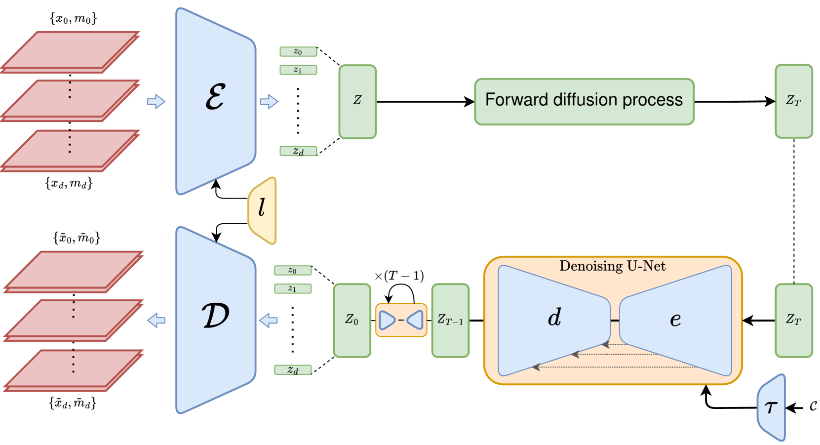
LatentArtiFusion is a tool designed to remove unwanted blemishes or imperfections from histological images. These types of images, used in medical research and diagnostics, can sometimes contain artifacts - things like dust, scratches, or other distortions that can interfere with analysis.
The LatentArtiFusion framework uses a special type of machine learning model called a latent diffusion model to identify and remove these artifacts. Latent diffusion models work by breaking down an image into fundamental building blocks, then reconstructing a clean version of the image without the unwanted elements.
The key innovations in LatentArtiFusion include a new loss function that helps the model focus on the most important parts of the image, and an attention-based architecture that allows the model to better understand the relationships between different regions of the image.
Technical Explanation
The LatentArtiFusion framework leverages latent diffusion models to effectively remove artifacts from histological images. Latent diffusion models work by decomposing an image into a series of latent representations, which are then gradually refined to produce a clean output.
The key innovations in LatentArtiFusion include:
-
A new loss function that combines information about the background and foreground of the image, helping the model focus on the most salient regions.
-
An attention-based architecture that allows the model to better understand the relationships between different parts of the image, further improving artifact removal performance.
The authors evaluate LatentArtiFusion on a range of histological image datasets, demonstrating its effectiveness at restoring images with various types of artifacts, such as dust, scratches, and staining irregularities.
Critical Analysis
The LatentArtiFusion framework presents a promising approach for addressing the challenge of histological artifact removal. The use of latent diffusion models, combined with the novel loss function and attention-based architecture, appears to be an effective and efficient solution.
However, the paper does not address potential limitations or caveats of the approach. For example, it is unclear how LatentArtiFusion would perform on more complex or severe types of artifacts, or how it might scale to larger datasets or higher-resolution images.
Additionally, the authors do not discuss potential ethical considerations or societal implications of their work, such as the impact on medical diagnostics or the importance of maintaining the integrity of histological data.
Conclusion
LatentArtiFusion presents a novel and effective framework for restoring histological images by leveraging latent diffusion models and innovative architectural and loss function design. This work has the potential to significantly improve the quality and reliability of histological analysis, with important implications for medical research and diagnostics.
While the paper demonstrates the efficacy of the approach, further research is needed to address potential limitations and explore the broader societal impact of this technology.
This summary was produced with help from an AI and may contain inaccuracies - check out the links to read the original source documents!
Related Papers
0
LatentArtiFusion: An Effective and Efficient Histological Artifacts Restoration Framework
Zhenqi He, Wenrui Liu, Minghao Yin, Kai Han
Histological artifacts pose challenges for both pathologists and Computer-Aided Diagnosis (CAD) systems, leading to errors in analysis. Current approaches for histological artifact restoration, based on Generative Adversarial Networks (GANs) and pixel-level Diffusion Models, suffer from performance limitations and computational inefficiencies. In this paper, we propose a novel framework, LatentArtiFusion, which leverages the latent diffusion model (LDM) to reconstruct histological artifacts with high performance and computational efficiency. Unlike traditional pixel-level diffusion frameworks, LatentArtiFusion executes the restoration process in a lower-dimensional latent space, significantly improving computational efficiency. Moreover, we introduce a novel regional artifact reconstruction algorithm in latent space to prevent mistransfer in non-artifact regions, distinguishing our approach from GAN-based methods. Through extensive experiments on real-world histology datasets, LatentArtiFusion demonstrates remarkable speed, outperforming state-of-the-art pixel-level diffusion frameworks by more than 30X. It also consistently surpasses GAN-based methods by at least 5% across multiple evaluation metrics. Furthermore, we evaluate the effectiveness of our proposed framework in downstream tissue classification tasks, showcasing its practical utility. Code is available at https://github.com/bugs-creator/LatentArtiFusion.
Read more7/30/2024

0
Equipping Computational Pathology Systems with Artifact Processing Pipelines: A Showcase for Computation and Performance Trade-offs
Neel Kanwal, Farbod Khoraminia, Umay Kiraz, Andres Mosquera-Zamudio, Carlos Monteagudo, Emiel A. M. Janssen, Tahlita C. M. Zuiverloon, Chunmig Rong, Kjersti Engan
Histopathology is a gold standard for cancer diagnosis under a microscopic examination. However, histological tissue processing procedures result in artifacts, which are ultimately transferred to the digitized version of glass slides, known as whole slide images (WSIs). Artifacts are diagnostically irrelevant areas and may result in wrong deep learning (DL) algorithms predictions. Therefore, detecting and excluding artifacts in the computational pathology (CPATH) system is essential for reliable automated diagnosis. In this paper, we propose a mixture of experts (MoE) scheme for detecting five notable artifacts, including damaged tissue, blur, folded tissue, air bubbles, and histologically irrelevant blood from WSIs. First, we train independent binary DL models as experts to capture particular artifact morphology. Then, we ensemble their predictions using a fusion mechanism. We apply probabilistic thresholding over the final probability distribution to improve the sensitivity of the MoE. We developed DL pipelines using two MoEs and two multiclass models of state-of-the-art deep convolutional neural networks (DCNNs) and vision transformers (ViTs). DCNNs-based MoE and ViTs-based MoE schemes outperformed simpler multiclass models and were tested on datasets from different hospitals and cancer types, where MoE using DCNNs yielded the best results. The proposed MoE yields 86.15% F1 and 97.93% sensitivity scores on unseen data, retaining less computational cost for inference than MoE using ViTs. This best performance of MoEs comes with relatively higher computational trade-offs than multiclass models. The proposed artifact detection pipeline will not only ensure reliable CPATH predictions but may also provide quality control.
Read more5/24/2024

0
F2FLDM: Latent Diffusion Models with Histopathology Pre-Trained Embeddings for Unpaired Frozen Section to FFPE Translation
Man M. Ho, Shikha Dubey, Yosep Chong, Beatrice Knudsen, Tolga Tasdizen
The Frozen Section (FS) technique is a rapid and efficient method, taking only 15-30 minutes to prepare slides for pathologists' evaluation during surgery, enabling immediate decisions on further surgical interventions. However, FS process often introduces artifacts and distortions like folds and ice-crystal effects. In contrast, these artifacts and distortions are absent in the higher-quality formalin-fixed paraffin-embedded (FFPE) slides, which require 2-3 days to prepare. While Generative Adversarial Network (GAN)-based methods have been used to translate FS to FFPE images (F2F), they may leave morphological inaccuracies with remaining FS artifacts or introduce new artifacts, reducing the quality of these translations for clinical assessments. In this study, we benchmark recent generative models, focusing on GANs and Latent Diffusion Models (LDMs), to overcome these limitations. We introduce a novel approach that combines LDMs with Histopathology Pre-Trained Embeddings to enhance restoration of FS images. Our framework leverages LDMs conditioned by both text and pre-trained embeddings to learn meaningful features of FS and FFPE histopathology images. Through diffusion and denoising techniques, our approach not only preserves essential diagnostic attributes like color staining and tissue morphology but also proposes an embedding translation mechanism to better predict the targeted FFPE representation of input FS images. As a result, this work achieves a significant improvement in classification performance, with the Area Under the Curve rising from 81.99% to 94.64%, accompanied by an advantageous CaseFD. This work establishes a new benchmark for FS to FFPE image translation quality, promising enhanced reliability and accuracy in histopathology FS image analysis. Our work is available at https://minhmanho.github.io/f2f_ldm/.
Read more4/22/2024
0
3D MRI Synthesis with Slice-Based Latent Diffusion Models: Improving Tumor Segmentation Tasks in Data-Scarce Regimes
Aghiles Kebaili, J'er^ome Lapuyade-Lahorgue, Pierre Vera, Su Ruan
Despite the increasing use of deep learning in medical image segmentation, the limited availability of annotated training data remains a major challenge due to the time-consuming data acquisition and privacy regulations. In the context of segmentation tasks, providing both medical images and their corresponding target masks is essential. However, conventional data augmentation approaches mainly focus on image synthesis. In this study, we propose a novel slice-based latent diffusion architecture designed to address the complexities of volumetric data generation in a slice-by-slice fashion. This approach extends the joint distribution modeling of medical images and their associated masks, allowing a simultaneous generation of both under data-scarce regimes. Our approach mitigates the computational complexity and memory expensiveness typically associated with diffusion models. Furthermore, our architecture can be conditioned by tumor characteristics, including size, shape, and relative position, thereby providing a diverse range of tumor variations. Experiments on a segmentation task using the BRATS2022 confirm the effectiveness of the synthesized volumes and masks for data augmentation.
Read more6/11/2024