F2FLDM: Latent Diffusion Models with Histopathology Pre-Trained Embeddings for Unpaired Frozen Section to FFPE Translation

0

Sign in to get full access
Overview
- Presents a new model called F2FLDM (Frozen Section to FFPE Latent Diffusion Model) for translating histopathology images between different sample preparation methods
- Uses a latent diffusion architecture with histopathology-specific pre-trained embeddings to enable unpaired translation between frozen section (FS) and formalin-fixed, paraffin-embedded (FFPE) tissue samples
- Aims to improve upon previous generative models for this task by leveraging more informative representations learned from large-scale histopathology data
Plain English Explanation
F2FLDM: Latent Diffusion Models with Histopathology Pre-Trained Embeddings for Unpaired Frozen Section to FFPE Translation describes a new machine learning model designed to automatically convert histopathology images from one type of tissue sample preparation (frozen section) to another (formalin-fixed, paraffin-embedded).
Histopathology is the study of diseased tissues under a microscope, and tissue samples can be prepared in different ways that affect their appearance. Frozen section samples provide fast results but may not fully preserve the tissue structure, while FFPE samples take longer to prepare but have better structural preservation. Being able to convert between these two types of images could be useful for applications like digital pathology, where images need to be standardized.
The key innovation in this work is the use of a "latent diffusion" architecture, which learns to generate new images by progressively refining a noisy input. The model is also pre-trained on a large dataset of histopathology images, allowing it to better understand the visual features that are important for this domain. This pre-training helps the model translate between FS and FFPE samples without requiring paired training data, which can be difficult to obtain.
By leveraging more powerful deep learning techniques and histopathology-specific knowledge, the authors aim to improve on previous approaches for this type of image-to-image translation task. The results demonstrate that the F2FLDM model is able to generate realistic FFPE images from FS inputs, suggesting it could be a useful tool for medical image analysis.
Technical Explanation
F2FLDM: Latent Diffusion Models with Histopathology Pre-Trained Embeddings for Unpaired Frozen Section to FFPE Translation introduces a new deep learning model for translating histopathology images between frozen section (FS) and formalin-fixed, paraffin-embedded (FFPE) tissue samples.
The core of the F2FLDM architecture is a latent diffusion model, which generates new images by progressively refining a noisy input representation. This diffusion-based approach has been shown to produce high-quality, diverse images in other domains. To adapt it for histopathology, the authors incorporate pre-trained embeddings that capture domain-specific visual features learned from a large dataset of histopathology images.
A key aspect of the work is the ability to perform unpaired translation between FS and FFPE samples. This is important because obtaining paired training data (i.e., FS and FFPE images of the same tissue) can be challenging in practice. By leveraging the pre-trained embeddings, the F2FLDM model can learn the underlying visual translation between the two modalities without requiring strictly matched input-output pairs.
The paper evaluates the F2FLDM model on a dataset of FS and FFPE breast cancer samples, demonstrating that it outperforms previous generative approaches for this task. Qualitative and quantitative results indicate that the generated FFPE images exhibit realistic tissue structures and staining patterns, suggesting the model has effectively learned the nuanced visual differences between the two tissue preparation methods.
Critical Analysis
The F2FLDM paper presents a promising approach for tackling the challenging problem of unpaired histopathology image translation. By leveraging a latent diffusion architecture and pre-trained histopathology embeddings, the authors have developed a model that can generate realistic FFPE images from FS inputs without requiring paired training data.
One potential limitation of the work is the reliance on a relatively small dataset of FS and FFPE breast cancer samples. While the authors demonstrate strong performance on this specific dataset, it would be valuable to evaluate the model's generalization to a wider range of histopathology use cases and tissue types. [Expanding the evaluation to additional medical image datasets could help validate the broader applicability of the F2FLDM approach.
Additionally, the authors do not explore the potential clinical utility of the generated FFPE images. It would be informative to understand how well these translated images would perform in downstream tasks like disease diagnosis or tissue analysis, compared to real FFPE samples. [Incorporating such multi-modal evaluation could further strengthen the practical relevance of the F2FLDM model.
Another area for further research could be investigating the [potential for coarse-to-fine latent diffusion to improve the F2FLDM architecture. By progressively refining the image generation at multiple scales, this approach could potentially lead to even higher-fidelity FFPE translations.
Overall, the F2FLDM paper presents an exciting step forward in the field of medical image translation, with the potential to significantly impact digital pathology workflows and analytical variability in medical imaging.
Conclusion
The F2FLDM paper introduces a novel deep learning model for translating histopathology images between frozen section (FS) and formalin-fixed, paraffin-embedded (FFPE) tissue samples. By leveraging a latent diffusion architecture and pre-trained histopathology embeddings, the model is able to generate realistic FFPE images from FS inputs without requiring paired training data.
This work represents an important advancement in the field of medical image translation, with potential applications in digital pathology and other areas of computational histology. The ability to automatically convert between FS and FFPE modalities could help standardize image analysis workflows and enable more seamless integration of different tissue preparation methods.
While the current evaluation is limited to a specific breast cancer dataset, the authors have laid the groundwork for further exploring the broader applicability and clinical utility of the F2FLDM approach. Expanding the model's capabilities, evaluating its performance on a wider range of histopathology use cases, and investigating its impact on downstream medical tasks could all be fruitful avenues for future research.
Overall, this paper demonstrates the power of combining state-of-the-art deep learning techniques with domain-specific knowledge to tackle challenging problems in medical imaging. The F2FLDM model represents an important step forward in bridging the gap between different histopathology image modalities, with the potential to improve diagnostic workflows and advance our understanding of disease pathology.
This summary was produced with help from an AI and may contain inaccuracies - check out the links to read the original source documents!
Related Papers
0
F2FLDM: Latent Diffusion Models with Histopathology Pre-Trained Embeddings for Unpaired Frozen Section to FFPE Translation
Man M. Ho, Shikha Dubey, Yosep Chong, Beatrice Knudsen, Tolga Tasdizen
The Frozen Section (FS) technique is a rapid and efficient method, taking only 15-30 minutes to prepare slides for pathologists' evaluation during surgery, enabling immediate decisions on further surgical interventions. However, FS process often introduces artifacts and distortions like folds and ice-crystal effects. In contrast, these artifacts and distortions are absent in the higher-quality formalin-fixed paraffin-embedded (FFPE) slides, which require 2-3 days to prepare. While Generative Adversarial Network (GAN)-based methods have been used to translate FS to FFPE images (F2F), they may leave morphological inaccuracies with remaining FS artifacts or introduce new artifacts, reducing the quality of these translations for clinical assessments. In this study, we benchmark recent generative models, focusing on GANs and Latent Diffusion Models (LDMs), to overcome these limitations. We introduce a novel approach that combines LDMs with Histopathology Pre-Trained Embeddings to enhance restoration of FS images. Our framework leverages LDMs conditioned by both text and pre-trained embeddings to learn meaningful features of FS and FFPE histopathology images. Through diffusion and denoising techniques, our approach not only preserves essential diagnostic attributes like color staining and tissue morphology but also proposes an embedding translation mechanism to better predict the targeted FFPE representation of input FS images. As a result, this work achieves a significant improvement in classification performance, with the Area Under the Curve rising from 81.99% to 94.64%, accompanied by an advantageous CaseFD. This work establishes a new benchmark for FS to FFPE image translation quality, promising enhanced reliability and accuracy in histopathology FS image analysis. Our work is available at https://minhmanho.github.io/f2f_ldm/.
Read more4/22/2024
0
Leveraging Pre-trained Models for FF-to-FFPE Histopathological Image Translation
Qilai Zhang, Jiawen Li, Peiran Liao, Jiali Hu, Tian Guan, Anjia Han, Yonghong He
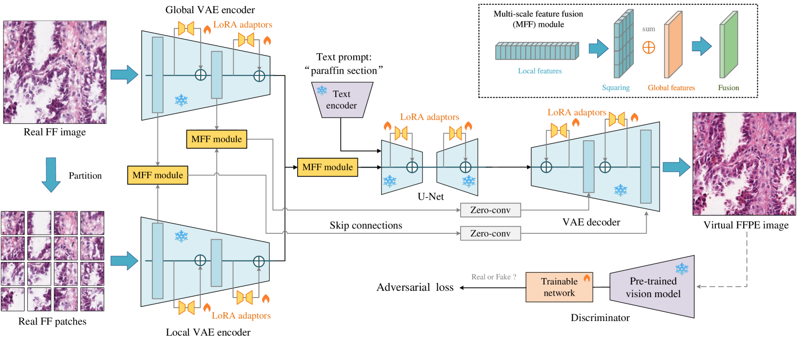
The two primary types of Hematoxylin and Eosin (H&E) slides in histopathology are Formalin-Fixed Paraffin-Embedded (FFPE) and Fresh Frozen (FF). FFPE slides offer high quality histopathological images but require a labor-intensive acquisition process. In contrast, FF slides can be prepared quickly, but the image quality is relatively poor. Our task is to translate FF images into FFPE style, thereby improving the image quality for diagnostic purposes. In this paper, we propose Diffusion-FFPE, a method for FF-to-FFPE histopathological image translation using a pre-trained diffusion model. Specifically, we employ a one-step diffusion model as the generator and fine-tune it with LoRA adapters using adversarial learning objectives. To ensure that the model effectively captures both global structural information and local details, we propose a multi-scale feature fusion (MFF) module. This module utilizes two VAE encoders to extract features of varying image sizes and performs feature fusion before feeding them into the UNet. Furthermore, we utilize a pre-trained vision-language model for histopathology as the backbone for the discriminator to further improve performance We conducted FF-to-FFPE translation experiments on the TCGA-NSCLC datasets, and our method achieved better performance compared to other methods. The code and models are released at https://github.com/QilaiZhang/Diffusion-FFPE.
Read more6/27/2024
🤷
0
FDDM: Unsupervised Medical Image Translation with a Frequency-Decoupled Diffusion Model
Yunxiang Li, Hua-Chieh Shao, Xiaoxue Qian, You Zhang
Diffusion models have demonstrated significant potential in producing high-quality images in medical image translation to aid disease diagnosis, localization, and treatment. Nevertheless, current diffusion models have limited success in achieving faithful image translations that can accurately preserve the anatomical structures of medical images, especially for unpaired datasets. The preservation of structural and anatomical details is essential to reliable medical diagnosis and treatment planning, as structural mismatches can lead to disease misidentification and treatment errors. In this study, we introduce the Frequency Decoupled Diffusion Model (FDDM) for MR-to-CT conversion. FDDM first obtains the anatomical information of the CT image from the MR image through an initial conversion module. This anatomical information then guides a subsequent diffusion model to generate high-quality CT images. Our diffusion model uses a dual-path reverse diffusion process for low-frequency and high-frequency information, achieving a better balance between image quality and anatomical accuracy. We extensively evaluated FDDM using public datasets for brain MR-to-CT and pelvis MR-to-CT translations, demonstrating its superior performance to other GAN-based, VAE-based, and diffusion-based models. The evaluation metrics included Frechet Inception Distance (FID), Peak Signal-to-Noise Ratio (PSNR), and Structural Similarity Index Measure (SSIM). FDDM achieved the best scores on all metrics for both datasets, particularly excelling in FID, with scores of 25.9 for brain data and 29.2 for pelvis data, significantly outperforming other methods. These results demonstrate that FDDM can generate high-quality target domain images while maintaining the accuracy of translated anatomical structures.
Read more6/28/2024

0
LatentArtiFusion: An Effective and Efficient Histological Artifacts Restoration Framework
Zhenqi He, Wenrui Liu, Minghao Yin, Kai Han
Histological artifacts pose challenges for both pathologists and Computer-Aided Diagnosis (CAD) systems, leading to errors in analysis. Current approaches for histological artifact restoration, based on Generative Adversarial Networks (GANs) and pixel-level Diffusion Models, suffer from performance limitations and computational inefficiencies. In this paper, we propose a novel framework, LatentArtiFusion, which leverages the latent diffusion model (LDM) to reconstruct histological artifacts with high performance and computational efficiency. Unlike traditional pixel-level diffusion frameworks, LatentArtiFusion executes the restoration process in a lower-dimensional latent space, significantly improving computational efficiency. Moreover, we introduce a novel regional artifact reconstruction algorithm in latent space to prevent mistransfer in non-artifact regions, distinguishing our approach from GAN-based methods. Through extensive experiments on real-world histology datasets, LatentArtiFusion demonstrates remarkable speed, outperforming state-of-the-art pixel-level diffusion frameworks by more than 30X. It also consistently surpasses GAN-based methods by at least 5% across multiple evaluation metrics. Furthermore, we evaluate the effectiveness of our proposed framework in downstream tissue classification tasks, showcasing its practical utility. Code is available at https://github.com/bugs-creator/LatentArtiFusion.
Read more7/30/2024