Model Ensemble for Brain Tumor Segmentation in Magnetic Resonance Imaging

0

Sign in to get full access
Overview
- This paper presents a model ensemble approach for brain tumor segmentation in magnetic resonance imaging (MRI) data.
- The ensemble combines multiple deep learning models to improve the accuracy of brain tumor segmentation, including pediatric brain tumors, meningioma, and metastases.
- The proposed method is evaluated on publicly available brain tumor datasets and demonstrates improved performance compared to individual models.
Plain English Explanation
Brain tumors are abnormal growths that can develop in the brain. Accurately identifying and segmenting brain tumors in medical images, such as MRI scans, is crucial for diagnosis and treatment planning. This paper introduces a novel approach to improve brain tumor segmentation using a model ensemble.
The researchers combined multiple deep learning models, each trained to identify different types of brain tumors, including those found in children as well as more common types like meningioma and metastases. By combining the predictions from these specialized models, the ensemble approach was able to achieve better overall accuracy in detecting and delineating brain tumors compared to using a single model alone.
This is important because accurately segmenting brain tumors can help doctors better understand the extent and location of the tumor, which informs the best treatment plan for the patient. The improved performance of the ensemble model could lead to more reliable and precise brain tumor diagnosis and management.
Technical Explanation
The researchers developed a model ensemble for brain tumor segmentation in MRI data. The ensemble combined multiple deep learning models, each trained to segment specific types of brain tumors, such as pediatric brain tumors, meningioma, and metastases.
The ensemble approach leverages the strengths of these specialized models to improve the overall segmentation accuracy. The individual models were trained on publicly available brain tumor datasets, and their predictions were combined using a weighted voting scheme to generate the final segmentation results.
The researchers evaluated the performance of the ensemble model on several brain tumor segmentation benchmarks and compared it to the individual models. The results showed that the ensemble approach outperformed the individual models, demonstrating the benefits of combining multiple, specialized deep learning models for this task.
Critical Analysis
The paper provides a comprehensive evaluation of the proposed ensemble model, including comparisons to individual models and state-of-the-art methods. However, the researchers acknowledge that the ensemble approach may be computationally more expensive than using a single model, as it requires running multiple models and combining their outputs.
Additionally, the paper does not explore the specific contributions of each individual model within the ensemble or the sensitivity of the ensemble's performance to the choice of individual models. Further research could investigate these aspects to provide deeper insights into the ensemble's behavior and potential areas for improvement.
Conclusion
This paper presents a promising approach for improving brain tumor segmentation in MRI data by leveraging a model ensemble. The combination of specialized deep learning models for different tumor types helps to enhance the overall segmentation accuracy, which can have significant implications for clinical diagnosis and treatment planning. While the ensemble approach may be computationally more demanding, the improved performance could make it a valuable tool for medical practitioners working with brain tumor imaging data.
This summary was produced with help from an AI and may contain inaccuracies - check out the links to read the original source documents!
Related Papers
0
Model Ensemble for Brain Tumor Segmentation in Magnetic Resonance Imaging
Daniel Capell'an-Mart'in, Zhifan Jiang, Abhijeet Parida, Xinyang Liu, Van Lam, Hareem Nisar, Austin Tapp, Sarah Elsharkawi, Maria J. Ledesma-Carbayo, Syed Muhammad Anwar, Marius George Linguraru
Segmenting brain tumors in multi-parametric magnetic resonance imaging enables performing quantitative analysis in support of clinical trials and personalized patient care. This analysis provides the potential to impact clinical decision-making processes, including diagnosis and prognosis. In 2023, the well-established Brain Tumor Segmentation (BraTS) challenge presented a substantial expansion with eight tasks and 4,500 brain tumor cases. In this paper, we present a deep learning-based ensemble strategy that is evaluated for newly included tumor cases in three tasks: pediatric brain tumors (PED), intracranial meningioma (MEN), and brain metastases (MET). In particular, we ensemble outputs from state-of-the-art nnU-Net and Swin UNETR models on a region-wise basis. Furthermore, we implemented a targeted post-processing strategy based on a cross-validated threshold search to improve the segmentation results for tumor sub-regions. The evaluation of our proposed method on unseen test cases for the three tasks resulted in lesion-wise Dice scores for PED: 0.653, 0.809, 0.826; MEN: 0.876, 0.867, 0.849; and MET: 0.555, 0.6, 0.58; for the enhancing tumor, tumor core, and whole tumor, respectively. Our method was ranked first for PED, third for MEN, and fourth for MET, respectively.
Read more9/14/2024
0
On Enhancing Brain Tumor Segmentation Across Diverse Populations with Convolutional Neural Networks
Fadillah Maani, Anees Ur Rehman Hashmi, Numan Saeed, Mohammad Yaqub
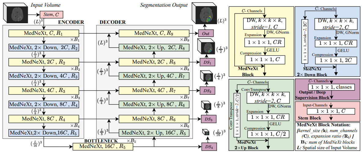
Brain tumor segmentation is a fundamental step in assessing a patient's cancer progression. However, manual segmentation demands significant expert time to identify tumors in 3D multimodal brain MRI scans accurately. This reliance on manual segmentation makes the process prone to intra- and inter-observer variability. This work proposes a brain tumor segmentation method as part of the BraTS-GoAT challenge. The task is to segment tumors in brain MRI scans automatically from various populations, such as adults, pediatrics, and underserved sub-Saharan Africa. We employ a recent CNN architecture for medical image segmentation, namely MedNeXt, as our baseline, and we implement extensive model ensembling and postprocessing for inference. Our experiments show that our method performs well on the unseen validation set with an average DSC of 85.54% and HD95 of 27.88. The code is available on https://github.com/BioMedIA-MBZUAI/BraTS2024_BioMedIAMBZ.
Read more5/7/2024
🤿
0
An Optimized Ensemble Deep Learning Model For Brain Tumor Classification
Md. Alamin Talukder, Md. Manowarul Islam, Md Ashraf Uddin
Brain tumors present a grave risk to human life, demanding precise and timely diagnosis for effective treatment. Inaccurate identification of brain tumors can significantly diminish life expectancy, underscoring the critical need for precise diagnostic methods. Manual identification of brain tumors within vast Magnetic Resonance Imaging (MRI) image datasets is arduous and time-consuming. Thus, the development of a reliable deep learning (DL) model is essential to enhance diagnostic accuracy and ultimately save lives. This study introduces an innovative optimization-based deep ensemble approach employing transfer learning (TL) to efficiently classify brain tumors. Our methodology includes meticulous preprocessing, reconstruction of TL architectures, fine-tuning, and ensemble DL models utilizing weighted optimization techniques such as Genetic Algorithm-based Weight Optimization (GAWO) and Grid Search-based Weight Optimization (GSWO). Experimentation is conducted on the Figshare Contrast-Enhanced MRI (CE-MRI) brain tumor dataset, comprising 3064 images. Our approach achieves notable accuracy scores, with Xception, ResNet50V2, ResNet152V2, InceptionResNetV2, GAWO, and GSWO attaining 99.42%, 98.37%, 98.22%, 98.26%, 99.71%, and 99.76% accuracy, respectively. Notably, GSWO demonstrates superior accuracy, averaging 99.76% accuracy across five folds on the Figshare CE-MRI brain tumor dataset. The comparative analysis highlights the significant performance enhancement of our proposed model over existing counterparts. In conclusion, our optimized deep ensemble model exhibits exceptional accuracy in swiftly classifying brain tumors. Furthermore, it has the potential to assist neurologists and clinicians in making accurate and immediate diagnostic decisions.
Read more5/7/2024
0
Segmentation of Brain Metastases in MRI: A Two-Stage Deep Learning Approach with Modality Impact Study
Yousef Sadegheih, Dorit Merhof
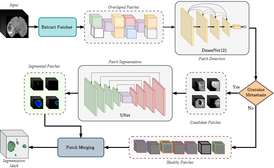
Brain metastasis segmentation poses a significant challenge in medical imaging due to the complex presentation and variability in size and location of metastases. In this study, we first investigate the impact of different imaging modalities on segmentation performance using a 3D U-Net. Through a comprehensive analysis, we determine that combining all available modalities does not necessarily enhance performance. Instead, the combination of T1-weighted with contrast enhancement (T1c), T1-weighted (T1), and FLAIR modalities yields superior results. Building on these findings, we propose a two-stage detection and segmentation model specifically designed to accurately segment brain metastases. Our approach demonstrates that leveraging three key modalities (T1c, T1, and FLAIR) achieves significantly higher accuracy compared to single-pass deep learning models. This targeted combination allows for precise segmentation, capturing even small metastases that other models often miss. Our model sets a new benchmark in brain metastasis segmentation, highlighting the importance of strategic modality selection and multi-stage processing in medical imaging. Our implementation is freely accessible to the research community on href{https://github.com/xmindflow/Met-Seg}{GitHub}.
Read more7/22/2024