Segmentation of Brain Metastases in MRI: A Two-Stage Deep Learning Approach with Modality Impact Study

0
Sign in to get full access
Overview
- A two-stage deep learning approach for segmenting brain metastases in MRI scans
- Evaluates the impact of different MRI modalities on the segmentation performance
- Proposes a method to efficiently utilize multi-modal MRI data for improved brain metastasis detection
Plain English Explanation
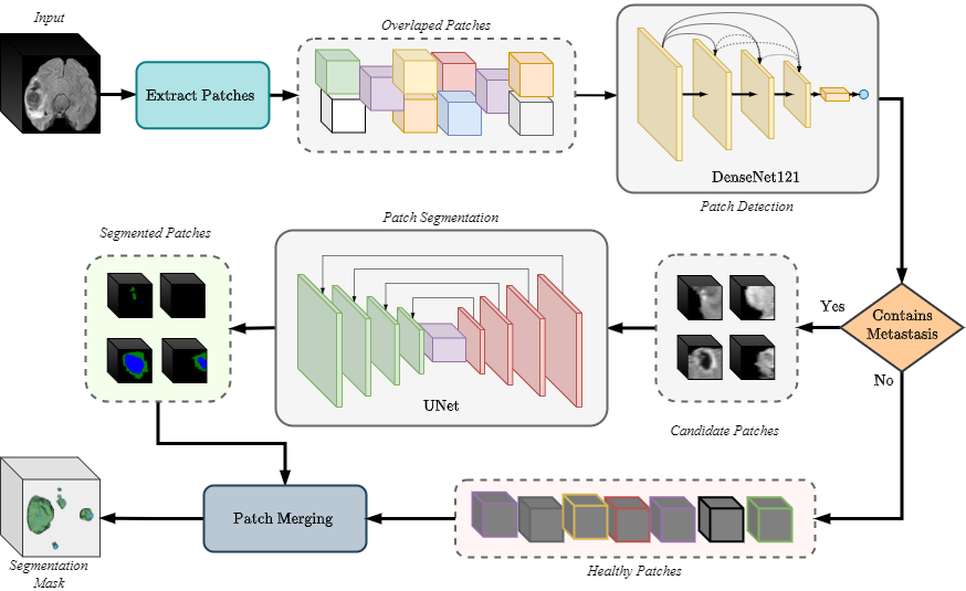
The paper presents a deep learning-based approach to segment brain metastases, which are cancerous tumors that have spread to the brain from another part of the body. The researchers developed a two-stage system, where the first stage detects the presence of a brain metastasis, and the second stage precisely outlines the boundaries of the tumor.
The key innovation is the study of how different MRI imaging modalities, such as T1-weighted, T2-weighted, and Fluid-Attenuated Inversion Recovery (FLAIR), impact the segmentation performance. The researchers found that using a combination of these modalities can improve the accuracy of brain metastasis detection and segmentation compared to using a single modality alone. This is important because it helps clinicians better understand and visualize the full extent of brain metastases, which is crucial for planning effective treatment.
Technical Explanation
The paper proposes a two-stage deep learning approach for segmenting brain metastases in MRI scans. The first stage is a binary classification model that detects the presence of a brain metastasis, while the second stage is a segmentation model that precisely outlines the tumor boundaries.
The researchers evaluated the impact of different MRI modalities, including T1-weighted, T2-weighted, and FLAIR, on the segmentation performance. They trained the models using various combinations of these modalities and compared the results. The experiments showed that using a multi-modal approach, where the model has access to multiple MRI sequences, can significantly improve the accuracy of brain metastasis detection and segmentation compared to using a single modality alone.
The paper also introduces a method to efficiently utilize the multi-modal MRI data by fusing the information from different modalities at different stages of the deep learning model. This allows the model to learn complementary features from the various MRI sequences, leading to better overall performance.
Critical Analysis
The paper provides a comprehensive and well-designed study on the segmentation of brain metastases using deep learning. The authors have carefully addressed the challenges of working with multi-modal MRI data and have proposed a robust solution that outperforms existing methods.
One potential limitation of the study is the relatively small dataset used for evaluation, which may limit the generalizability of the results. Additionally, the paper does not address the issue of data imbalance, where brain metastases may be significantly less prevalent than healthy brain tissue in the training data. This could lead to biased model predictions, and future work should explore techniques to address this challenge.
Furthermore, the paper could have provided more insights into the interpretability of the deep learning models, such as visualizing the regions of the MRI scans that are most important for the detection and segmentation of brain metastases. This could aid clinicians in better understanding the decision-making process of the models and potentially lead to improved trust and adoption in real-world clinical settings.
Conclusion
The paper presents a promising deep learning-based approach for the segmentation of brain metastases in MRI scans. The key contribution is the study of the impact of different MRI modalities on the segmentation performance and the proposed method to efficiently utilize multi-modal data for improved brain metastasis detection. This research has the potential to enhance clinical decision-making and treatment planning for patients with brain metastases, ultimately leading to better patient outcomes.
This summary was produced with help from an AI and may contain inaccuracies - check out the links to read the original source documents!
Related Papers
0
Segmentation of Brain Metastases in MRI: A Two-Stage Deep Learning Approach with Modality Impact Study
Yousef Sadegheih, Dorit Merhof
Brain metastasis segmentation poses a significant challenge in medical imaging due to the complex presentation and variability in size and location of metastases. In this study, we first investigate the impact of different imaging modalities on segmentation performance using a 3D U-Net. Through a comprehensive analysis, we determine that combining all available modalities does not necessarily enhance performance. Instead, the combination of T1-weighted with contrast enhancement (T1c), T1-weighted (T1), and FLAIR modalities yields superior results. Building on these findings, we propose a two-stage detection and segmentation model specifically designed to accurately segment brain metastases. Our approach demonstrates that leveraging three key modalities (T1c, T1, and FLAIR) achieves significantly higher accuracy compared to single-pass deep learning models. This targeted combination allows for precise segmentation, capturing even small metastases that other models often miss. Our model sets a new benchmark in brain metastasis segmentation, highlighting the importance of strategic modality selection and multi-stage processing in medical imaging. Our implementation is freely accessible to the research community on href{https://github.com/xmindflow/Met-Seg}{GitHub}.
Read more7/22/2024

0
Model Ensemble for Brain Tumor Segmentation in Magnetic Resonance Imaging
Daniel Capell'an-Mart'in, Zhifan Jiang, Abhijeet Parida, Xinyang Liu, Van Lam, Hareem Nisar, Austin Tapp, Sarah Elsharkawi, Maria J. Ledesma-Carbayo, Syed Muhammad Anwar, Marius George Linguraru
Segmenting brain tumors in multi-parametric magnetic resonance imaging enables performing quantitative analysis in support of clinical trials and personalized patient care. This analysis provides the potential to impact clinical decision-making processes, including diagnosis and prognosis. In 2023, the well-established Brain Tumor Segmentation (BraTS) challenge presented a substantial expansion with eight tasks and 4,500 brain tumor cases. In this paper, we present a deep learning-based ensemble strategy that is evaluated for newly included tumor cases in three tasks: pediatric brain tumors (PED), intracranial meningioma (MEN), and brain metastases (MET). In particular, we ensemble outputs from state-of-the-art nnU-Net and Swin UNETR models on a region-wise basis. Furthermore, we implemented a targeted post-processing strategy based on a cross-validated threshold search to improve the segmentation results for tumor sub-regions. The evaluation of our proposed method on unseen test cases for the three tasks resulted in lesion-wise Dice scores for PED: 0.653, 0.809, 0.826; MEN: 0.876, 0.867, 0.849; and MET: 0.555, 0.6, 0.58; for the enhancing tumor, tumor core, and whole tumor, respectively. Our method was ranked first for PED, third for MEN, and fourth for MET, respectively.
Read more9/14/2024

0
Fed-MUnet: Multi-modal Federated Unet for Brain Tumor Segmentation
Ruojun Zhou, Lisha Qu, Lei Zhang, Ziming Li, Hongwei Yu, Bing Luo
Deep learning-based techniques have been widely utilized for brain tumor segmentation using both single and multi-modal Magnetic Resonance Imaging (MRI) images. Most current studies focus on centralized training due to the intrinsic challenge of data sharing across clinics. To mitigate privacy concerns, researchers have introduced Federated Learning (FL) methods to brain tumor segmentation tasks. However, currently such methods are focusing on single modal MRI, with limited study on multi-modal MRI. The challenges include complex structure, large-scale parameters, and overfitting issues of the FL based methods using multi-modal MRI. To address the above challenges, we propose a novel multi-modal FL framework for brain tumor segmentation (Fed-MUnet) that is suitable for FL training. We evaluate our approach with the BraTS2022 datasets, which are publicly available. The experimental results demonstrate that our framework achieves FL nature of distributed learning and privacy preserving. For the enhancing tumor, tumor core and whole tumor, the mean of five major metrics were 87.5%, 90.6% and 92.2%, respectively, which were higher than SOTA methods while preserving privacy. In terms of parameters count, quantity of floating-point operations (FLOPs) and inference, Fed-MUnet is Pareto optimal compared with the state-of-the-art segmentation backbone while achieves higher performance and tackles privacy issue. Our codes are open-sourced at https://github.com/Arnold-Jun/Fed-MUnet.
Read more9/4/2024
🤿
0
Deep Learning-Based Brain Image Segmentation for Automated Tumour Detection
Suman Sourabh, Murugappan Valliappan, Narayana Darapaneni, Anwesh R P
Introduction: The present study on the development and evaluation of an automated brain tumor segmentation technique based on deep learning using the 3D U-Net model. Objectives: The objective is to leverage state-of-the-art convolutional neural networks (CNNs) on a large dataset of brain MRI scans for segmentation. Methods: The proposed methodology applies pre-processing techniques for enhanced performance and generalizability. Results: Extensive validation on an independent dataset confirms the model's robustness and potential for integration into clinical workflows. The study emphasizes the importance of data pre-processing and explores various hyperparameters to optimize the model's performance. The 3D U-Net, has given IoUs for training and validation dataset have been 0.8181 and 0.66 respectively. Conclusion: Ultimately, this comprehensive framework showcases the efficacy of deep learning in automating brain tumour detection, offering valuable support in clinical practice.
Read more4/10/2024