ResNCT: A Deep Learning Model for the Synthesis of Nephrographic Phase Images in CT Urography

0
🤿
Sign in to get full access
Overview
- This research paper presents a transformer-based deep learning model, called the Residual transformer model for Nephrographic phase CT image synthesis (ResNCT), for synthesizing nephrographic phase images in CT urography (CTU) examinations from unenhanced and urographic phase images.
- The model was developed and evaluated on a dataset of 119 patients with three-phase CT urography studies.
- The synthesized nephrographic phase images were compared to the corresponding ground truth nephrographic phase images using various performance metrics.
Plain English Explanation
In medical imaging, CT urography (CTU) examinations typically involve taking multiple scans of the patient at different stages of a contrast agent moving through the body. This includes an unenhanced scan, a urographic phase scan, and a nephrographic phase scan. The nephrographic phase scan is important for diagnosing certain conditions, but it also exposes the patient to additional radiation.
The researchers in this study developed a deep learning model, called ResNCT, that can generate the nephrographic phase images from just the unenhanced and urographic phase scans. This means the nephrographic phase scan could potentially be skipped, reducing the radiation exposure for the patient by 33%.
The model was trained on a dataset of 119 patients and was able to generate synthetic nephrographic phase images that were very similar to the actual ground truth nephrographic phase images, as measured by various image quality metrics. This suggests the model could be a useful tool for reducing radiation exposure in CTU exams without sacrificing image quality.
Technical Explanation
The researchers used a dataset of 119 patients with three-phase CT urography studies to develop and evaluate the ResNCT model. The three phases for each patient were first aligned using an affine registration algorithm.
The ResNCT model was then trained to take the unenhanced and urographic phase images as inputs and generate the corresponding nephrographic phase images. The model architecture incorporates residual connections and transformer-based components, similar to techniques used in models like WituNet and CT image denoising methods.
The synthesized nephrographic phase images were compared to the ground truth nephrographic phase images using several performance metrics, including peak signal-to-noise ratio (PSNR), structural similarity index (SSIM), normalized cross-correlation coefficient (NCC), mean absolute error (MAE), and root mean squared error (RMSE). The model achieved high scores on these metrics, indicating the synthesized images were very similar to the actual nephrographic phase images.
Critical Analysis
The researchers mentioned that the ResNCT model provides a way to eliminate the acquisition of the nephrographic phase in CTU exams, resulting in a 33% reduction in radiation dose for patients. This is a significant potential benefit, as reducing radiation exposure is an important goal in medical imaging.
However, the research did not address potential limitations or challenges in deploying such a model in a clinical setting. For example, the model was trained and evaluated on a relatively small dataset of 119 patients, so its performance on a larger and more diverse patient population is still unknown. Additionally, the researchers did not discuss how the model would need to be integrated into existing clinical workflows or how its outputs would be validated by radiologists.
Further research would be needed to address these practical considerations and ensure the ResNCT model can be safely and effectively implemented in real-world CTU examinations. Nonetheless, the promising results presented in this paper suggest the model has the potential to significantly improve the patient experience and reduce radiation exposure in CT urography.
Conclusion
This research paper presents a novel transformer-based deep learning model, ResNCT, that can generate nephrographic phase CT images from unenhanced and urographic phase inputs. The model was able to produce synthetic nephrographic images that were highly similar to the ground truth images, as demonstrated by various performance metrics.
If successfully implemented in clinical practice, the ResNCT model could potentially eliminate the need for the nephrographic phase scan in CT urography examinations, reducing the radiation exposure for patients by 33%. This would be a significant advancement in medical imaging that could improve the patient experience and safety while maintaining diagnostic image quality.
This summary was produced with help from an AI and may contain inaccuracies - check out the links to read the original source documents!
Related Papers
🤿
0
ResNCT: A Deep Learning Model for the Synthesis of Nephrographic Phase Images in CT Urography
Syed Jamal Safdar Gardezi (Department of Radiology, University of Wisconsin School of Medicine & Public Health, Madison, WI, USA), Lucas Aronson (Department of Radiology, University of Wisconsin School of Medicine & Public Health, Madison, WI, USA), Peter Wawrzyn (Department of Biomedical Engineering, University of Wisconsin Madison, Madison, WI, USA), Hongkun Yu (Department of Biomedical Engineering, University of Wisconsin Madison, Madison, WI, USA), E. Jason Abel (Department of Urology, University of Wisconsin School of Medicine & Public Health, Madison, WI, USA), Daniel D. Shapiro (Department of Urology, University of Wisconsin School of Medicine & Public Health, Madison, WI, USA), Meghan G. Lubner (Department of Radiology, University of Wisconsin School of Medicine & Public Health, Madison, WI, USA), Joshua Warner (Department of Radiology, University of Wisconsin School of Medicine & Public Health, Madison, WI, USA), Giuseppe Toia (Department of Radiology, University of Wisconsin School of Medicine & Public Health, Madison, WI, USA), Lu Mao (Department of Biostatistics, University of Wisconsin School of Medicine & Public Health, Madison, WI, USA), Pallavi Tiwari (Department of Radiology, University of Wisconsin School of Medicine & Public Health, Madison, WI, USA, Department of Biomedical Engineering, University of Wisconsin Madison, Madison, WI, USA), Andrew L. Wentland (Department of Radiology, University of Wisconsin School of Medicine & Public Health, Madison, WI, USA, Department of Biomedical Engineering, University of Wisconsin Madison, Madison, WI, USA, Department of Medical Physics, University of Wisconsin School of Medicine & Public Health, Madison, WI, USA)
Purpose: To develop and evaluate a transformer-based deep learning model for the synthesis of nephrographic phase images in CT urography (CTU) examinations from the unenhanced and urographic phases. Materials and Methods: This retrospective study was approved by the local Institutional Review Board. A dataset of 119 patients (mean $pm$ SD age, 65 $pm$ 12 years; 75/44 males/females) with three-phase CT urography studies was curated for deep learning model development. The three phases for each patient were aligned with an affine registration algorithm. A custom model, coined Residual transformer model for Nephrographic phase CT image synthesis (ResNCT), was developed and implemented with paired inputs of non-contrast and urographic sets of images trained to produce the nephrographic phase images, that were compared with the corresponding ground truth nephrographic phase images. The synthesized images were evaluated with multiple performance metrics, including peak signal to noise ratio (PSNR), structural similarity index (SSIM), normalized cross correlation coefficient (NCC), mean absolute error (MAE), and root mean squared error (RMSE). Results: The ResNCT model successfully generated synthetic nephrographic images from non-contrast and urographic image inputs. With respect to ground truth nephrographic phase images, the images synthesized by the model achieved high PSNR (27.8 $pm$ 2.7 dB), SSIM (0.88 $pm$ 0.05), and NCC (0.98 $pm$ 0.02), and low MAE (0.02 $pm$ 0.005) and RMSE (0.042 $pm$ 0.016). Conclusion: The ResNCT model synthesized nephrographic phase CT images with high similarity to ground truth images. The ResNCT model provides a means of eliminating the acquisition of the nephrographic phase with a resultant 33% reduction in radiation dose for CTU examinations.
Read more5/30/2024
0
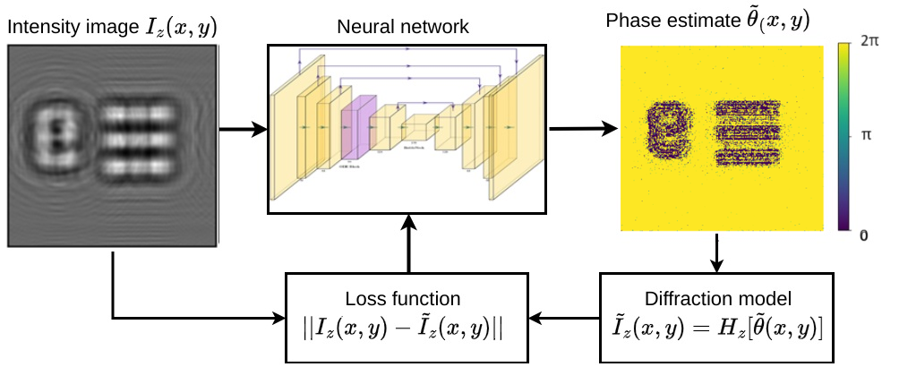
Res-U2Net: Untrained Deep Learning for Phase Retrieval and Image Reconstruction
Carlos Osorio Quero, Daniel Leykam, Irving Rondon Ojeda
Conventional deep learning-based image reconstruction methods require a large amount of training data which can be hard to obtain in practice. Untrained deep learning methods overcome this limitation by training a network to invert a physical model of the image formation process. Here we present a novel untrained Res-U2Net model for phase retrieval. We use the extracted phase information to determine changes in an object's surface and generate a mesh representation of its 3D structure. We compare the performance of Res-U2Net phase retrieval against UNet and U2Net using images from the GDXRAY dataset.
Read more4/11/2024
🧠
0
Automatic Segmentation of the Kidneys and Cystic Renal Lesions on Non-Contrast CT Using a Convolutional Neural Network
Lucas Aronson (Department of Radiology, University of Wisconsin School of Medicine & Public Health, Madison, WI, USA), Ruben Ngnitewe Massaa (Department of Radiology, University of Wisconsin School of Medicine & Public Health, Madison, WI, USA), Syed Jamal Safdar Gardezi (Department of Radiology, University of Wisconsin School of Medicine & Public Health, Madison, WI, USA), Andrew L. Wentland (Department of Radiology, University of Wisconsin School of Medicine & Public Health, Madison, WI, USA, Department of Medical Physics, University of Wisconsin School of Medicine & Public Health, Madison, WI, USA, Department of Biomedical Engineering, University of Wisconsin School of Medicine & Public Health, Madison, WI, USA)
Objective: Automated segmentation tools are useful for calculating kidney volumes rapidly and accurately. Furthermore, these tools have the power to facilitate large-scale image-based artificial intelligence projects by generating input labels, such as for image registration algorithms. Prior automated segmentation models have largely ignored non-contrast computed tomography (CT) imaging. This work aims to implement and train a deep learning (DL) model to segment the kidneys and cystic renal lesions (CRLs) from non-contrast CT scans. Methods: Manual segmentation of the kidneys and CRLs was performed on 150 non-contrast abdominal CT scans. The data were divided into an 80/20 train/test split and a deep learning (DL) model was trained to segment the kidneys and CRLs. Various scoring metrics were used to assess model performance, including the Dice Similarity Coefficient (DSC), Jaccard Index (JI), and absolute and percent error kidney volume and lesion volume. Bland-Altman (B-A) analysis was performed to compare manual versus DL-based kidney volumes. Results: The DL model achieved a median kidney DSC of 0.934, median CRL DSC of 0.711, and total median study DSC of 0.823. Average volume errors were 0.9% for renal parenchyma, 37.0% for CRLs, and 2.2% overall. B-A analysis demonstrated that DL-based volumes tended to be greater than manual volumes, with a mean bias of +3.0 ml (+/- 2 SD of +/- 50.2 ml). Conclusion: A deep learning model trained to segment kidneys and cystic renal lesions on non-contrast CT examinations was able to provide highly accurate segmentations, with a median kidney Dice Similarity Coefficient of 0.934. Keywords: deep learning; kidney segmentation; artificial intelligence; convolutional neural networks.
Read more5/15/2024
🌿
0
Enhancing Super-Resolution Networks through Realistic Thick-Slice CT Simulation
Zeyu Tang, Xiaodan Xing, Guang Yang
Deep learning-based Generative Models have the potential to convert low-resolution CT images into high-resolution counterparts without long acquisition times and increased radiation exposure in thin-slice CT imaging. However, procuring appropriate training data for these Super-Resolution (SR) models is challenging. Previous SR research has simulated thick-slice CT images from thin-slice CT images to create training pairs. However, these methods either rely on simplistic interpolation techniques that lack realism or sinogram reconstruction, which require the release of raw data and complex reconstruction algorithms. Thus, we introduce a simple yet realistic method to generate thick CT images from thin-slice CT images, facilitating the creation of training pairs for SR algorithms. The training pairs produced by our method closely resemble real data distributions (PSNR=49.74 vs. 40.66, p$<$0.05). A multivariate Cox regression analysis involving thick slice CT images with lung fibrosis revealed that only the radiomics features extracted using our method demonstrated a significant correlation with mortality (HR=1.19 and HR=1.14, p$<$0.005). This paper represents the first to identify and address the challenge of generating appropriate paired training data for Deep Learning-based CT SR models, which enhances the efficacy and applicability of SR models in real-world scenarios.
Read more6/4/2024