Functional Imaging Constrained Diffusion for Brain PET Synthesis from Structural MRI
2405.02504
0
0
Abstract
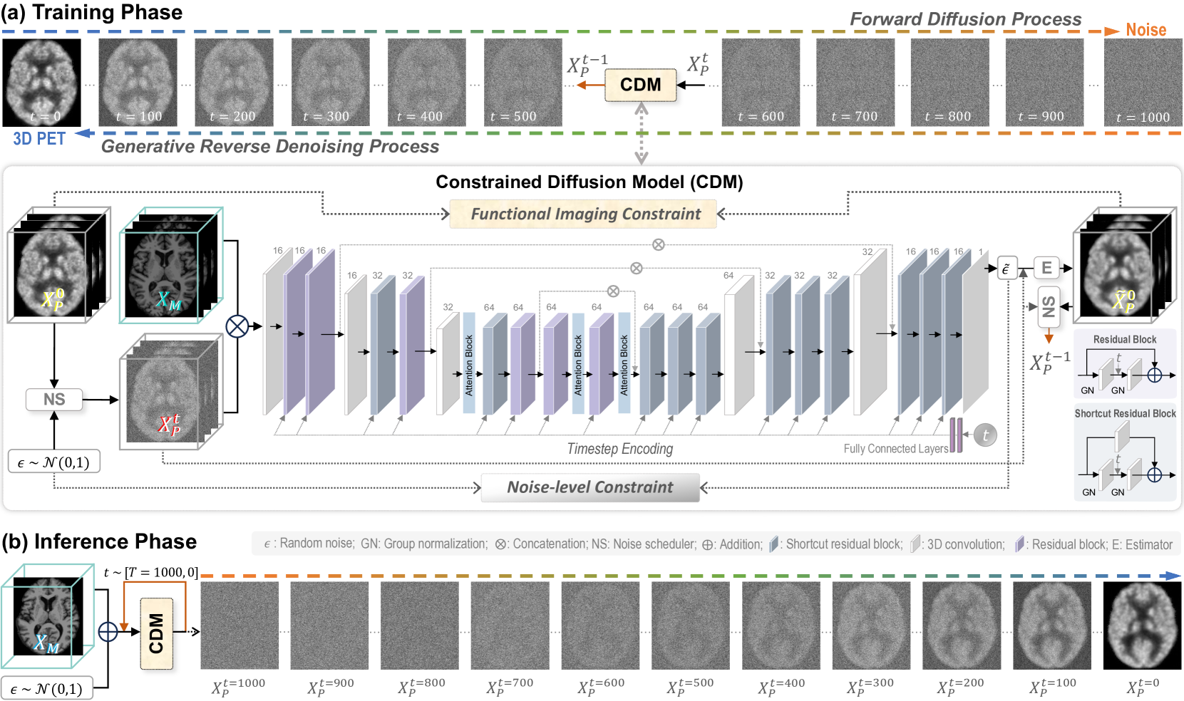
Magnetic resonance imaging (MRI) and positron emission tomography (PET) are increasingly used in multimodal analysis of neurodegenerative disorders. While MRI is broadly utilized in clinical settings, PET is less accessible. Many studies have attempted to use deep generative models to synthesize PET from MRI scans. However, they often suffer from unstable training and inadequately preserve brain functional information conveyed by PET. To this end, we propose a functional imaging constrained diffusion (FICD) framework for 3D brain PET image synthesis with paired structural MRI as input condition, through a new constrained diffusion model (CDM). The FICD introduces noise to PET and then progressively removes it with CDM, ensuring high output fidelity throughout a stable training phase. The CDM learns to predict denoised PET with a functional imaging constraint introduced to ensure voxel-wise alignment between each denoised PET and its ground truth. Quantitative and qualitative analyses conducted on 293 subjects with paired T1-weighted MRI and 18F-fluorodeoxyglucose (FDG)-PET scans suggest that FICD achieves superior performance in generating FDG-PET data compared to state-of-the-art methods. We further validate the effectiveness of the proposed FICD on data from a total of 1,262 subjects through three downstream tasks, with experimental results suggesting its utility and generalizability.
Create account to get full access
Overview
• This research paper presents a method for synthesizing brain positron emission tomography (PET) images from structural magnetic resonance imaging (MRI) data, using a diffusion model constrained by functional imaging information.
• The proposed approach aims to address the challenges of acquiring PET data, which is more complex and expensive than MRI, by leveraging the wealth of structural MRI data to generate realistic PET-like images.
• The method incorporates functional imaging constraints, such as functional MRI (fMRI) or amyloid PET, to guide the synthesis process and improve the accuracy of the generated PET-like images.
Plain English Explanation
Brain imaging is an important tool for studying the structure and function of the human brain. Positron emission tomography (PET) is a type of brain imaging that can provide information about brain activity and various brain-related disorders, such as Alzheimer's disease. However, PET scans are more complex and expensive to acquire compared to magnetic resonance imaging (MRI) scans, which are more widely available and commonly used.
This research proposes a method to generate realistic PET-like images from structural MRI data, by using a diffusion model that is constrained by functional imaging information. The key idea is to leverage the wealth of existing MRI data to synthesize PET-like images, which can then be used for various brain research and clinical applications, without the need for additional PET scans.
The researchers incorporate functional imaging data, such as functional MRI (fMRI) or amyloid PET, to guide the synthesis process and ensure that the generated PET-like images are more accurate and realistic. This approach aims to provide a more accessible and cost-effective way to obtain PET-like information from widely available MRI scans.
Technical Explanation
The researchers present a method for synthesizing brain PET images from structural MRI data, using a diffusion model that is constrained by functional imaging information. The proposed approach, called Functional Imaging Constrained Diffusion (FICD), aims to address the challenges of acquiring PET data, which is more complex and expensive than MRI.
The FICD model consists of a diffusion-based synthesis process that is guided by functional imaging constraints, such as fMRI or amyloid PET data. The diffusion model learns the underlying relationship between the structural MRI and PET data, while the functional imaging constraints help to ensure that the generated PET-like images are more accurate and physiologically plausible.
The researchers evaluate their method on a dataset of brain MRI and PET scans, and compare the synthesized PET-like images to the ground truth PET scans. They demonstrate that the FICD method outperforms other state-of-the-art approaches in terms of both image quality and accuracy, as measured by various quantitative metrics.
Critical Analysis
The researchers acknowledge several limitations and areas for further research in their paper. One key limitation is the reliance on the availability of functional imaging data, such as fMRI or amyloid PET, which may not be readily accessible in all clinical settings. Additionally, the performance of the FICD method may be sensitive to the quality and alignment of the functional imaging data used as constraints.
Further research could explore ways to reduce the dependency on functional imaging data, or to leverage alternative sources of information, such as full-dose whole-body PET or semantic 3D brain MRI, to guide the PET synthesis process. Investigating the generalizability of the FICD method to different brain imaging modalities and applications would also be a valuable direction for future work.
Conclusion
The Functional Imaging Constrained Diffusion (FICD) method presented in this research paper offers a promising approach for synthesizing brain PET images from structural MRI data. By leveraging functional imaging constraints, the method can generate PET-like images that are more accurate and physiologically plausible than previous state-of-the-art approaches.
This work has the potential to significantly improve the accessibility and cost-effectiveness of brain PET imaging, which is crucial for various neuroscience research and clinical applications, such as 3D amyloid-beta PET synthesis and full-dose whole-body PET synthesis. Further advancements in this area could lead to more widespread adoption of PET-like imaging capabilities and enable more comprehensive understanding and diagnosis of brain-related disorders.
This summary was produced with help from an AI and may contain inaccuracies - check out the links to read the original source documents!
Related Papers
📈
Synthesizing PET images from High-field and Ultra-high-field MR images Using Joint Diffusion Attention Model
Taofeng Xie, Chentao Cao, Zhuoxu Cui, Yu Guo, Caiying Wu, Xuemei Wang, Qingneng Li, Zhanli Hu, Tao Sun, Ziru Sang, Yihang Zhou, Yanjie Zhu, Dong Liang, Qiyu Jin, Hongwu Zeng, Guoqing Chen, Haifeng Wang
0
0
MRI and PET are crucial diagnostic tools for brain diseases, as they provide complementary information on brain structure and function. However, PET scanning is costly and involves radioactive exposure, resulting in a lack of PET. Moreover, simultaneous PET and MRI at ultra-high-field are currently hardly infeasible. Ultra-high-field imaging has unquestionably proven valuable in both clinical and academic settings, especially in the field of cognitive neuroimaging. These motivate us to propose a method for synthetic PET from high-filed MRI and ultra-high-field MRI. From a statistical perspective, the joint probability distribution (JPD) is the most direct and fundamental means of portraying the correlation between PET and MRI. This paper proposes a novel joint diffusion attention model which has the joint probability distribution and attention strategy, named JDAM. JDAM has a diffusion process and a sampling process. The diffusion process involves the gradual diffusion of PET to Gaussian noise by adding Gaussian noise, while MRI remains fixed. JPD of MRI and noise-added PET was learned in the diffusion process. The sampling process is a predictor-corrector. PET images were generated from MRI by JPD of MRI and noise-added PET. The predictor is a reverse diffusion process and the corrector is Langevin dynamics. Experimental results on the public Alzheimer's Disease Neuroimaging Initiative (ADNI) dataset demonstrate that the proposed method outperforms state-of-the-art CycleGAN for high-field MRI (3T MRI). Finally, synthetic PET images from the ultra-high-field (5T MRI and 7T MRI) be attempted, providing a possibility for ultra-high-field PET-MRI imaging.
6/21/2024
Revolutionizing Disease Diagnosis with simultaneous functional PET/MR and Deeply Integrated Brain Metabolic, Hemodynamic, and Perfusion Networks
Luoyu Wang, Yitian Tao, Qing Yang, Yan Liang, Siwei Liu, Hongcheng Shi, Dinggang Shen, Han Zhang
0
0
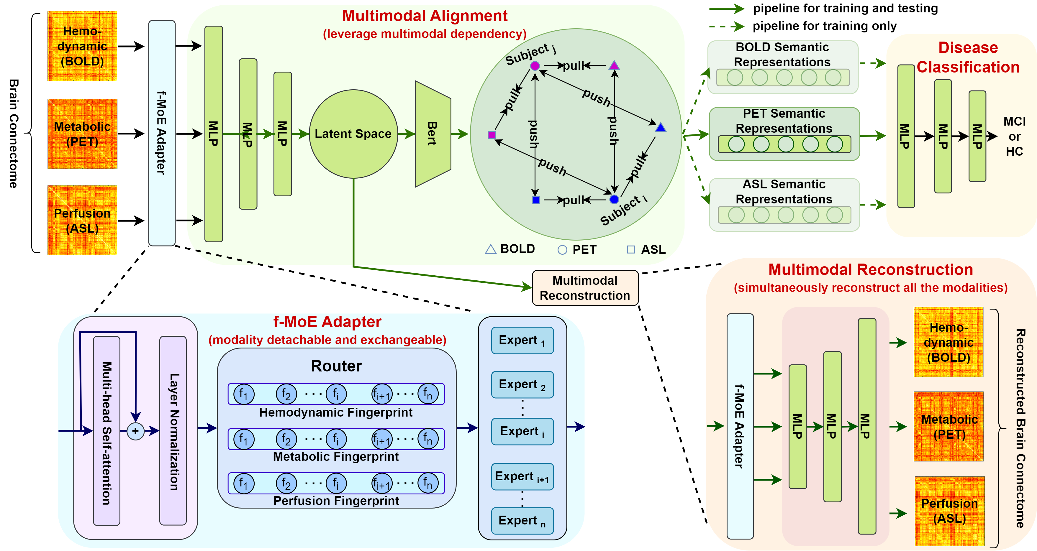
Simultaneous functional PET/MR (sf-PET/MR) presents a cutting-edge multimodal neuroimaging technique. It provides an unprecedented opportunity for concurrently monitoring and integrating multifaceted brain networks built by spatiotemporally covaried metabolic activity, neural activity, and cerebral blood flow (perfusion). Albeit high scientific/clinical values, short in hardware accessibility of PET/MR hinders its applications, let alone modern AI-based PET/MR fusion models. Our objective is to develop a clinically feasible AI-based disease diagnosis model trained on comprehensive sf-PET/MR data with the power of, during inferencing, allowing single modality input (e.g., PET only) as well as enforcing multimodal-based accuracy. To this end, we propose MX-ARM, a multimodal MiXture-of-experts Alignment and Reconstruction Model. It is modality detachable and exchangeable, allocating different multi-layer perceptrons dynamically (mixture of experts) through learnable weights to learn respective representations from different modalities. Such design will not sacrifice model performance in uni-modal situation. To fully exploit the inherent complex and nonlinear relation among modalities while producing fine-grained representations for uni-modal inference, we subsequently add a modal alignment module to line up a dominant modality (e.g., PET) with representations of auxiliary modalities (MR). We further adopt multimodal reconstruction to promote the quality of learned features. Experiments on precious multimodal sf-PET/MR data for Mild Cognitive Impairment diagnosis showcase the efficacy of our model toward clinically feasible precision medicine.
4/1/2024

PASTA: Pathology-Aware MRI to PET Cross-Modal Translation with Diffusion Models
Yitong Li, Igor Yakushev, Dennis M. Hedderich, Christian Wachinger
0
0
Positron emission tomography (PET) is a well-established functional imaging technique for diagnosing brain disorders. However, PET's high costs and radiation exposure limit its widespread use. In contrast, magnetic resonance imaging (MRI) does not have these limitations. Although it also captures neurodegenerative changes, MRI is a less sensitive diagnostic tool than PET. To close this gap, we aim to generate synthetic PET from MRI. Herewith, we introduce PASTA, a novel pathology-aware image translation framework based on conditional diffusion models. Compared to the state-of-the-art methods, PASTA excels in preserving both structural and pathological details in the target modality, which is achieved through its highly interactive dual-arm architecture and multi-modal condition integration. A cycle exchange consistency and volumetric generation strategy elevate PASTA's capability to produce high-quality 3D PET scans. Our qualitative and quantitative results confirm that the synthesized PET scans from PASTA not only reach the best quantitative scores but also preserve the pathology correctly. For Alzheimer's classification, the performance of synthesized scans improves over MRI by 4%, almost reaching the performance of actual PET. Code is available at https://github.com/ai-med/PASTA.
5/28/2024
🤯
Three-Dimensional Amyloid-Beta PET Synthesis from Structural MRI with Conditional Generative Adversarial Networks
Fernando Vega, Abdoljalil Addeh, M. Ethan MacDonald
0
0
Motivation: Alzheimer's Disease hallmarks include amyloid-beta deposits and brain atrophy, detectable via PET and MRI scans, respectively. PET is expensive, invasive and exposes patients to ionizing radiation. MRI is cheaper, non-invasive, and free from ionizing radiation but limited to measuring brain atrophy. Goal: To develop an 3D image translation model that synthesizes amyloid-beta PET images from T1-weighted MRI, exploiting the known relationship between amyloid-beta and brain atrophy. Approach: The model was trained on 616 PET/MRI pairs and validated with 264 pairs. Results: The model synthesized amyloid-beta PET images from T1-weighted MRI with high-degree of similarity showing high SSIM and PSNR metrics (SSIM>0.95&PSNR=28). Impact: Our model proves the feasibility of synthesizing amyloid-beta PET images from structural MRI ones, significantly enhancing accessibility for large-cohort studies and early dementia detection, while also reducing cost, invasiveness, and radiation exposure.
5/6/2024