Robust image segmentation model based on binary level set

0
Sign in to get full access
Overview
- Presents a robust image segmentation model based on binary level sets
- Introduces a novel energy functional that combines regional and edge information for improved segmentation
- Demonstrates superior performance compared to existing level set-based methods on challenging datasets
Plain English Explanation
The paper describes a new approach for automatically identifying and separating different objects or regions within an image, a process known as image segmentation. The researchers developed a mathematical model that uses a technique called "binary level sets" to efficiently partition the image into meaningful segments.
The key innovation is that this model combines information about the visual boundaries of objects (edge information) with information about the distinct regions or areas within the image (regional information). This allows the segmentation to be more accurate and robust even when the images have complex or varying backgrounds.
The researchers demonstrate that their approach outperforms other level set-based segmentation methods on a variety of challenging datasets, including images with clutter, occlusions, and varying illumination. This suggests the new model is a promising advance for applications that require efficient and reliable image analysis, such as medical imaging, autonomous driving, or video surveillance.
Technical Explanation
The paper introduces a novel energy functional for binary level set-based image segmentation that integrates both regional and edge information. The regional term encourages the level set to align with distinct intensity or texture patterns in the image, while the edge term drives the level set toward the boundaries between objects.
This combined energy functional is minimized using an efficient numerical scheme based on the split Bregman method. The researchers demonstrate that this optimization approach is less sensitive to parameter settings and initialization compared to previous level set methods.
Extensive experiments on benchmark datasets for cell image segmentation show that the proposed model achieves state-of-the-art performance, particularly in cases with complex backgrounds, occlusions, and intensity inhomogeneities. The authors attribute this improved robustness to the complementary nature of the regional and edge terms in the energy functional.
Critical Analysis
The paper presents a technically sound and well-designed image segmentation model that addresses known limitations of previous level set methods. The combination of regional and edge information is a clever strategy that enhances the model's ability to accurately delineate object boundaries, even in challenging scenarios.
One potential concern is the computational complexity of the optimization process, which may limit the real-time applicability of the method, especially for high-resolution images or video streams. The authors do not provide a detailed analysis of the runtime performance of their approach.
Additionally, the paper focuses on demonstrating the model's effectiveness on a specific set of benchmark datasets, primarily in the biomedical imaging domain. It would be helpful to see further evaluations on a broader range of image types and applications to better assess the generalizability of the approach.
Overall, this research represents a valuable contribution to the field of image segmentation, and the proposed model shows promise for improving the robustness and accuracy of visual analysis tasks. However, as with any research, there are opportunities for further refinement and exploration to address the potential limitations mentioned.
Conclusion
The paper presents a robust and effective image segmentation model based on binary level sets that combines regional and edge information to achieve superior performance on challenging datasets. This work advances the state-of-the-art in level set-based segmentation methods and has the potential to benefit a wide range of applications requiring accurate and reliable image analysis, such as medical imaging, autonomous navigation, and video surveillance.
This summary was produced with help from an AI and may contain inaccuracies - check out the links to read the original source documents!
Related Papers
0
Robust image segmentation model based on binary level set
Wenqi Zhao
In order to improve the robustness of traditional image segmentation models to noise, this paper models the illumination term in intensity inhomogeneity images. Additionally, to enhance the model's robustness to noisy images, we incorporate the binary level set model into the proposed model. Compared to the traditional level set, the binary level set eliminates the need for continuous reinitialization. Moreover, by introducing the variational operator GL, our model demonstrates better capability in segmenting noisy images. Finally, we employ the three-step splitting operator method for solving, and the effectiveness of the proposed model is demonstrated on various images.
Read more4/16/2024
🖼️
0
Transparency Distortion Robustness for SOTA Image Segmentation Tasks
Volker Knauthe, Arne Rak, Tristan Wirth, Thomas Pollabauer, Simon Metzler, Arjan Kuijper, Dieter W. Fellner
Semantic Image Segmentation facilitates a multitude of real-world applications ranging from autonomous driving over industrial process supervision to vision aids for human beings. These models are usually trained in a supervised fashion using example inputs. Distribution Shifts between these examples and the inputs in operation may cause erroneous segmentations. The robustness of semantic segmentation models against distribution shifts caused by differing camera or lighting setups, lens distortions, adversarial inputs and image corruptions has been topic of recent research. However, robustness against spatially varying radial distortion effects that can be caused by uneven glass structures (e.g. windows) or the chaotic refraction in heated air has not been addressed by the research community yet. We propose a method to synthetically augment existing datasets with spatially varying distortions. Our experiments show, that these distortion effects degrade the performance of state-of-the-art segmentation models. Pretraining and enlarged model capacities proof to be suitable strategies for mitigating performance degradation to some degree, while fine-tuning on distorted images only leads to marginal performance improvements.
Read more5/22/2024
📈
0
DiffSeg: A Segmentation Model for Skin Lesions Based on Diffusion Difference
Zhihao Shuai, Yinan Chen, Shunqiang Mao, Yihan Zho, Xiaohong Zhang
Weakly supervised medical image segmentation (MIS) using generative models is crucial for clinical diagnosis. However, the accuracy of the segmentation results is often limited by insufficient supervision and the complex nature of medical imaging. Existing models also only provide a single outcome, which does not allow for the measurement of uncertainty. In this paper, we introduce DiffSeg, a segmentation model for skin lesions based on diffusion difference which exploits diffusion model principles to ex-tract noise-based features from images with diverse semantic information. By discerning difference between these noise features, the model identifies diseased areas. Moreover, its multi-output capability mimics doctors' annotation behavior, facilitating the visualization of segmentation result consistency and ambiguity. Additionally, it quantifies output uncertainty using Generalized Energy Distance (GED), aiding interpretability and decision-making for physicians. Finally, the model integrates outputs through the Dense Conditional Random Field (DenseCRF) algorithm to refine the segmentation boundaries by considering inter-pixel correlations, which improves the accuracy and optimizes the segmentation results. We demonstrate the effectiveness of DiffSeg on the ISIC 2018 Challenge dataset, outperforming state-of-the-art U-Net-based methods.
Read more4/26/2024
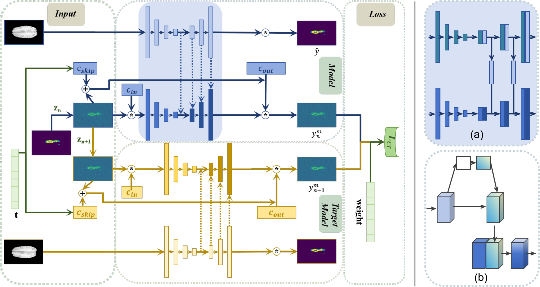
0
CTS: A Consistency-Based Medical Image Segmentation Model
Kejia Zhang, Lan Zhang, Haiwei Pan, Baolong Yu
In medical image segmentation tasks, diffusion models have shown significant potential. However, mainstream diffusion models suffer from drawbacks such as multiple sampling times and slow prediction results. Recently, consistency models, as a standalone generative network, have resolved this issue. Compared to diffusion models, consistency models can reduce the sampling times to once, not only achieving similar generative effects but also significantly speeding up training and prediction. However, they are not suitable for image segmentation tasks, and their application in the medical imaging field has not yet been explored. Therefore, this paper applies the consistency model to medical image segmentation tasks, designing multi-scale feature signal supervision modes and loss function guidance to achieve model convergence. Experiments have verified that the CTS model can obtain better medical image segmentation results with a single sampling during the test phase.
Read more5/16/2024