Segmentation-Free Outcome Prediction in Head and Neck Cancer: Deep Learning-based Feature Extraction from Multi-Angle Maximum Intensity Projections (MA-MIPs) of PET Images

0

Sign in to get full access
Overview
- This paper explores a deep learning-based approach for predicting treatment outcomes in head and neck cancer patients using PET image data, without requiring segmentation of the tumor.
- The key innovation is the use of multi-angle maximum intensity projections (MA-MIPs) of the PET images as input to the deep learning model, which captures 3D information without the need for explicit tumor segmentation.
- The proposed method aims to provide a more automated and efficient alternative to traditional segmentation-based approaches for outcome prediction in head and neck cancer.
Plain English Explanation
When treating head and neck cancer, doctors need to predict how well a patient will respond to their treatment plan. This is an important step in providing the best possible care. However, the traditional approach to making these predictions often involves a time-consuming process of manually outlining, or "segmenting," the tumor in medical images.
The researchers behind this study explored a new way to make these predictions without needing to segment the tumor. Instead, they used a deep learning model that could extract useful information directly from the 3D medical images, in a process called "feature extraction."
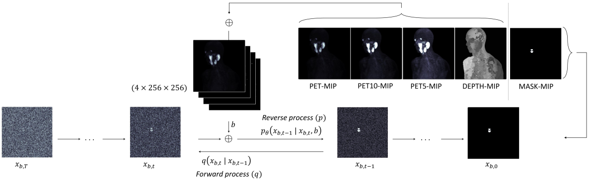
The key innovation was their use of "multi-angle maximum intensity projections" (MA-MIPs) of the PET images. These are essentially 2D "snapshots" of the 3D image data, taken from multiple angles. By feeding these MA-MIPs into their deep learning model, the researchers were able to capture the 3D structure of the tumor without the need for manual segmentation.
This segmentation-free approach could provide a more automated and efficient way to predict treatment outcomes in head and neck cancer patients, potentially leading to better-informed treatment decisions and improved patient care.
Technical Explanation
The researchers developed a deep learning-based approach for predicting treatment outcomes in head and neck cancer patients using PET image data, without requiring explicit tumor segmentation.
The key component of their method was the use of multi-angle maximum intensity projections (MA-MIPs) as input to the deep learning model. MA-MIPs are 2D representations of 3D PET image data, created by projecting the maximum pixel values along multiple viewing angles. This allowed the model to capture 3D information about the tumor without the need for segmentation.
The deep learning model itself was a convolutional neural network (CNN) that was trained to predict treatment outcomes, such as overall survival or locoregional control, directly from the MA-MIP inputs. The researchers explored different CNN architectures and training strategies to optimize the model's performance.
Through extensive experiments on a large dataset of head and neck cancer patients, the proposed segmentation-free approach demonstrated competitive or superior performance compared to traditional segmentation-based methods. The ability to bypass the segmentation step resulted in a more automated and efficient workflow for outcome prediction.
This work builds on previous efforts in using deep learning for medical image analysis without explicit segmentation, and highlights the potential of such approaches to improve clinical decision-making in oncology.
Critical Analysis
The key strength of this research is the demonstration of a segmentation-free approach for outcome prediction in head and neck cancer, which can potentially streamline the clinical workflow and reduce the burden on healthcare providers.
However, the paper does not provide a detailed analysis of the limitations of the proposed method. For example, it is unclear how the performance of the segmentation-free approach compares to human expert segmentation, or how the method would perform on more heterogeneous patient populations or different imaging modalities.
Additionally, the paper does not discuss the interpretability of the deep learning model's predictions. While the segmentation-free approach may be more efficient, it is important to understand the underlying factors driving the model's predictions to build trust and facilitate clinical adoption.
Further research is needed to address these limitations and explore the broader applicability of the segmentation-free approach to other cancer types and clinical settings. Incorporating multimodal data, such as clinical and genomic information, may also improve the predictive power of the model.
Conclusion
This paper presents a novel deep learning-based approach for predicting treatment outcomes in head and neck cancer patients using PET image data, without requiring explicit tumor segmentation. The key innovation is the use of multi-angle maximum intensity projections (MA-MIPs) as input to the deep learning model, which captures 3D information about the tumor while avoiding the need for manual segmentation.
The proposed segmentation-free method has the potential to streamline the clinical workflow and improve the efficiency of outcome prediction in head and neck cancer. While further research is needed to address the limitations and explore the broader applicability of this approach, this work represents an important step towards more automated and data-driven decision-making in oncology.
This summary was produced with help from an AI and may contain inaccuracies - check out the links to read the original source documents!
Related Papers
0
Segmentation-Free Outcome Prediction in Head and Neck Cancer: Deep Learning-based Feature Extraction from Multi-Angle Maximum Intensity Projections (MA-MIPs) of PET Images
Amirhosein Toosi, Isaac Shiri, Habib Zaidi, Arman Rahmim
We introduce an innovative, simple, effective segmentation-free approach for outcome prediction in head & neck cancer (HNC) patients. By harnessing deep learning-based feature extraction techniques and multi-angle maximum intensity projections (MA-MIPs) applied to Fluorodeoxyglucose Positron Emission Tomography (FDG-PET) volumes, our proposed method eliminates the need for manual segmentations of regions-of-interest (ROIs) such as primary tumors and involved lymph nodes. Instead, a state-of-the-art object detection model is trained to perform automatic cropping of the head and neck region on the PET volumes. A pre-trained deep convolutional neural network backbone is then utilized to extract deep features from MA-MIPs obtained from 72 multi-angel axial rotations of the cropped PET volumes. These deep features extracted from multiple projection views of the PET volumes are then aggregated and fused, and employed to perform recurrence-free survival analysis on a cohort of 489 HNC patients. The proposed approach outperforms the best performing method on the target dataset for the task of recurrence-free survival analysis. By circumventing the manual delineation of the malignancies on the FDG PET-CT images, our approach eliminates the dependency on subjective interpretations and highly enhances the reproducibility of the proposed survival analysis method.
Read more5/6/2024

0
Deep Learning-Based Segmentation of Tumors in PET/CT Volumes: Benchmark of Different Architectures and Training Strategies
Monika G'orka, Daniel Jaworek, Marek Wodzinski
Cancer is one of the leading causes of death globally, and early diagnosis is crucial for patient survival. Deep learning algorithms have great potential for automatic cancer analysis. Artificial intelligence has achieved high performance in recognizing and segmenting single lesions. However, diagnosing multiple lesions remains a challenge. This study examines and compares various neural network architectures and training strategies for automatically segmentation of cancer lesions using PET/CT images from the head, neck, and whole body. The authors analyzed datasets from the AutoPET and HECKTOR challenges, exploring popular single-step segmentation architectures and presenting a two-step approach. The results indicate that the V-Net and nnU-Net models were the most effective for their respective datasets. The results for the HECKTOR dataset ranged from 0.75 to 0.76 for the aggregated Dice coefficient. Eliminating cancer-free cases from the AutoPET dataset was found to improve the performance of most models. In the case of AutoPET data, the average segmentation efficiency after training only on images containing cancer lesions increased from 0.55 to 0.66 for the classic Dice coefficient and from 0.65 to 0.73 for the aggregated Dice coefficient. The research demonstrates the potential of artificial intelligence in precise oncological diagnostics and may contribute to the development of more targeted and effective cancer assessment techniques.
Read more4/16/2024
0
How To Segment in 3D Using 2D Models: Automated 3D Segmentation of Prostate Cancer Metastatic Lesions on PET Volumes Using Multi-Angle Maximum Intensity Projections and Diffusion Models
Amirhosein Toosi, Sara Harsini, Franc{c}ois B'enard, Carlos Uribe, Arman Rahmim
Prostate specific membrane antigen (PSMA) positron emission tomography/computed tomography (PET/CT) imaging provides a tremendously exciting frontier in visualization of prostate cancer (PCa) metastatic lesions. However, accurate segmentation of metastatic lesions is challenging due to low signal-to-noise ratios and variable sizes, shapes, and locations of the lesions. This study proposes a novel approach for automated segmentation of metastatic lesions in PSMA PET/CT 3D volumetric images using 2D denoising diffusion probabilistic models (DDPMs). Instead of 2D trans-axial slices or 3D volumes, the proposed approach segments the lesions on generated multi-angle maximum intensity projections (MA-MIPs) of the PSMA PET images, then obtains the final 3D segmentation masks from 3D ordered subset expectation maximization (OSEM) reconstruction of 2D MA-MIPs segmentations. Our proposed method achieved superior performance compared to state-of-the-art 3D segmentation approaches in terms of accuracy and robustness in detecting and segmenting small metastatic PCa lesions. The proposed method has significant potential as a tool for quantitative analysis of metastatic burden in PCa patients.
Read more7/29/2024

0
New!Enhancing Lesion Segmentation in PET/CT Imaging with Deep Learning and Advanced Data Preprocessing Techniques
Jiayi Liu, Qiaoyi Xue, Youdan Feng, Tianming Xu, Kaixin Shen, Chuyun Shen, Yuhang Shi
The escalating global cancer burden underscores the critical need for precise diagnostic tools in oncology. This research employs deep learning to enhance lesion segmentation in PET/CT imaging, utilizing a dataset of 900 whole-body FDG-PET/CT and 600 PSMA-PET/CT studies from the AutoPET challenge III. Our methodical approach includes robust preprocessing and data augmentation techniques to ensure model robustness and generalizability. We investigate the influence of non-zero normalization and modifications to the data augmentation pipeline, such as the introduction of RandGaussianSharpen and adjustments to the Gamma transform parameter. This study aims to contribute to the standardization of preprocessing and augmentation strategies in PET/CT imaging, potentially improving the diagnostic accuracy and the personalized management of cancer patients. Our code will be open-sourced and available at https://github.com/jiayiliu-pku/DC2024.
Read more9/17/2024