Segmentation-Guided Knee Radiograph Generation using Conditional Diffusion Models
2404.03541
0
0

Abstract
Deep learning-based medical image processing algorithms require representative data during development. In particular, surgical data might be difficult to obtain, and high-quality public datasets are limited. To overcome this limitation and augment datasets, a widely adopted solution is the generation of synthetic images. In this work, we employ conditional diffusion models to generate knee radiographs from contour and bone segmentations. Remarkably, two distinct strategies are presented by incorporating the segmentation as a condition into the sampling and training process, namely, conditional sampling and conditional training. The results demonstrate that both methods can generate realistic images while adhering to the conditioning segmentation. The conditional training method outperforms the conditional sampling method and the conventional U-Net.
Create account to get full access
Overview
- This paper proposes a method for generating realistic knee radiographs using conditional diffusion models, with the goal of improving medical image synthesis and analysis.
- The approach leverages segmentation maps as a conditioning input to guide the generation process and produce more anatomically accurate radiographs.
- Experiments on a large dataset of knee radiographs demonstrate the effectiveness of the proposed method in generating high-quality synthetic images that can aid tasks like model pretraining and data augmentation.
Plain English Explanation
This research focuses on a technique for creating artificial knee X-ray images using a type of machine learning model called a diffusion model. Diffusion models work by gradually adding noise to an image and then learning to remove that noise in a controlled way, allowing them to generate new images from scratch.
The key innovation in this work is that the diffusion model is "conditioned" on segmentation maps of the knee joint. Segmentation maps are images that outline the different anatomical structures, like bones and soft tissues. By incorporating this additional information, the model is able to generate X-ray images that are more anatomically realistic and true to the underlying anatomy.
The researchers tested their approach on a large dataset of real knee X-rays and found that the synthetic images produced by their model were of high visual quality and could potentially be useful for tasks like training other AI models or creating more diverse training data for medical imaging analysis.
Technical Explanation
The authors propose a conditional diffusion model for generating realistic knee radiographs, with segmentation maps as the conditioning input. Diffusion models work by gradually adding noise to an image and then learning to reverse this process to generate new images.
The segmentation maps provide the model with important anatomical information about the structure of the knee joint, which helps it produce radiographs that are more anatomically accurate. The model is trained on a large dataset of real knee X-rays and their corresponding segmentation maps.
Experiments show that the proposed approach outperforms baseline image-to-image translation models in terms of visual quality and fidelity to the underlying anatomy. The synthetic radiographs generated by the model can potentially be used for data augmentation or as pretraining for other medical imaging models, improving their performance on tasks like stress testing or open-vocabulary segmentation.
Critical Analysis
The authors acknowledge several limitations of their work, such as the need for high-quality segmentation maps as input and the potential for bias in the training data. Additionally, the model's ability to generalize to diverse patient populations and imaging conditions remains an open question.
While the results are promising, further research is needed to fully understand the capabilities and limitations of this approach. Potential areas for improvement include incorporating additional conditioning information, such as demographic data or clinical metadata, and exploring ways to make the model more robust to variations in imaging protocols and patient characteristics.
It will also be important to carefully evaluate the ethical implications of using synthetic medical images, particularly around issues of privacy, data provenance, and the potential for misuse or misinterpretation by medical professionals.
Conclusion
This paper presents a novel approach for generating realistic knee radiographs using conditional diffusion models. By leveraging segmentation maps as a conditioning input, the model is able to produce synthetic images that are more anatomically accurate and visually compelling than those generated by baseline image-to-image translation methods.
The potential applications of this work include data augmentation for medical imaging analysis, pretraining of AI models, and the creation of diverse training datasets to improve the robustness and generalization of biomedical vision systems. However, further research is needed to address the limitations and ethical considerations surrounding the use of synthetic medical data.
Overall, this research represents an important step forward in the field of medical image synthesis, with the ability to generate high-quality radiographs that could have a significant impact on the development of more accurate and reliable AI-driven tools for healthcare.
This summary was produced with help from an AI and may contain inaccuracies - check out the links to read the original source documents!
Related Papers
Anatomically-Controllable Medical Image Generation with Segmentation-Guided Diffusion Models
Nicholas Konz, Yuwen Chen, Haoyu Dong, Maciej A. Mazurowski
0
0
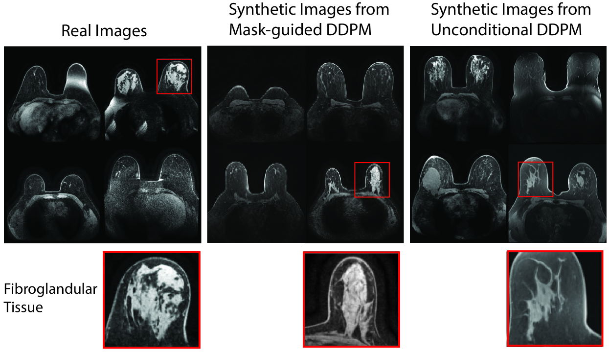
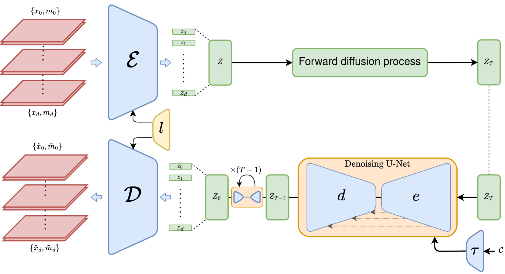
Diffusion models have enabled remarkably high-quality medical image generation, yet it is challenging to enforce anatomical constraints in generated images. To this end, we propose a diffusion model-based method that supports anatomically-controllable medical image generation, by following a multi-class anatomical segmentation mask at each sampling step. We additionally introduce a random mask ablation training algorithm to enable conditioning on a selected combination of anatomical constraints while allowing flexibility in other anatomical areas. We compare our method (SegGuidedDiff) to existing methods on breast MRI and abdominal/neck-to-pelvis CT datasets with a wide range of anatomical objects. Results show that our method reaches a new state-of-the-art in the faithfulness of generated images to input anatomical masks on both datasets, and is on par for general anatomical realism. Finally, our model also enjoys the extra benefit of being able to adjust the anatomical similarity of generated images to real images of choice through interpolation in its latent space. SegGuidedDiff has many applications, including cross-modality translation, and the generation of paired or counterfactual data. Our code is available at https://github.com/mazurowski-lab/segmentation-guided-diffusion.
6/21/2024
📊
Using Diffusion Models to Generate Synthetic Labelled Data for Medical Image Segmentation
Daniel Saragih, Atsuhiro Hibi, Pascal Tyrrell
0
0
Medical image analysis has become a prominent area where machine learning has been applied. However, high quality, publicly available data is limited either due to patient privacy laws or the time and cost required for experts to annotate images. In this retrospective study, we designed and evaluated a pipeline to generate synthetic labeled polyp images for augmenting medical image segmentation models with the aim of reducing this data scarcity. In particular, we trained diffusion models on the HyperKvasir dataset, comprising 1000 images of polyps in the human GI tract from 2008 to 2016. Qualitative expert review, Fr'echet Inception Distance (FID), and Multi-Scale Structural Similarity (MS-SSIM) were tested for evaluation. Additionally, various segmentation models were trained with the generated data and evaluated using Dice score and Intersection over Union. We found that our pipeline produced images more akin to real polyp images based on FID scores, and segmentation performance also showed improvements over GAN methods when trained entirely, or partially, with synthetic data, despite requiring less compute for training. Moreover, the improvement persists when tested on different datasets, showcasing the transferability of the generated images.
5/13/2024
3D MRI Synthesis with Slice-Based Latent Diffusion Models: Improving Tumor Segmentation Tasks in Data-Scarce Regimes
Aghiles Kebaili, J'er^ome Lapuyade-Lahorgue, Pierre Vera, Su Ruan
0
0
Despite the increasing use of deep learning in medical image segmentation, the limited availability of annotated training data remains a major challenge due to the time-consuming data acquisition and privacy regulations. In the context of segmentation tasks, providing both medical images and their corresponding target masks is essential. However, conventional data augmentation approaches mainly focus on image synthesis. In this study, we propose a novel slice-based latent diffusion architecture designed to address the complexities of volumetric data generation in a slice-by-slice fashion. This approach extends the joint distribution modeling of medical images and their associated masks, allowing a simultaneous generation of both under data-scarce regimes. Our approach mitigates the computational complexity and memory expensiveness typically associated with diffusion models. Furthermore, our architecture can be conditioned by tumor characteristics, including size, shape, and relative position, thereby providing a diverse range of tumor variations. Experiments on a segmentation task using the BRATS2022 confirm the effectiveness of the synthesized volumes and masks for data augmentation.
6/11/2024
🖼️
An Automated Real-Time Approach for Image Processing and Segmentation of Fluoroscopic Images and Videos Using a Single Deep Learning Network
Viet Dung Nguyen, Michael T. LaCour, Richard D. Komistek
0
0
Image segmentation in total knee arthroplasty is crucial for precise preoperative planning and accurate implant positioning, leading to improved surgical outcomes and patient satisfaction. The biggest challenges of image segmentation in total knee arthroplasty include accurately delineating complex anatomical structures, dealing with image artifacts and noise, and developing robust algorithms that can handle anatomical variations and pathologies commonly encountered in patients. The potential of using machine learning for image segmentation in total knee arthroplasty lies in its ability to improve segmentation accuracy, automate the process, and provide real-time assistance to surgeons, leading to enhanced surgical planning, implant placement, and patient outcomes. This paper proposes a methodology to use deep learning for robust and real-time total knee arthroplasty image segmentation. The deep learning model, trained on a large dataset, demonstrates outstanding performance in accurately segmenting both the implanted femur and tibia, achieving an impressive mean-Average-Precision (mAP) of 88.83 when compared to the ground truth while also achieving a real-time segmented speed of 20 frames per second (fps). We have introduced a novel methodology for segmenting implanted knee fluoroscopic or x-ray images that showcases remarkable levels of accuracy and speed, paving the way for various potential extended applications.
5/28/2024