Solving the inverse problem of microscopy deconvolution with a residual Beylkin-Coifman-Rokhlin neural network

0

Sign in to get full access
Overview
- This research paper proposes a novel neural network architecture, called the Residual Beylkin-Coifman-Rokhlin (RBC) neural network, for solving the inverse problem of microscopy deconvolution.
- Microscopy deconvolution is the process of recovering a sharp image from a blurred microscope image, which is important for high-resolution imaging in fields like biology and materials science.
- The RBC neural network combines the benefits of physics-informed models and deep learning to efficiently and accurately perform microscopy deconvolution.
Plain English Explanation
Microscopes are essential tools for studying small-scale structures, like cells or materials, in great detail. However, the images captured by microscopes can sometimes be blurry or distorted, making it difficult to see the fine details. This blurring is caused by a complex optical phenomenon called the "point spread function," which describes how light is scattered as it passes through the microscope's lenses.
The researchers in this paper have developed a new type of artificial intelligence (AI) system, called the Residual Beylkin-Coifman-Rokhlin (RBC) neural network, to help solve this problem. The RBC neural network is trained to analyze the blurry microscope image and "deconvolve" it - that is, to remove the blurring effect and recover the original sharp image. This is done by incorporating mathematical models of the optical physics involved in microscopy, which helps the AI system understand the underlying causes of the blurring.
By combining the strengths of physics-based models and deep learning, the RBC neural network can deconvolve microscope images more efficiently and accurately than previous methods. This could lead to significant improvements in the quality and resolution of microscopic imaging, which would be valuable for researchers studying everything from biological cells to advanced materials.
Technical Explanation
The researchers propose a novel neural network architecture called the Residual Beylkin-Coifman-Rokhlin (RBC) neural network to solve the inverse problem of microscopy deconvolution. The RBC neural network integrates physics-informed models, specifically the Beylkin-Coifman-Rokhlin (BCR) method, into a deep learning framework to efficiently and accurately perform microscopy deconvolution.
The BCR method is a fast algorithm for applying convolution and deconvolution operations, which are central to the microscopy deconvolution problem. The researchers incorporate the BCR method into the neural network's architecture, allowing it to learn the inverse mapping from blurred microscope images to their corresponding sharp images.
The RBC neural network is designed as a residual network, with skip connections that allow the model to better preserve important details in the deconvolved images. The researchers also introduce a custom loss function that combines the traditional mean squared error (MSE) loss with a term that encourages the network to learn the correct physics-based model.
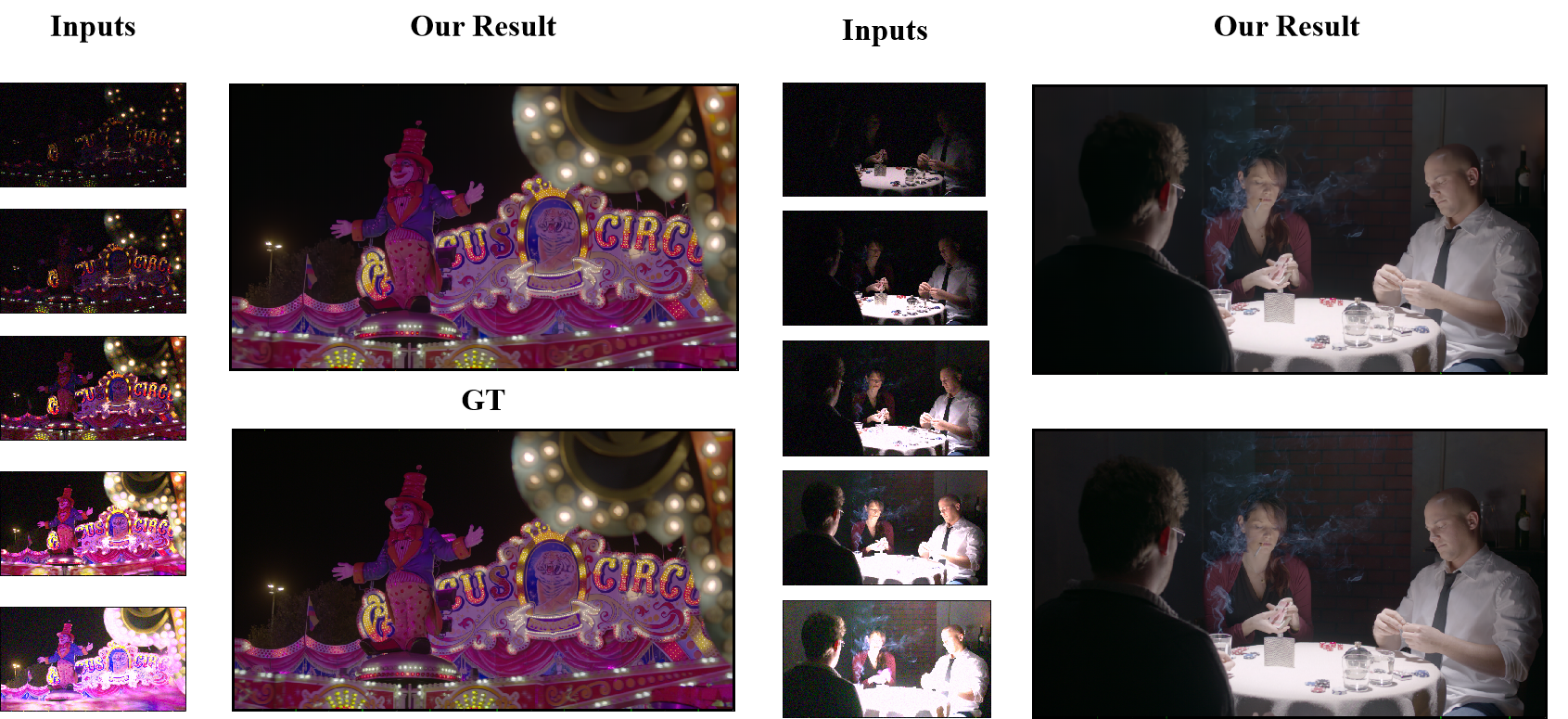
The proposed RBC neural network is evaluated on both simulated and real microscopy datasets, demonstrating superior performance compared to existing deep learning and optimization-based deconvolution methods. The model is able to effectively remove blur and recover fine details in the recovered images, while also being computationally efficient.
Critical Analysis
The research paper presents a well-designed and thorough investigation of the RBC neural network for microscopy deconvolution. The incorporation of the physics-based BCR method into the neural network architecture is a key strength, as it allows the model to leverage domain-specific knowledge to improve its performance.
However, the paper does not address some potential limitations of the RBC neural network. For example, the model may still struggle with certain types of complex or non-linear blur, which can occur in real-world microscopy scenarios. Additionally, the reliance on the BCR method may limit the network's flexibility and ability to adapt to new types of blur or imaging modalities.
Further research could explore ways to make the RBC neural network more robust and adaptable, such as by incorporating additional physics-informed components or exploring alternative neural network architectures. Additionally, the researchers could investigate the RBC network's performance on a wider range of microscopy data, including more challenging samples or imaging conditions.
Overall, the RBC neural network represents an interesting and promising approach to the microscopy deconvolution problem, but there may be opportunities to further improve its capabilities and broaden its applicability. As with any research in this field, it is important to think critically about the findings and consider how they fit into the broader context of microscopy and image restoration techniques.
Conclusion
The Residual Beylkin-Coifman-Rokhlin (RBC) neural network proposed in this research paper offers a novel and effective solution to the inverse problem of microscopy deconvolution. By integrating physics-informed models into a deep learning framework, the RBC network is able to efficiently and accurately recover sharp images from blurred microscope data.
This work demonstrates the potential for combining the strengths of physics-based approaches and data-driven machine learning to tackle complex imaging challenges. The RBC network's success in microscopy deconvolution suggests that similar techniques could be applied to [other image restoration tasks, such as solar telescope deblurring or low-rank tensor restoration.]
Overall, the RBC neural network represents an important step forward in the field of microscopy imaging and could have significant implications for researchers and scientists working with high-resolution microscopic data. [As with any new technology, it will be important to continue exploring its capabilities and limitations, as well as how it can be integrated with other state-of-the-art techniques like Swin Transformer-based deconvolution.]
This summary was produced with help from an AI and may contain inaccuracies - check out the links to read the original source documents!
Related Papers
0
Solving the inverse problem of microscopy deconvolution with a residual Beylkin-Coifman-Rokhlin neural network
Rui Li, Mikhail Kudryashev, Artur Yakimovich
Optic deconvolution in light microscopy (LM) refers to recovering the object details from images, revealing the ground truth of samples. Traditional explicit methods in LM rely on the point spread function (PSF) during image acquisition. Yet, these approaches often fall short due to inaccurate PSF models and noise artifacts, hampering the overall restoration quality. In this paper, we approached the optic deconvolution as an inverse problem. Motivated by the nonstandard-form compression scheme introduced by Beylkin, Coifman, and Rokhlin (BCR), we proposed an innovative physics-informed neural network Multi-Stage Residual-BCR Net (m-rBCR) to approximate the optic deconvolution. We validated the m-rBCR model on four microscopy datasets - two simulated microscopy datasets from ImageNet and BioSR, real dSTORM microscopy images, and real widefield microscopy images. In contrast to the explicit deconvolution methods (e.g. Richardson-Lucy) and other state-of-the-art NN models (U-Net, DDPM, CARE, DnCNN, ESRGAN, RCAN, Noise2Noise, MPRNet, and MIMO-U-Net), the m-rBCR model demonstrates superior performance to other candidates by PSNR and SSIM in two real microscopy datasets and the simulated BioSR dataset. In the simulated ImageNet dataset, m-rBCR ranks the second-best place (right after MIMO-U-Net). With the backbone from the optical physics, m-rBCR exploits the trainable parameters with better performances (from ~30 times fewer than the benchmark MIMO-U-Net to ~210 times than ESRGAN). This enables m-rBCR to achieve a shorter runtime (from ~3 times faster than MIMO-U-Net to ~300 times faster than DDPM). To summarize, by leveraging physics constraints our model reduced potentially redundant parameters significantly in expertise-oriented NN candidates and achieved high efficiency with superior performance.
Read more7/16/2024
0
CRNet: A Detail-Preserving Network for Unified Image Restoration and Enhancement Task
Kangzhen Yang, Tao Hu, Kexin Dai, Genggeng Chen, Yu Cao, Wei Dong, Peng Wu, Yanning Zhang, Qingsen Yan
In real-world scenarios, images captured often suffer from blurring, noise, and other forms of image degradation, and due to sensor limitations, people usually can only obtain low dynamic range images. To achieve high-quality images, researchers have attempted various image restoration and enhancement operations on photographs, including denoising, deblurring, and high dynamic range imaging. However, merely performing a single type of image enhancement still cannot yield satisfactory images. In this paper, to deal with the challenge above, we propose the Composite Refinement Network (CRNet) to address this issue using multiple exposure images. By fully integrating information-rich multiple exposure inputs, CRNet can perform unified image restoration and enhancement. To improve the quality of image details, CRNet explicitly separates and strengthens high and low-frequency information through pooling layers, using specially designed Multi-Branch Blocks for effective fusion of these frequencies. To increase the receptive field and fully integrate input features, CRNet employs the High-Frequency Enhancement Module, which includes large kernel convolutions and an inverted bottleneck ConvFFN. Our model secured third place in the first track of the Bracketing Image Restoration and Enhancement Challenge, surpassing previous SOTA models in both testing metrics and visual quality.
Read more4/23/2024

0
PHOCUS: Physics-Based Deconvolution for Ultrasound Resolution Enhancement
Felix Duelmer, Walter Simson, Mohammad Farid Azampour, Magdalena Wysocki, Angelos Karlas, Nassir Navab
Ultrasound is widely used in medical diagnostics allowing for accessible and powerful imaging but suffers from resolution limitations due to diffraction and the finite aperture of the imaging system, which restricts diagnostic use. The impulse function of an ultrasound imaging system is called the point spread function (PSF), which is convolved with the spatial distribution of reflectors in the image formation process. Recovering high-resolution reflector distributions by removing image distortions induced by the convolution process improves image clarity and detail. Conventionally, deconvolution techniques attempt to rectify the imaging system's dependent PSF, working directly on the radio-frequency (RF) data. However, RF data is often not readily accessible. Therefore, we introduce a physics-based deconvolution process using a modeled PSF, working directly on the more commonly available B-mode images. By leveraging Implicit Neural Representations (INRs), we learn a continuous mapping from spatial locations to their respective echogenicity values, effectively compensating for the discretized image space. Our contribution consists of a novel methodology for retrieving a continuous echogenicity map directly from a B-mode image through a differentiable physics-based rendering pipeline for ultrasound resolution enhancement. We qualitatively and quantitatively evaluate our approach on synthetic data, demonstrating improvements over traditional methods in metrics such as PSNR and SSIM. Furthermore, we show qualitative enhancements on an ultrasound phantom and an in-vivo acquisition of a carotid artery.
Read more8/9/2024

0
A Unified Framework for Microscopy Defocus Deblur with Multi-Pyramid Transformer and Contrastive Learning
Yuelin Zhang, Pengyu Zheng, Wanquan Yan, Chengyu Fang, Shing Shin Cheng
Defocus blur is a persistent problem in microscope imaging that poses harm to pathology interpretation and medical intervention in cell microscopy and microscope surgery. To address this problem, a unified framework including the multi-pyramid transformer (MPT) and extended frequency contrastive regularization (EFCR) is proposed to tackle two outstanding challenges in microscopy deblur: longer attention span and data deficiency. The MPT employs an explicit pyramid structure at each network stage that integrates the cross-scale window attention (CSWA), the intra-scale channel attention (ISCA), and the feature-enhancing feed-forward network (FEFN) to capture long-range cross-scale spatial interaction and global channel context. The EFCR addresses the data deficiency problem by exploring latent deblur signals from different frequency bands. It also enables deblur knowledge transfer to learn cross-domain information from extra data, improving deblur performance for labeled and unlabeled data. Extensive experiments and downstream task validation show the framework achieves state-of-the-art performance across multiple datasets. Project page: https://github.com/PieceZhang/MPT-CataBlur.
Read more6/5/2024