PHOCUS: Physics-Based Deconvolution for Ultrasound Resolution Enhancement

0

Sign in to get full access
Overview
- The paper introduces PHOCUS, a physics-based deconvolution method for enhancing the resolution of ultrasound images.
- It uses an implicit neural representation of the point-spread-function (PSF) to perform deconvolution and improve image quality.
- The method can be applied to various ultrasound imaging modalities, including B-mode, Doppler, and contrast-enhanced imaging.
Plain English Explanation
Ultrasound imaging is a widely used diagnostic tool, but the images it produces can be blurry and lack fine detail. This is because the sound waves used in ultrasound have a natural spreading effect, known as the point-spread-function, which reduces the sharpness of the image.
The PHOCUS method addresses this problem by deconvolving the ultrasound images to undo the blurring effect of the point-spread-function. It uses a special type of neural network to model the point-spread-function, which allows it to be inverted and applied to the raw ultrasound data to produce a sharper, more detailed image.
This approach can be used with different types of ultrasound imaging, such as B-mode, Doppler, and contrast-enhanced imaging, making it a versatile tool for improving image quality in a variety of medical applications.
Technical Explanation
The PHOCUS method models the point-spread-function (PSF) of the ultrasound imaging system using an implicit neural representation. This means that the PSF is represented as a continuous function, rather than a discrete set of values, which allows it to be inverted and applied to the raw ultrasound data to perform deconvolution.
The PSF model is trained on a dataset of simulated ultrasound images, which are generated using a physics-based simulation of the ultrasound imaging process. This ensures that the PSF model accurately captures the physical characteristics of the imaging system, enabling effective deconvolution.
The deconvolution process involves applying the inverted PSF model to the raw ultrasound data, which effectively reverses the blurring effect and enhances the image resolution. The method can be applied to various ultrasound modalities, including B-mode, Doppler, and contrast-enhanced imaging, by adapting the PSF model to the specific imaging characteristics of each modality.
Critical Analysis
The PHOCUS method represents a promising approach for enhancing the resolution of ultrasound images, but it does have some limitations that should be considered. The reliance on simulated training data means that the method may not fully capture the complexities and variabilities of real-world ultrasound imaging, which could limit its performance in certain clinical scenarios.
Additionally, the computational complexity of the implicit neural representation and deconvolution process may pose challenges for real-time clinical implementation, particularly in resource-constrained settings. Further research and optimization may be needed to address these practical concerns.
Conclusion
The PHOCUS method demonstrates the potential of physics-based deconvolution techniques to improve the resolution and quality of ultrasound images. By modeling the underlying point-spread-function using an implicit neural representation, the method can be applied to a wide range of ultrasound modalities, potentially enhancing the diagnostic capabilities of this widely used imaging technology. While further research is needed to address some of the practical limitations, the PHOCUS approach represents an exciting step forward in the field of ultrasound image enhancement.
This summary was produced with help from an AI and may contain inaccuracies - check out the links to read the original source documents!
Related Papers
0
PHOCUS: Physics-Based Deconvolution for Ultrasound Resolution Enhancement
Felix Duelmer, Walter Simson, Mohammad Farid Azampour, Magdalena Wysocki, Angelos Karlas, Nassir Navab
Ultrasound is widely used in medical diagnostics allowing for accessible and powerful imaging but suffers from resolution limitations due to diffraction and the finite aperture of the imaging system, which restricts diagnostic use. The impulse function of an ultrasound imaging system is called the point spread function (PSF), which is convolved with the spatial distribution of reflectors in the image formation process. Recovering high-resolution reflector distributions by removing image distortions induced by the convolution process improves image clarity and detail. Conventionally, deconvolution techniques attempt to rectify the imaging system's dependent PSF, working directly on the radio-frequency (RF) data. However, RF data is often not readily accessible. Therefore, we introduce a physics-based deconvolution process using a modeled PSF, working directly on the more commonly available B-mode images. By leveraging Implicit Neural Representations (INRs), we learn a continuous mapping from spatial locations to their respective echogenicity values, effectively compensating for the discretized image space. Our contribution consists of a novel methodology for retrieving a continuous echogenicity map directly from a B-mode image through a differentiable physics-based rendering pipeline for ultrasound resolution enhancement. We qualitatively and quantitatively evaluate our approach on synthetic data, demonstrating improvements over traditional methods in metrics such as PSNR and SSIM. Furthermore, we show qualitative enhancements on an ultrasound phantom and an in-vivo acquisition of a carotid artery.
Read more8/9/2024

0
Inverse Problem Approach to Aberration Correction for in vivo Transcranial Imaging Based on a Sparse Representation of Contrast-enhanced Ultrasound Data
Paul Xing, Antoine Malescot, Eric Martineau, Ravi Rungta, Jean Provost
Transcranial ultrasound imaging is currently limited by attenuation and aberration induced by the skull. First used in contrast-enhanced ultrasound (CEUS), highly echoic microbubbles allowed for the development of novel imaging modalities such as ultrasound localization microscopy (ULM). Herein, we develop an inverse problem approach to aberration correction (IPAC) that leverages the sparsity of microbubble signals. We propose to use the textit{a priori} knowledge of the medium based upon microbubble localization and wave propagation to build a forward model to link the measured signals directly to the aberration function. A standard least-squares inversion is then used to retrieve the aberration function. We first validated IPAC on simulated data of a vascular network using plane wave as well as divergent wave emissions. We then evaluated the reproducibility of IPAC textit{in vivo} in 5 mouse brains. We showed that aberration correction improved the contrast of CEUS images by 4.6 dB. For ULM images, IPAC yielded sharper vessels, reduced vessel duplications, and improved the resolution from 21.1 $mu$m to 18.3 $mu$m. Aberration correction also improved hemodynamic quantification for velocity magnitude and flow direction.
Read more5/15/2024
0
Learning Point Spread Function Invertibility Assessment for Image Deconvolution
Romario Gualdr'on-Hurtado, Roman Jacome, Sergio Urrea, Henry Arguello, Luis Gonzalez
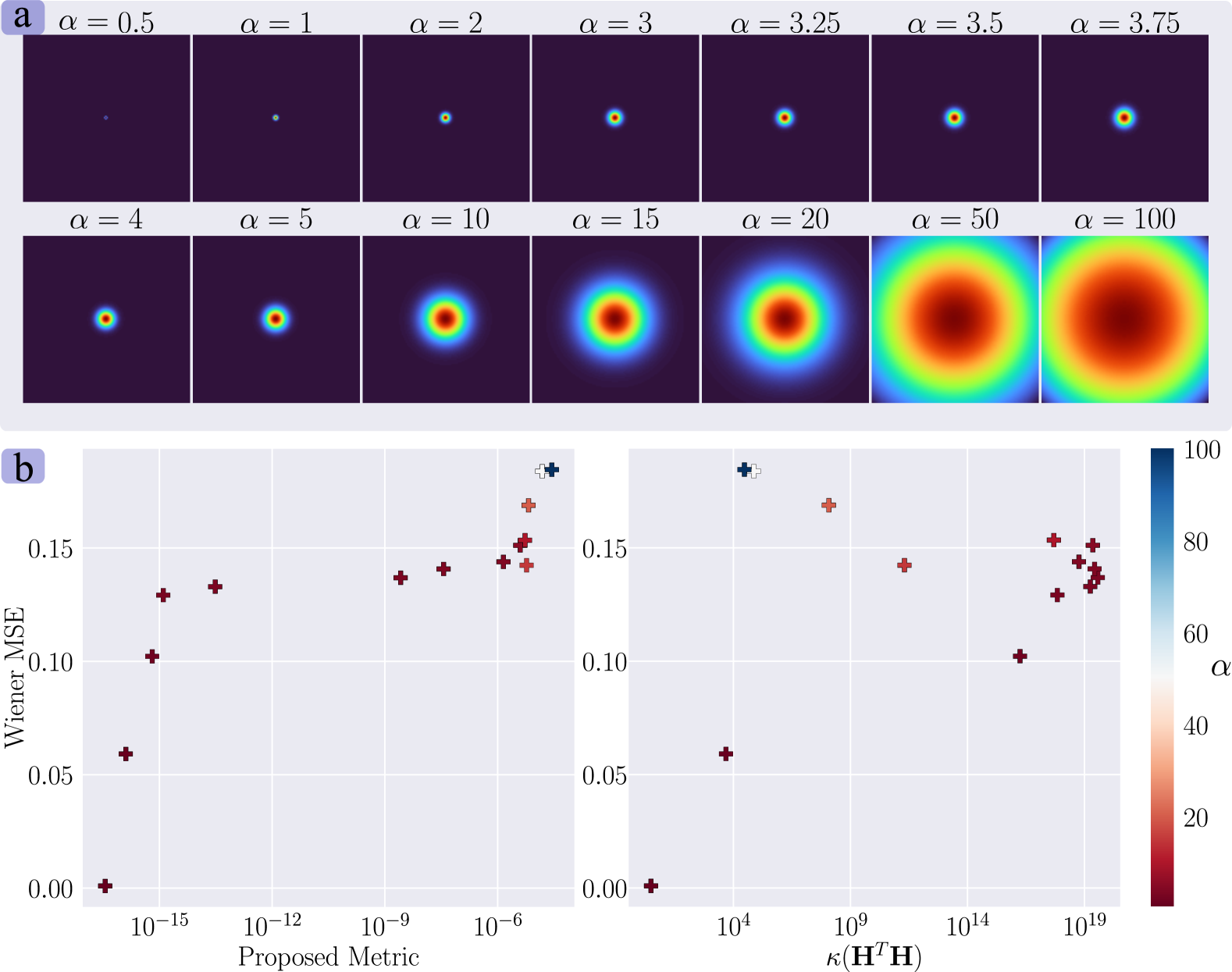
Deep-learning (DL)-based image deconvolution (ID) has exhibited remarkable recovery performance, surpassing traditional linear methods. However, unlike traditional ID approaches that rely on analytical properties of the point spread function (PSF) to achieve high recovery performance - such as specific spectrum properties or small conditional numbers in the convolution matrix - DL techniques lack quantifiable metrics for evaluating PSF suitability for DL-assisted recovery. Aiming to enhance deconvolution quality, we propose a metric that employs a non-linear approach to learn the invertibility of an arbitrary PSF using a neural network by mapping it to a unit impulse. A lower discrepancy between the mapped PSF and a unit impulse indicates a higher likelihood of successful inversion by a DL network. Our findings reveal that this metric correlates with high recovery performance in DL and traditional methods, thereby serving as an effective regularizer in deconvolution tasks. This approach reduces the computational complexity over conventional condition number assessments and is a differentiable process. These useful properties allow its application in designing diffractive optical elements through end-to-end (E2E) optimization, achieving invertible PSFs, and outperforming the E2E baseline framework.
Read more6/27/2024
👨🏫
0
Enhancing super-resolution ultrasound localisation through multi-frame deconvolution exploiting spatiotemporal coherence
Su Yan, Clotilde Vi'e, Marcelo Lerendegui, Herman Verinaz-Jadan, Jipeng Yan, Martina Tashkova, James Burn, Bingxue Wang, Gary Frost, Kevin G. Murphy, Meng-Xing Tang
Super-resolution ultrasound imaging through microbubble (MB) localisation and tracking, also known as ultrasound localisation microscopy, allows non-invasive sub-diffraction resolution imaging of microvasculature in animals and humans. The number of MBs localised from the acquired contrast-enhanced ultrasound (CEUS) images and the localisation precision directly influence the quality of the resulting super-resolution microvasculature images. However, non-negligible noise present in the CEUS images can make localising MBs challenging. To enhance the MB localisation performance, we propose a Multi-Frame Deconvolution (MF-Decon) framework that can exploit the spatiotemporal coherence inherent in the CEUS data, with new spatial and temporal regularisers designed based on total variation (TV) and regularisation by denoising (RED). Based on the MF-Decon framework, we introduce two novel methods: MF-Decon with spatial and temporal TVs (MF-Decon+3DTV) and MF-Decon with spatial RED and temporal TV (MF-Decon+RED+TV). Results from in silico simulations indicate that our methods outperform two widely used methods using deconvolution or normalised cross-correlation across all evaluation metrics, including precision, recall, $F_1$ score, mean and standard localisation errors. In particular, our methods improve MB localisation precision by up to 39% and recall by up to 12%. Super-resolution microvasculature maps generated with our methods on a publicly available in vivo rat brain dataset show less noise, better contrast, higher resolution and more vessel structures.
Read more7/10/2024