A Survey on Cell Nuclei Instance Segmentation and Classification: Leveraging Context and Attention

0

Sign in to get full access
Overview
- This research paper provides a comprehensive survey on the topic of cell nuclei instance segmentation and classification.
- The paper examines how recent deep learning approaches leverage context and attention mechanisms to improve performance in these tasks.
- The survey covers a wide range of related research, highlighting key insights and advancements.
Plain English Explanation
The paper discusses techniques for automatically identifying and classifying individual cell nuclei in microscope images. This is an important task in biomedical image analysis and digital pathology.
The key ideas are:
- Context: Analyzing the surrounding cells and tissue can provide useful information to improve segmentation and classification.
- Attention: Focusing the model's "attention" on the most relevant parts of the image can enhance its performance.
By incorporating these ideas, recent deep learning approaches have achieved impressive results in tasks like identifying different types of cells from microscope images. The survey examines how these techniques work and their potential applications in fields like cancer research and drug development.
Technical Explanation
The paper provides a comprehensive review of the latest research on cell nuclei instance segmentation and classification. It focuses on how deep learning models can leverage contextual information and attention mechanisms to improve performance in these tasks.
The survey covers a range of deep learning architectures, including U-Net, ResNet, and attention-based models. It discusses how these models can leverage contextual cues, such as the spatial relationships between cells, as well as attention mechanisms that focus the model's "gaze" on the most relevant parts of the input image.
The paper also examines various datasets and evaluation metrics used in this domain, providing a comprehensive overview of the state-of-the-art and identifying promising directions for future research.
Critical Analysis
The paper provides a thorough and well-researched survey of the field, highlighting key advancements and the potential of leveraging context and attention for cell nuclei segmentation and classification. However, some potential limitations or areas for further research include:
- Generalization: While the reviewed techniques have shown promising results, it's important to assess their ability to generalize to diverse cell types, imaging modalities, and clinical settings.
- Interpretability: Many of the deep learning models used in this domain are complex "black boxes." Improving the interpretability of these models could help build trust and facilitate their adoption in clinical applications.
- Real-world Deployment: The survey focuses primarily on academic research. Bridging the gap between these techniques and successful real-world deployment in clinical workflows deserves further investigation.
Overall, the paper provides a valuable resource for researchers and practitioners working in the field of biomedical image analysis, offering insights into the state-of-the-art and promising directions for future work.
Conclusion
This comprehensive survey examines how recent deep learning approaches can leverage context and attention mechanisms to improve cell nuclei instance segmentation and classification. By incorporating information about the spatial relationships between cells and focusing the model's attention on the most relevant image regions, these techniques have demonstrated significant advancements in this important field of biomedical image analysis.
The paper offers a thorough overview of the latest research, covering a wide range of architectures and datasets. While the reviewed methods have shown promising results, the survey also identifies areas for further investigation, such as model generalization, interpretability, and real-world deployment. Overall, this work provides a valuable resource for researchers and practitioners working to advance the state-of-the-art in cell nuclei analysis and its applications in fields like cancer research and drug development.
This summary was produced with help from an AI and may contain inaccuracies - check out the links to read the original source documents!
Related Papers
0
A Survey on Cell Nuclei Instance Segmentation and Classification: Leveraging Context and Attention
Jo~ao D. Nunes, Diana Montezuma, Domingos Oliveira, Tania Pereira, Jaime S. Cardoso
Manually annotating nuclei from the gigapixel Hematoxylin and Eosin (H&E)-stained Whole Slide Images (WSIs) is a laborious and costly task, meaning automated algorithms for cell nuclei instance segmentation and classification could alleviate the workload of pathologists and clinical researchers and at the same time facilitate the automatic extraction of clinically interpretable features. But due to high intra- and inter-class variability of nuclei morphological and chromatic features, as well as H&E-stains susceptibility to artefacts, state-of-the-art algorithms cannot correctly detect and classify instances with the necessary performance. In this work, we hypothesise context and attention inductive biases in artificial neural networks (ANNs) could increase the generalization of algorithms for cell nuclei instance segmentation and classification. We conduct a thorough survey on context and attention methods for cell nuclei instance segmentation and classification from H&E-stained microscopy imaging, while providing a comprehensive discussion of the challenges being tackled with context and attention. Besides, we illustrate some limitations of current approaches and present ideas for future research. As a case study, we extend both a general instance segmentation and classification method (Mask-RCNN) and a tailored cell nuclei instance segmentation and classification model (HoVer-Net) with context- and attention-based mechanisms, and do a comparative analysis on a multi-centre colon nuclei identification and counting dataset. Although pathologists rely on context at multiple levels while paying attention to specific Regions of Interest (RoIs) when analysing and annotating WSIs, our findings suggest translating that domain knowledge into algorithm design is no trivial task, but to fully exploit these mechanisms, the scientific understanding of these methods should be addressed.
Read more7/29/2024
🧠
0
Channel Boosted CNN-Transformer-based Multi-Level and Multi-Scale Nuclei Segmentation
Zunaira Rauf, Abdul Rehman Khan, Asifullah Khan
Accurate nuclei segmentation is an essential foundation for various applications in computational pathology, including cancer diagnosis and treatment planning. Even slight variations in nuclei representations can significantly impact these downstream tasks. However, achieving accurate segmentation remains challenging due to factors like clustered nuclei, high intra-class variability in size and shape, resemblance to other cells, and color or contrast variations between nuclei and background. Despite the extensive utilization of Convolutional Neural Networks (CNNs) in medical image segmentation, they may have trouble capturing long-range dependencies crucial for accurate nuclei delineation. Transformers address this limitation but might miss essential low-level features. To overcome these limitations, we utilized CNN-Transformer-based techniques for nuclei segmentation in H&E stained histology images. In this work, we proposed two CNN-Transformer architectures, Nuclei Hybrid Vision Transformer (NucleiHVT) and Channel Boosted Nuclei Hybrid Vision Transformer (CB-NucleiHVT), that leverage the strengths of both CNNs and Transformers to effectively learn nuclei boundaries in multi-organ histology images. The first architecture, NucleiHVT is inspired by the UNet architecture and incorporates the dual attention mechanism to capture both multi-level and multi-scale context effectively. The CB-NucleiHVT network, on the other hand, utilizes the concept of channel boosting to learn diverse feature spaces, enhancing the model's ability to distinguish subtle variations in nuclei characteristics. Detailed evaluation of two medical image segmentation datasets shows that the proposed architectures outperform existing CNN-based, Transformer-based, and hybrid methods. The proposed networks demonstrated effective results both in terms of quantitative metrics, and qualitative visual assessment.
Read more7/30/2024

0
Attention Is Not What You Need: Revisiting Multi-Instance Learning for Whole Slide Image Classification
Xin Liu, Weijia Zhang, Min-Ling Zhang
Although attention-based multi-instance learning algorithms have achieved impressive performances on slide-level whole slide image (WSI) classification tasks, they are prone to mistakenly focus on irrelevant patterns such as staining conditions and tissue morphology, leading to incorrect patch-level predictions and unreliable interpretability. Moreover, these attention-based MIL algorithms tend to focus on salient instances and struggle to recognize hard-to-classify instances. In this paper, we first demonstrate that attention-based WSI classification methods do not adhere to the standard MIL assumptions. From the standard MIL assumptions, we propose a surprisingly simple yet effective instance-based MIL method for WSI classification (FocusMIL) based on max-pooling and forward amortized variational inference. We argue that synergizing the standard MIL assumption with variational inference encourages the model to focus on tumour morphology instead of spurious correlations. Our experimental evaluations show that FocusMIL significantly outperforms the baselines in patch-level classification tasks on the Camelyon16 and TCGA-NSCLC benchmarks. Visualization results show that our method also achieves better classification boundaries for identifying hard instances and mitigates the effect of spurious correlations between bags and labels.
Read more8/20/2024
0
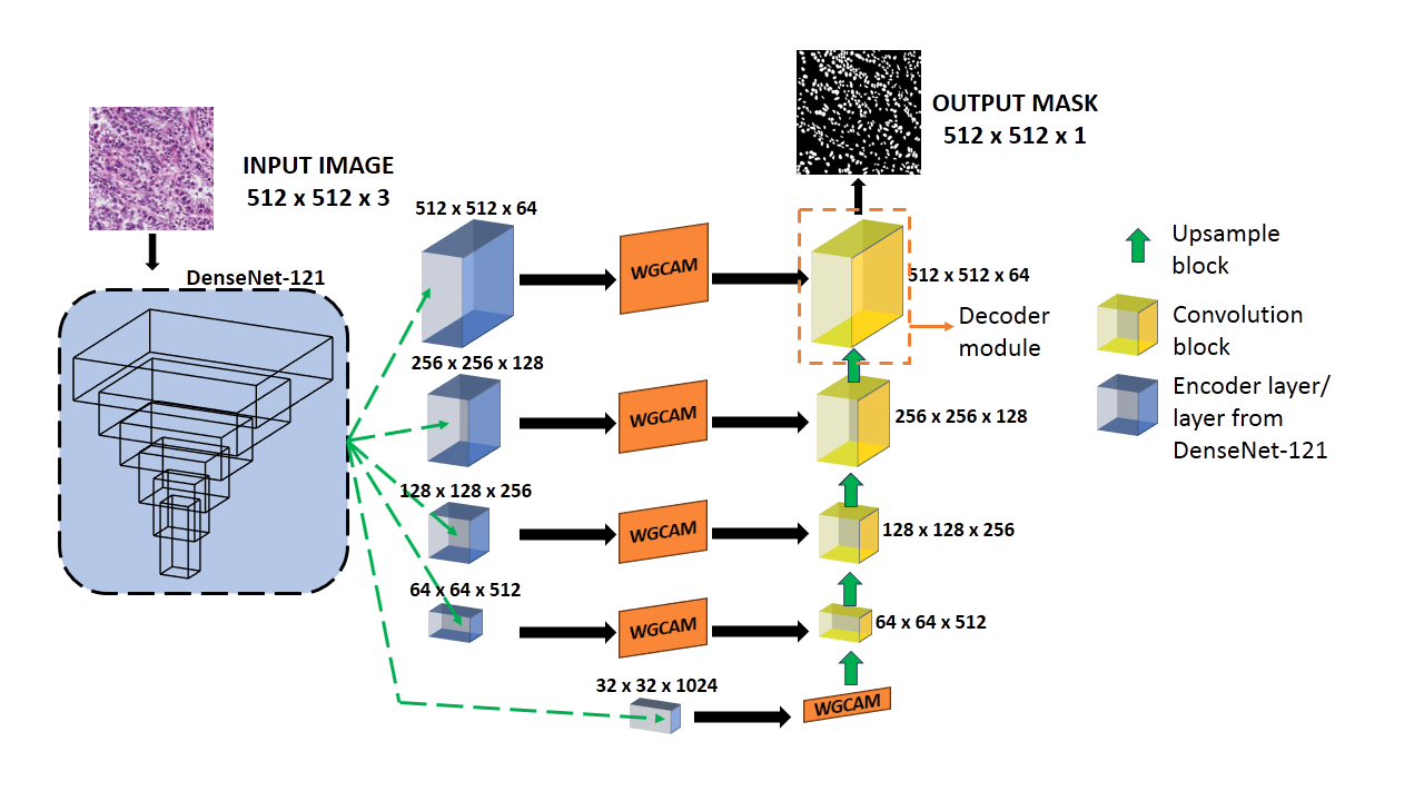
AWGUNET: Attention-Aided Wavelet Guided U-Net for Nuclei Segmentation in Histopathology Images
Ayush Roy, Payel Pramanik, Dmitrii Kaplun, Sergei Antonov, Ram Sarkar
Accurate nuclei segmentation in histopathological images is crucial for cancer diagnosis. Automating this process offers valuable support to clinical experts, as manual annotation is time-consuming and prone to human errors. However, automating nuclei segmentation presents challenges due to uncertain cell boundaries, intricate staining, and diverse structures. In this paper, we present a segmentation approach that combines the U-Net architecture with a DenseNet-121 backbone, harnessing the strengths of both to capture comprehensive contextual and spatial information. Our model introduces the Wavelet-guided channel attention module to enhance cell boundary delineation, along with a learnable weighted global attention module for channel-specific attention. The decoder module, composed of an upsample block and convolution block, further refines segmentation in handling staining patterns. The experimental results conducted on two publicly accessible histopathology datasets, namely Monuseg and TNBC, underscore the superiority of our proposed model, demonstrating its potential to advance histopathological image analysis and cancer diagnosis. The code is made available at: https://github.com/AyushRoy2001/AWGUNET.
Read more6/13/2024