Synthesizing PET images from High-field and Ultra-high-field MR images Using Joint Diffusion Attention Model
2305.03901
0
0
📈
Abstract
MRI and PET are crucial diagnostic tools for brain diseases, as they provide complementary information on brain structure and function. However, PET scanning is costly and involves radioactive exposure, resulting in a lack of PET. Moreover, simultaneous PET and MRI at ultra-high-field are currently hardly infeasible. Ultra-high-field imaging has unquestionably proven valuable in both clinical and academic settings, especially in the field of cognitive neuroimaging. These motivate us to propose a method for synthetic PET from high-filed MRI and ultra-high-field MRI. From a statistical perspective, the joint probability distribution (JPD) is the most direct and fundamental means of portraying the correlation between PET and MRI. This paper proposes a novel joint diffusion attention model which has the joint probability distribution and attention strategy, named JDAM. JDAM has a diffusion process and a sampling process. The diffusion process involves the gradual diffusion of PET to Gaussian noise by adding Gaussian noise, while MRI remains fixed. JPD of MRI and noise-added PET was learned in the diffusion process. The sampling process is a predictor-corrector. PET images were generated from MRI by JPD of MRI and noise-added PET. The predictor is a reverse diffusion process and the corrector is Langevin dynamics. Experimental results on the public Alzheimer's Disease Neuroimaging Initiative (ADNI) dataset demonstrate that the proposed method outperforms state-of-the-art CycleGAN for high-field MRI (3T MRI). Finally, synthetic PET images from the ultra-high-field (5T MRI and 7T MRI) be attempted, providing a possibility for ultra-high-field PET-MRI imaging.
Create account to get full access
Overview
- MRI and PET are important tools for diagnosing brain diseases, providing complementary information on brain structure and function
- PET scanning is expensive and exposes patients to radiation, leading to a lack of PET data
- Ultra-high-field MRI has proven valuable in research and clinical settings, especially for cognitive neuroscience
- The paper proposes a method to synthesize PET images from high-field and ultra-high-field MRI data
Plain English Explanation
Doctors and researchers use two main imaging techniques to study the brain: MRI (magnetic resonance imaging) and PET (positron emission tomography). MRI provides detailed information about the brain's structure, while PET shows how the brain is functioning. Together, these techniques give a comprehensive view of the brain.
However, PET scans have some drawbacks. They are expensive to perform and expose patients to small amounts of radiation. As a result, there is often a lack of PET data available for research and clinical use. In contrast, MRI is more widely used and can be performed at higher magnetic field strengths, providing even more detailed brain images.
The researchers in this paper wanted to find a way to generate synthetic PET images from MRI data. This would allow researchers and doctors to get the benefits of PET imaging without the downsides. The key idea is to use a statistical model to learn the relationship between MRI and PET data, and then use that model to predict PET images from MRI scans.
The researchers tested their approach on a dataset of brain scans from Alzheimer's disease patients. They found that their method outperformed previous techniques for generating PET images from high-field 3T MRI. They also demonstrated that their approach could be used to generate PET images from ultra-high-field 5T and 7T MRI, which opens up new possibilities for brain imaging research and clinical applications.
Technical Explanation
The key contribution of this paper is a novel deep learning model called the Joint Diffusion Attention Model (JDAM) for synthesizing PET images from MRI data. JDAM has two main components:
-
Diffusion Process: This involves gradually adding Gaussian noise to the PET data while keeping the MRI data fixed. This allows the model to learn the joint probability distribution (JPD) between the MRI and the noise-corrupted PET data.
-
Sampling Process: This is a predictor-corrector process that uses the learned JPD to generate new PET images from MRI inputs. The predictor is a reverse diffusion process that removes the Gaussian noise, and the corrector uses Langevin dynamics to refine the generated PET images.
The researchers evaluated JDAM on the Alzheimer's Disease Neuroimaging Initiative (ADNI) dataset, comparing it to the state-of-the-art CycleGAN approach for 3T MRI to PET synthesis. They found that JDAM outperformed CycleGAN, demonstrating the benefits of the joint diffusion and attention mechanisms.
Additionally, the researchers showed that JDAM can be used to synthesize PET images from ultra-high-field 5T and 7T MRI, which is a significant advancement in the field of brain imaging.
Critical Analysis
The paper makes a compelling case for the potential of JDAM to address the limitations of PET imaging by synthesizing PET images from MRI data. The joint diffusion and attention mechanisms seem to be a promising approach for capturing the complex relationships between these two modalities.
However, the paper does not address some important caveats and limitations. For example, it is unclear how well JDAM would perform on datasets from different populations or with different brain pathologies. Additionally, the paper does not discuss the potential for JDAM to introduce artifacts or biases into the generated PET images, which could be a concern for clinical applications.
Further research is needed to better understand the limitations and edge cases of the JDAM approach, as well as to explore ways to improve its robustness and reliability. Rigorous validation on diverse datasets and comparison to other state-of-the-art methods would also help to establish the true capabilities and limitations of this approach.
Conclusion
This paper presents a novel deep learning model, JDAM, for synthesizing PET images from MRI data. By leveraging the joint probability distribution and attention mechanisms, JDAM demonstrates promising results in generating PET images from both high-field and ultra-high-field MRI scans.
The ability to generate PET-like images from widely available MRI data could have significant implications for brain imaging research and clinical practice. It could improve access to PET imaging, reduce radiation exposure, and enable new applications of ultra-high-field MRI. However, further research is needed to fully understand the strengths, limitations, and potential biases of this approach.
Overall, this work represents an important step forward in the field of cross-modal brain imaging synthesis, and the JDAM model could pave the way for more efficient and accessible brain imaging in the future.
This summary was produced with help from an AI and may contain inaccuracies - check out the links to read the original source documents!
Related Papers
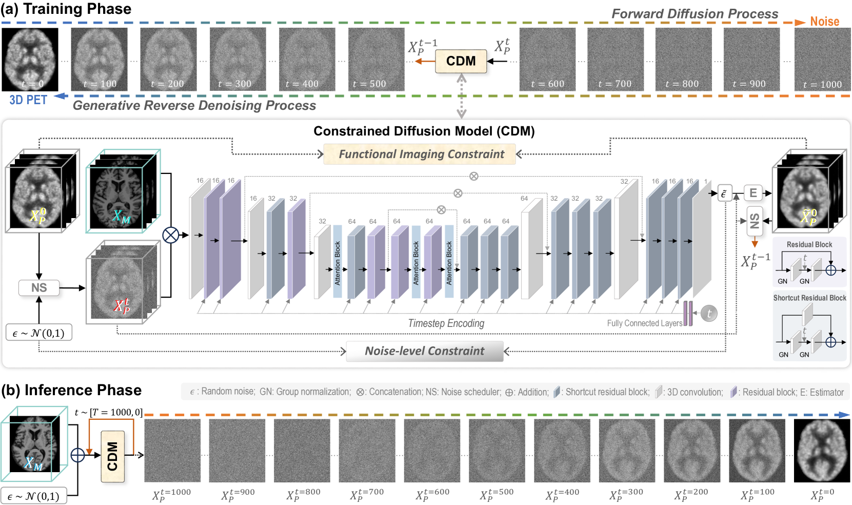
Functional Imaging Constrained Diffusion for Brain PET Synthesis from Structural MRI
Minhui Yu, Mengqi Wu, Ling Yue, Andrea Bozoki, Mingxia Liu
0
0
Magnetic resonance imaging (MRI) and positron emission tomography (PET) are increasingly used in multimodal analysis of neurodegenerative disorders. While MRI is broadly utilized in clinical settings, PET is less accessible. Many studies have attempted to use deep generative models to synthesize PET from MRI scans. However, they often suffer from unstable training and inadequately preserve brain functional information conveyed by PET. To this end, we propose a functional imaging constrained diffusion (FICD) framework for 3D brain PET image synthesis with paired structural MRI as input condition, through a new constrained diffusion model (CDM). The FICD introduces noise to PET and then progressively removes it with CDM, ensuring high output fidelity throughout a stable training phase. The CDM learns to predict denoised PET with a functional imaging constraint introduced to ensure voxel-wise alignment between each denoised PET and its ground truth. Quantitative and qualitative analyses conducted on 293 subjects with paired T1-weighted MRI and 18F-fluorodeoxyglucose (FDG)-PET scans suggest that FICD achieves superior performance in generating FDG-PET data compared to state-of-the-art methods. We further validate the effectiveness of the proposed FICD on data from a total of 1,262 subjects through three downstream tasks, with experimental results suggesting its utility and generalizability.
5/10/2024
📈
New!Diffusion Transformer Model With Compact Prior for Low-dose PET Reconstruction
Bin Huang, Xubiao Liu, Lei Fang, Qiegen Liu, Bingxuan Li
0
0
Positron emission tomography (PET) is an advanced medical imaging technique that plays a crucial role in non-invasive clinical diagnosis. However, while reducing radiation exposure through low-dose PET scans is beneficial for patient safety, it often results in insufficient statistical data. This scarcity of data poses significant challenges for accurately reconstructing high-quality images, which are essential for reliable diagnostic outcomes. In this research, we propose a diffusion transformer model (DTM) guided by joint compact prior (JCP) to enhance the reconstruction quality of low-dose PET imaging. In light of current research findings, we present a pioneering PET reconstruction model that integrates diffusion and transformer models for joint optimization. This model combines the powerful distribution mapping abilities of diffusion models with the capacity of transformers to capture long-range dependencies, offering significant advantages for low-dose PET reconstruction. Additionally, the incorporation of the lesion refining block and penalized weighted least squares (PWLS) enhance the recovery capability of lesion regions and preserves detail information, solving blurring problems in lesion areas and texture details of most deep learning frameworks. Experimental results demonstrate the effectiveness of DTM in enhancing image quality and preserving critical clinical information for low-dose PET scans. Our approach not only reduces radiation exposure risks but also provides a more reliable PET imaging tool for early disease detection and patient management.
7/2/2024

PASTA: Pathology-Aware MRI to PET Cross-Modal Translation with Diffusion Models
Yitong Li, Igor Yakushev, Dennis M. Hedderich, Christian Wachinger
0
0
Positron emission tomography (PET) is a well-established functional imaging technique for diagnosing brain disorders. However, PET's high costs and radiation exposure limit its widespread use. In contrast, magnetic resonance imaging (MRI) does not have these limitations. Although it also captures neurodegenerative changes, MRI is a less sensitive diagnostic tool than PET. To close this gap, we aim to generate synthetic PET from MRI. Herewith, we introduce PASTA, a novel pathology-aware image translation framework based on conditional diffusion models. Compared to the state-of-the-art methods, PASTA excels in preserving both structural and pathological details in the target modality, which is achieved through its highly interactive dual-arm architecture and multi-modal condition integration. A cycle exchange consistency and volumetric generation strategy elevate PASTA's capability to produce high-quality 3D PET scans. Our qualitative and quantitative results confirm that the synthesized PET scans from PASTA not only reach the best quantitative scores but also preserve the pathology correctly. For Alzheimer's classification, the performance of synthesized scans improves over MRI by 4%, almost reaching the performance of actual PET. Code is available at https://github.com/ai-med/PASTA.
5/28/2024
📈
Full-dose Whole-body PET Synthesis from Low-dose PET Using High-efficiency Denoising Diffusion Probabilistic Model: PET Consistency Model
Shaoyan Pan, Elham Abouei, Junbo Peng, Joshua Qian, Jacob F Wynne, Tonghe Wang, Chih-Wei Chang, Justin Roper, Jonathon A Nye, Hui Mao, Xiaofeng Yang
0
0
Objective: Positron Emission Tomography (PET) has been a commonly used imaging modality in broad clinical applications. One of the most important tradeoffs in PET imaging is between image quality and radiation dose: high image quality comes with high radiation exposure. Improving image quality is desirable for all clinical applications while minimizing radiation exposure is needed to reduce risk to patients. Approach: We introduce PET Consistency Model (PET-CM), an efficient diffusion-based method for generating high-quality full-dose PET images from low-dose PET images. It employs a two-step process, adding Gaussian noise to full-dose PET images in the forward diffusion, and then denoising them using a PET Shifted-window Vision Transformer (PET-VIT) network in the reverse diffusion. The PET-VIT network learns a consistency function that enables direct denoising of Gaussian noise into clean full-dose PET images. PET-CM achieves state-of-the-art image quality while requiring significantly less computation time than other methods. Results: In experiments comparing eighth-dose to full-dose images, PET-CM demonstrated impressive performance with NMAE of 1.278+/-0.122%, PSNR of 33.783+/-0.824dB, SSIM of 0.964+/-0.009, NCC of 0.968+/-0.011, HRS of 4.543, and SUV Error of 0.255+/-0.318%, with an average generation time of 62 seconds per patient. This is a significant improvement compared to the state-of-the-art diffusion-based model with PET-CM reaching this result 12x faster. Similarly, in the quarter-dose to full-dose image experiments, PET-CM delivered competitive outcomes, achieving an NMAE of 0.973+/-0.066%, PSNR of 36.172+/-0.801dB, SSIM of 0.984+/-0.004, NCC of 0.990+/-0.005, HRS of 4.428, and SUV Error of 0.151+/-0.192% using the same generation process, which underlining its high quantitative and clinical precision in both denoising scenario.
4/10/2024