Triage of 3D pathology data via 2.5D multiple-instance learning to guide pathologist assessments

0

Sign in to get full access
Overview
- This paper presents a 2.5D multiple-instance learning approach to triage 3D pathology data and guide pathologist assessments.
- The method leverages the contextual information in 3D pathology images to predict the presence of disease, without requiring full 3D segmentation.
- This can help prioritize cases for pathologist review and potentially improve the efficiency of digital pathology workflows.
Plain English Explanation
In the field of digital pathology, doctors analyze microscope images of tissue samples to detect and diagnose diseases like cancer. However, these tissue samples are 3D structures, but the images are typically 2D slices. This paper explores a way to use the 3D context of these samples to make better predictions, without having to fully reconstruct the 3D structure.
The key idea is to use a "multiple-instance learning" approach. Instead of analyzing a single 2D slice, the model looks at a collection or "bag" of 2D slices from the 3D sample. By considering this 3D context, the model can better identify patterns that indicate the presence of disease, even if the full 3D structure is not reconstructed.
This 2.5D approach builds on previous work in leveraging partial 3D information to improve medical image analysis. The key benefit is that it can help prioritize cases for pathologists to review, potentially making digital pathology workflows more efficient.
Technical Explanation
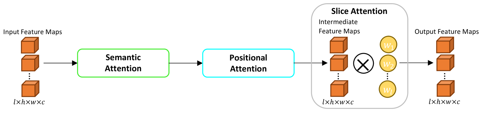
The paper proposes a 2.5D multiple-instance learning (MI-MIL) framework to triage 3D pathology data. Instead of directly predicting disease from individual 2D slices, the model takes a "bag" of 2D slices from a 3D sample as input. It then learns to predict the overall disease state of the 3D sample based on the collective information in the slice "bag".
This allows the model to capture relevant 3D contextual information, without requiring full 3D reconstruction. The 2.5D approach balances the benefits of 3D analysis with the computational efficiency of 2D processing.
The model uses a convolutional neural network backbone to extract features from each 2D slice. These slice-level features are then aggregated using a multiple-instance learning pooling module to predict the overall disease state of the 3D sample.
The authors evaluate their approach on two digital pathology datasets, showing improved performance compared to 2D baselines. They also demonstrate that the model can effectively triage cases, highlighting samples that are most likely to contain disease for prioritized pathologist review.
Critical Analysis
The paper presents a novel and promising approach to leveraging 3D context for digital pathology, without the full complexity of 3D reconstruction. The 2.5D multiple-instance learning framework is an interesting technical contribution that could have broader applications beyond just digital pathology.
However, the paper does not extensively explore the limitations of this approach. For example, it is unclear how the performance would scale with larger 3D sample sizes, or how robust the method is to variations in tissue preparation and imaging. Additionally, the paper does not provide a detailed analysis of the types of 3D patterns the model is able to detect.
Further research could investigate the interpretability of the model's predictions, to understand what 3D visual cues it is using to identify disease. This could lead to additional insights and potentially guide the development of more self-supervised approaches.
Conclusion
This paper presents a novel 2.5D multiple-instance learning approach to triage 3D pathology data, leveraging the contextual information in 3D tissue samples to predict disease presence. By considering a collection of 2D slices, the model can capture relevant 3D patterns without the full complexity of 3D reconstruction.
The proposed method shows promising results in prioritizing cases for pathologist review, potentially improving the efficiency of digital pathology workflows. While the paper does not fully explore the limitations of this approach, it represents an interesting technical contribution that could have broader applications in medical image analysis.
This summary was produced with help from an AI and may contain inaccuracies - check out the links to read the original source documents!
Related Papers
0
Triage of 3D pathology data via 2.5D multiple-instance learning to guide pathologist assessments
Gan Gao, Andrew H. Song, Fiona Wang, David Brenes, Rui Wang, Sarah S. L. Chow, Kevin W. Bishop, Lawrence D. True, Faisal Mahmood, Jonathan T. C. Liu
Accurate patient diagnoses based on human tissue biopsies are hindered by current clinical practice, where pathologists assess only a limited number of thin 2D tissue slices sectioned from 3D volumetric tissue. Recent advances in non-destructive 3D pathology, such as open-top light-sheet microscopy, enable comprehensive imaging of spatially heterogeneous tissue morphologies, offering the feasibility to improve diagnostic determinations. A potential early route towards clinical adoption for 3D pathology is to rely on pathologists for final diagnosis based on viewing familiar 2D H&E-like image sections from the 3D datasets. However, manual examination of the massive 3D pathology datasets is infeasible. To address this, we present CARP3D, a deep learning triage approach that automatically identifies the highest-risk 2D slices within 3D volumetric biopsy, enabling time-efficient review by pathologists. For a given slice in the biopsy, we estimate its risk by performing attention-based aggregation of 2D patches within each slice, followed by pooling of the neighboring slices to compute a context-aware 2.5D risk score. For prostate cancer risk stratification, CARP3D achieves an area under the curve (AUC) of 90.4% for triaging slices, outperforming methods relying on independent analysis of 2D sections (AUC=81.3%). These results suggest that integrating additional depth context enhances the model's discriminative capabilities. In conclusion, CARP3D has the potential to improve pathologist diagnosis via accurate triage of high-risk slices within large-volume 3D pathology datasets.
Read more6/12/2024

0
New!Digital Volumetric Biopsy Cores Improve Gleason Grading of Prostate Cancer Using Deep Learning
Ekaterina Redekop, Mara Pleasure, Zichen Wang, Anthony Sisk, Yang Zong, Kimberly Flores, William Speier, Corey W. Arnold
Prostate cancer (PCa) was the most frequently diagnosed cancer among American men in 2023. The histological grading of biopsies is essential for diagnosis, and various deep learning-based solutions have been developed to assist with this task. Existing deep learning frameworks are typically applied to individual 2D cross-sections sliced from 3D biopsy tissue specimens. This process impedes the analysis of complex tissue structures such as glands, which can vary depending on the tissue slice examined. We propose a novel digital pathology data source called a volumetric core, obtained via the extraction and co-alignment of serially sectioned tissue sections using a novel morphology-preserving alignment framework. We trained an attention-based multiple-instance learning (ABMIL) framework on deep features extracted from volumetric patches to automatically classify the Gleason Grade Group (GGG). To handle volumetric patches, we used a modified video transformer with a deep feature extractor pretrained using self-supervised learning. We ran our morphology-preserving alignment framework to construct 10,210 volumetric cores, leaving out 30% for pretraining. The rest of the dataset was used to train ABMIL, which resulted in a 0.958 macro-average AUC, 0.671 F1 score, 0.661 precision, and 0.695 recall averaged across all five GGG significantly outperforming the 2D baselines.
Read more9/16/2024
0
Cross-Slice Attention and Evidential Critical Loss for Uncertainty-Aware Prostate Cancer Detection
Alex Ling Yu Hung, Haoxin Zheng, Kai Zhao, Kaifeng Pang, Demetri Terzopoulos, Kyunghyun Sung
Current deep learning-based models typically analyze medical images in either 2D or 3D albeit disregarding volumetric information or suffering sub-optimal performance due to the anisotropic resolution of MR data. Furthermore, providing an accurate uncertainty estimation is beneficial to clinicians, as it indicates how confident a model is about its prediction. We propose a novel 2.5D cross-slice attention model that utilizes both global and local information, along with an evidential critical loss, to perform evidential deep learning for the detection in MR images of prostate cancer, one of the most common cancers and a leading cause of cancer-related death in men. We perform extensive experiments with our model on two different datasets and achieve state-of-the-art performance in prostate cancer detection along with improved epistemic uncertainty estimation. The implementation of the model is available at https://github.com/aL3x-O-o-Hung/GLCSA_ECLoss.
Read more7/2/2024

0
Deep Learning-Based Segmentation of Tumors in PET/CT Volumes: Benchmark of Different Architectures and Training Strategies
Monika G'orka, Daniel Jaworek, Marek Wodzinski
Cancer is one of the leading causes of death globally, and early diagnosis is crucial for patient survival. Deep learning algorithms have great potential for automatic cancer analysis. Artificial intelligence has achieved high performance in recognizing and segmenting single lesions. However, diagnosing multiple lesions remains a challenge. This study examines and compares various neural network architectures and training strategies for automatically segmentation of cancer lesions using PET/CT images from the head, neck, and whole body. The authors analyzed datasets from the AutoPET and HECKTOR challenges, exploring popular single-step segmentation architectures and presenting a two-step approach. The results indicate that the V-Net and nnU-Net models were the most effective for their respective datasets. The results for the HECKTOR dataset ranged from 0.75 to 0.76 for the aggregated Dice coefficient. Eliminating cancer-free cases from the AutoPET dataset was found to improve the performance of most models. In the case of AutoPET data, the average segmentation efficiency after training only on images containing cancer lesions increased from 0.55 to 0.66 for the classic Dice coefficient and from 0.65 to 0.73 for the aggregated Dice coefficient. The research demonstrates the potential of artificial intelligence in precise oncological diagnostics and may contribute to the development of more targeted and effective cancer assessment techniques.
Read more4/16/2024