Unsupervised Detection of Fetal Brain Anomalies using Denoising Diffusion Models

0

Sign in to get full access
Overview
- This paper presents an unsupervised method for detecting fetal brain anomalies in ultrasound images using denoising diffusion models.
- The proposed approach does not require labeled training data, making it suitable for medical imaging tasks where annotated data is scarce.
- The model is trained to learn the distribution of normal fetal brain images and can then identify anomalies that deviate from this learned distribution.
Plain English Explanation
Detecting abnormalities in fetal brains during pregnancy is an important medical task, but it can be challenging because there is often a lack of labeled training data. This paper introduces a new way to tackle this problem using a machine learning technique called denoising diffusion models.
The idea is to train the model on a large number of normal, healthy fetal brain images. The model learns to recognize the typical patterns and characteristics of a normal fetal brain. Then, when presented with a new image, the model can identify any parts of the brain that look different or unusual compared to the normal patterns it has learned.
This approach is "unsupervised," which means the model doesn't need to be given labeled examples of abnormal brains during training. It can learn to detect anomalies on its own by identifying deviations from the normal brain structure. This is helpful because it's often difficult and time-consuming to label medical images with the exact locations of abnormalities.
By using this unsupervised anomaly detection method, the researchers hope to create a tool that can assist clinicians in screening for potential fetal brain issues during pregnancy, without requiring a large dataset of labeled training examples.
Technical Explanation
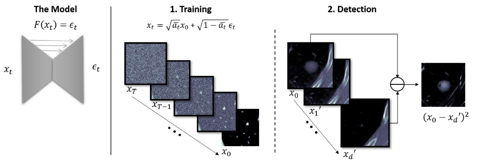
The authors propose using a denoising diffusion probabilistic model (DDPM) as the core of their anomaly detection approach. DDPMs are a type of generative model that can learn the distribution of normal data (in this case, healthy fetal brain images) in an unsupervised manner.
The training process involves gradually adding noise to the input images and then training the model to learn how to "denoise" them, i.e., remove the noise and recover the original clean image. By learning this denoising process, the model builds an internal representation of the normal image distribution.
At inference time, the model can be used to detect anomalies by comparing the "denoising score" of a new input image to the scores of the normal images seen during training. If the denoising score for a particular region of the new image is significantly lower than the scores of the normal images, the model flags that region as potentially anomalous.
The authors evaluate their approach on a dataset of fetal brain ultrasound images, and show that it can effectively detect various types of brain abnormalities without requiring any labeled training data.
Critical Analysis
The proposed method presents a promising approach to the challenge of fetal brain anomaly detection, particularly in settings where labeled training data is scarce. The use of unsupervised denoising diffusion models is an interesting and novel application of this emerging technique.
One potential limitation of the approach is that it may be sensitive to the quality and diversity of the training data. If the dataset of normal fetal brain images does not adequately capture the full range of natural variation, the model may struggle to generalize to all types of abnormalities. Further research could explore techniques to improve the robustness of the anomaly detection, such as leveraging Mahalanobis distance or combining it with other anomaly detection methods.
Additionally, while the paper demonstrates the effectiveness of the approach on ultrasound images, it would be interesting to see how well it generalizes to other medical imaging modalities, such as MRI or CT scans, which may have different characteristics and artifacts.
Conclusion
This paper introduces a novel unsupervised approach to detecting fetal brain anomalies in ultrasound images using denoising diffusion models. The key advantage of this method is that it does not require labeled training data, which can be scarce for medical imaging tasks.
By learning the distribution of normal fetal brain images, the model is able to identify regions that deviate from this learned "baseline," potentially flagging areas of concern for further clinical investigation. While the approach shows promising results, there are opportunities to explore its robustness and generalization to other medical imaging modalities.
Overall, this research represents an interesting application of emerging machine learning techniques to an important problem in prenatal care, with the potential to assist clinicians in early detection of fetal brain abnormalities.
This summary was produced with help from an AI and may contain inaccuracies - check out the links to read the original source documents!
Related Papers
0
Unsupervised Detection of Fetal Brain Anomalies using Denoising Diffusion Models
Markus Ditlev Sj{o}gren Olsen, Jakob Ambsdorf, Manxi Lin, Caroline Taks{o}e-Vester, Morten Bo S{o}ndergaard Svendsen, Anders Nymark Christensen, Mads Nielsen, Martin Gr{o}nneb{ae}k Tolsgaard, Aasa Feragen, Paraskevas Pegios
Congenital malformations of the brain are among the most common fetal abnormalities that impact fetal development. Previous anomaly detection methods on ultrasound images are based on supervised learning, rely on manual annotations, and risk missing underrepresented categories. In this work, we frame fetal brain anomaly detection as an unsupervised task using diffusion models. To this end, we employ an inpainting-based Noise Agnostic Anomaly Detection approach that identifies the abnormality using diffusion-reconstructed fetal brain images from multiple noise levels. Our approach only requires normal fetal brain ultrasound images for training, addressing the limited availability of abnormal data. Our experiments on a real-world clinical dataset show the potential of using unsupervised methods for fetal brain anomaly detection. Additionally, we comprehensively evaluate how different noise types affect diffusion models in the fetal anomaly detection domain.
Read more8/9/2024

0
Diffusion Models for Unsupervised Anomaly Detection in Fetal Brain Ultrasound
Hanna Mykula, Lisa Gasser, Silvia Lobmaier, Julia A. Schnabel, Veronika Zimmer, Cosmin I. Bercea
Ultrasonography is an essential tool in mid-pregnancy for assessing fetal development, appreciated for its non-invasive and real-time imaging capabilities. Yet, the interpretation of ultrasound images is often complicated by acoustic shadows, speckle noise, and other artifacts that obscure crucial diagnostic details. To address these challenges, our study presents a novel unsupervised anomaly detection framework specifically designed for fetal ultrasound imaging. This framework incorporates gestational age filtering, precise identification of fetal standard planes, and targeted segmentation of brain regions to enhance diagnostic accuracy. Furthermore, we introduce the use of denoising diffusion probabilistic models in this context, marking a significant innovation in detecting previously unrecognized anomalies. We rigorously evaluated the framework using various diffusion-based anomaly detection methods, noise types, and noise levels. Notably, AutoDDPM emerged as the most effective, achieving an area under the precision-recall curve of 79.8% in detecting anomalies. This advancement holds promise for improving the tools available for nuanced and effective prenatal diagnostics.
Read more7/23/2024

0
Detailed delineation of the fetal brain in diffusion MRI via multi-task learning
Davood Karimi, Camilo Calixto, Haykel Snoussi, Maria Camila Cortes-Albornoz, Clemente Velasco-Annis, Caitlin Rollins, Camilo Jaimes, Ali Gholipour, Simon K. Warfield
Diffusion-weighted MRI is increasingly used to study the normal and abnormal development of fetal brain in-utero. Recent studies have shown that dMRI can offer invaluable insights into the neurodevelopmental processes in the fetal stage. However, because of the low data quality and rapid brain development, reliable analysis of fetal dMRI data requires dedicated computational methods that are currently unavailable. The lack of automated methods for fast, accurate, and reproducible data analysis has seriously limited our ability to tap the potential of fetal brain dMRI for medical and scientific applications. In this work, we developed and validated a unified computational framework to (1) segment the brain tissue into white matter, cortical/subcortical gray matter, and cerebrospinal fluid, (2) segment 31 distinct white matter tracts, and (3) parcellate the brain's cortex and delineate the deep gray nuclei and white matter structures into 96 anatomically meaningful regions. We utilized a set of manual, semi-automatic, and automatic approaches to annotate 97 fetal brains. Using these labels, we developed and validated a multi-task deep learning method to perform the three computations. Our evaluations show that the new method can accurately carry out all three tasks, achieving a mean Dice similarity coefficient of 0.865 on tissue segmentation, 0.825 on white matter tract segmentation, and 0.819 on parcellation. The proposed method can greatly advance the field of fetal neuroimaging as it can lead to substantial improvements in fetal brain tractography, tract-specific analysis, and structural connectivity assessment.
Read more9/14/2024
0
Back-in-Time Diffusion: Unsupervised Detection of Medical Deepfakes
Fred Grabovski, Lior Yasur, Guy Amit, Yuval Elovici, Yisroel Mirsky
Recent progress in generative models has made it easier for a wide audience to edit and create image content, raising concerns about the proliferation of deepfakes, especially in healthcare. Despite the availability of numerous techniques for detecting manipulated images captured by conventional cameras, their applicability to medical images is limited. This limitation stems from the distinctive forensic characteristics of medical images, a result of their imaging process. In this work we propose a novel anomaly detector for medical imagery based on diffusion models. Normally, diffusion models are used to generate images. However, we show how a similar process can be used to detect synthetic content by making a model reverse the diffusion on a suspected image. We evaluate our method on the task of detecting fake tumors injected and removed from CT and MRI scans. Our method significantly outperforms other state of the art unsupervised detectors with an increased AUC of 0.9 from 0.79 for injection and of 0.96 from 0.91 for removal on average.
Read more7/23/2024