White Matter Geometry-Guided Score-Based Diffusion Model for Tissue Microstructure Imputation in Tractography Imaging

0
Sign in to get full access
Overview
- The paper presents a novel diffusion model for imputing missing tissue microstructure information in tractography imaging.
- The model incorporates the geometry of white matter tracts to guide the diffusion process and improve the accuracy of tissue microstructure estimation.
- The proposed approach aims to address the challenge of missing data in tractography imaging, which can limit the utility of this important neuroimaging technique.
Plain English Explanation
The human brain is a complex organ, and understanding its structure and function is crucial for various medical and scientific applications. One important tool for studying the brain is tractography imaging, which uses magnetic resonance imaging (MRI) to map the connections between different brain regions.
However, tractography imaging can sometimes have missing data, which can make it difficult to get a complete picture of the brain's structure. The researchers in this paper have developed a new diffusion model that can help fill in these gaps by using information about the geometry of the white matter tracts in the brain.
The key idea is that the shape and orientation of the white matter tracts can provide important clues about the underlying tissue microstructure, such as the density and organization of nerve fibers. The researchers' model uses this information to guide the diffusion process, which allows it to estimate the missing tissue microstructure data more accurately.
Technical Explanation
The researchers propose a score-based diffusion model that incorporates the geometry of white matter tracts to improve the imputation of missing tissue microstructure information in tractography imaging. The model leverages the relationship between the geometry of white matter tracts and the underlying tissue microstructure to guide the diffusion process and generate more accurate estimates of the missing data.
The key components of the model include:
-
Geometry-Guided Score Network: This network learns a score function that captures the relationship between the white matter tract geometry and the tissue microstructure, using a combination of diffusion-weighted MRI data and tractography information.
-
Diffusion-Based Imputation: The score function is then used to guide a diffusion-based process that generates plausible estimates of the missing tissue microstructure data, such as fractional anisotropy (FA), a measure of the directional organization of nerve fibers.
The researchers evaluate the performance of their model on several publicly available datasets and compare it to other state-of-the-art approaches for tissue microstructure imputation. Their results demonstrate that the proposed geometry-guided score-based diffusion model outperforms existing methods in terms of accuracy and robustness to missing data.
Critical Analysis
The researchers acknowledge several limitations of their work. First, the model relies on the availability of high-quality tractography data, which may not always be the case in practical scenarios. Second, the performance of the model may be sensitive to the choice of hyperparameters and the specific architecture of the score network.
Additionally, the researchers do not fully explore the impact of the imputed tissue microstructure data on downstream applications, such as connectivity analysis or disease diagnosis. Further research is needed to understand the practical implications of their approach in real-world clinical and neuroscience settings.
Overall, the proposed geometry-guided score-based diffusion model represents an interesting and promising approach to addressing the challenge of missing data in tractography imaging. However, additional validation and exploration of its practical applications would be valuable to fully assess the potential impact of this research.
Conclusion
The paper presents a novel diffusion model that leverages the geometry of white matter tracts to improve the imputation of missing tissue microstructure information in tractography imaging. By incorporating the relationship between tract geometry and tissue microstructure, the proposed model generates more accurate estimates of the missing data, which can enhance the utility of tractography imaging for various applications in neuroscience and medicine.
While the model has some limitations, this research demonstrates the potential of using physics-guided diffusion models to address the challenge of missing data in medical imaging. Further development and validation of this approach could lead to important advancements in our understanding of the human brain and its complex structure and function.
This summary was produced with help from an AI and may contain inaccuracies - check out the links to read the original source documents!
Related Papers
0
White Matter Geometry-Guided Score-Based Diffusion Model for Tissue Microstructure Imputation in Tractography Imaging
Yui Lo, Yuqian Chen, Fan Zhang, Dongnan Liu, Leo Zekelman, Suheyla Cetin-Karayumak, Yogesh Rathi, Weidong Cai, Lauren J. O'Donnell
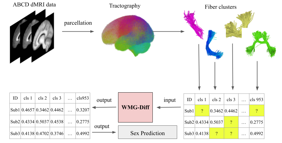
Parcellation of white matter tractography provides anatomical features for disease prediction, anatomical tract segmentation, surgical brain mapping, and non-imaging phenotype classifications. However, parcellation does not always reach 100% accuracy due to various factors, including inter-individual anatomical variability and the quality of neuroimaging scan data. The failure to identify parcels causes a problem of missing microstructure data values, which is especially challenging for downstream tasks that analyze large brain datasets. In this work, we propose a novel deep-learning model to impute tissue microstructure: the White Matter Geometry-guided Diffusion (WMG-Diff) model. Specifically, we first propose a deep score-based guided diffusion model to impute tissue microstructure for diffusion magnetic resonance imaging (dMRI) tractography fiber clusters. Second, we propose a white matter atlas geometric relationship-guided denoising function to guide the reverse denoising process at the subject-specific level. Third, we train and evaluate our model on a large dataset with 9342 subjects. Comprehensive experiments for tissue microstructure imputation and a downstream non-imaging phenotype prediction task demonstrate that our proposed WMG-Diff outperforms state-of-the-art methods.
Read more7/30/2024

0
Streamline tractography of the fetal brain in utero with machine learning
Weide Liu, Camilo Calixto, Simon K. Warfield, Davood Karimi
Diffusion-weighted magnetic resonance imaging (dMRI) is the only non-invasive tool for studying white matter tracts and structural connectivity of the brain. These assessments rely heavily on tractography techniques, which reconstruct virtual streamlines representing white matter fibers. Much effort has been devoted to improving tractography methodology for adult brains, while tractography of the fetal brain has been largely neglected. Fetal tractography faces unique difficulties due to low dMRI signal quality, immature and rapidly developing brain structures, and paucity of reference data. This work presents the first machine learning model for fetal tractography. The model input consists of five sources of information: (1) Fiber orientation, inferred from a diffusion tensor fit to the dMRI signal; (2) Directions of recent propagation steps; (3) Global spatial information, encoded as distances to keypoints in the brain cortex; (4) Tissue segmentation information; and (5) Prior information about the expected local fiber orientations supplied with an atlas. In order to mitigate the local tensor estimation error, a large spatial context around the current point in the diffusion tensor image is encoded using convolutional and attention neural network modules. Moreover, the diffusion tensor information at a hypothetical next point is included in the model input. Filtering rules based on anatomically constrained tractography are applied to prune implausible streamlines. We trained the model on manually-refined whole-brain fetal tractograms and validated the trained model on an independent set of 11 test scans with gestational ages between 23 and 36 weeks. Results show that our proposed method achieves superior performance across all evaluated tracts. The new method can significantly advance the capabilities of dMRI for studying normal and abnormal brain development in utero.
Read more8/27/2024

0
When Diffusion MRI Meets Diffusion Model: A Novel Deep Generative Model for Diffusion MRI Generation
Xi Zhu, Wei Zhang, Yijie Li, Lauren J. O'Donnell, Fan Zhang
Diffusion MRI (dMRI) is an advanced imaging technique characterizing tissue microstructure and white matter structural connectivity of the human brain. The demand for high-quality dMRI data is growing, driven by the need for better resolution and improved tissue contrast. However, acquiring high-quality dMRI data is expensive and time-consuming. In this context, deep generative modeling emerges as a promising solution to enhance image quality while minimizing acquisition costs and scanning time. In this study, we propose a novel generative approach to perform dMRI generation using deep diffusion models. It can generate high dimension (4D) and high resolution data preserving the gradients information and brain structure. We demonstrated our method through an image mapping task aimed at enhancing the quality of dMRI images from 3T to 7T. Our approach demonstrates highly enhanced performance in generating dMRI images when compared to the current state-of-the-art (SOTA) methods. This achievement underscores a substantial progression in enhancing dMRI quality, highlighting the potential of our novel generative approach to revolutionize dMRI imaging standards.
Read more8/26/2024
↗️
0
Diffusion MRI with Machine Learning
Davood Karimi
Diffusion-weighted magnetic resonance imaging (dMRI) offers unique capabilities including noninvasive probing of brain's tissue microstructure and structural connectivity. It is widely used for clinical assessment of brain pathologies and for neuroscience research. Analyzing the dMRI data to extract useful information for medical and scientific purposes can be challenging. The dMRI measurements often suffer from strong noise and artifacts, there is usually high inter-session and inter-scanner variability in the data, and considerable inter-subject heterogeneity in brain structure. Moreover, the relationship between measurements and the phenomena of interest can be highly complex. Recent years have witnessed increasing use of machine learning methods for dMRI analysis. This manuscript aims to assess these efforts, with a focus on methods that have addressed data preprocessing and harmonization, microstructure mapping, tractography, and white matter tract analysis. We study the main findings, strengths, and weaknesses of the existing methods and suggest topics for future research. We find that machine learning may be exceptionally suited to tackle some of the difficult tasks in dMRI analysis. However, for this to happen, several shortcomings of existing methods and critical unresolved issues need to be addressed. These include deficient evaluation practices, lack of rich training datasets and validation benchmarks, as well as model generalizability, reliability, and explainability concerns.
Read more7/29/2024