BMapOpt: Optimization of Brain Tissue Probability Maps using a Differentiable MRI Simulator

0
Sign in to get full access
Overview
- This research paper presents a method called BMapOpt for optimizing brain tissue probability maps using a differentiable MRI simulator.
- The goal is to improve the accuracy of brain tissue segmentation in MRI data by optimizing the underlying tissue probability maps.
- The approach leverages a differentiable MRI simulator to enable end-to-end optimization of the tissue probability maps.
Plain English Explanation
MRI scans are a powerful tool for studying the human brain, but accurately identifying different brain tissues in these images can be challenging. BMapOpt: Optimization of Brain Tissue Probability Maps using a Differentiable MRI Simulator introduces a new method to improve this process.
The key idea is to use a "differentiable MRI simulator" - a computer model that can accurately mimic how an MRI machine would capture an image of the brain. By coupling this simulator with an optimization algorithm, the researchers were able to fine-tune the underlying "tissue probability maps" that describe the composition of the brain. This allows the system to learn the optimal tissue maps that best match real MRI scans, leading to more accurate brain segmentation.
The differentiable MRI simulator acts like a virtual MRI machine, taking tissue maps as input and outputting simulated MRI images. By comparing these simulated images to real MRI scans, the system can figure out how to adjust the tissue maps to improve the match. It's a bit like having a flight simulator to test and refine aircraft designs before building the real thing.
This optimization-based approach contrasts with traditional brain segmentation methods that rely more on hand-crafted rules or machine learning models trained on limited datasets. BMapOpt's use of a differentiable simulator allows it to learn the tissue maps in a more flexible and data-driven way, potentially leading to better results.
Technical Explanation
The key components of the BMapOpt method are:
-
Differentiable MRI Simulator: The researchers developed a differentiable simulator that can model the physics of MRI image formation. This allows gradients to be computed through the simulation process, enabling end-to-end optimization.
-
Tissue Probability Map Optimization: BMapOpt takes an initial set of tissue probability maps as input. It then uses gradient-based optimization to iteratively update these maps, minimizing the discrepancy between the simulated MRI images and real MRI scans.
-
Architecture: The optimization is performed using a convolutional neural network that operates on the 3D tissue probability maps. This network learns to predict the adjustments needed to the tissue maps to better match the observed MRI data.
The key insight behind BMapOpt is that by coupling a differentiable MRI simulator with an optimization framework, the tissue probability maps can be refined in a principled, data-driven manner. This contrasts with traditional segmentation approaches that rely more on heuristics or limited training data.
Critical Analysis
The authors acknowledge several limitations and areas for future work:
- The current simulator models only the basic MRI physics and does not capture all sources of image variability. More sophisticated simulators could potentially improve the optimization.
- The method was evaluated on a single dataset, and its generalization to other brain imaging modalities or populations is not yet established.
- The computational cost of the optimization process may limit its practical applicability, especially for large-scale studies.
Additionally, one could question whether the learned tissue probability maps truly capture the underlying ground truth, or if they are merely optimizing for a better match to the observed MRI data. Further research would be needed to validate the biological plausibility of the optimized maps.
Conclusion
Overall, the BMapOpt method represents an interesting step towards more principled, data-driven approaches to brain tissue segmentation in MRI. By leveraging a differentiable MRI simulator, the system can optimize the underlying tissue probability maps to better match real-world imaging data. While further research is needed to address the current limitations, this work demonstrates the potential of differentiable simulators to enable new optimization-based techniques in medical imaging analysis.
This summary was produced with help from an AI and may contain inaccuracies - check out the links to read the original source documents!
Related Papers
0
BMapOpt: Optimization of Brain Tissue Probability Maps using a Differentiable MRI Simulator
Utkarsh Gupta, Emmanouil Nikolakakis, Moritz Zaiss, Razvan Marinescu
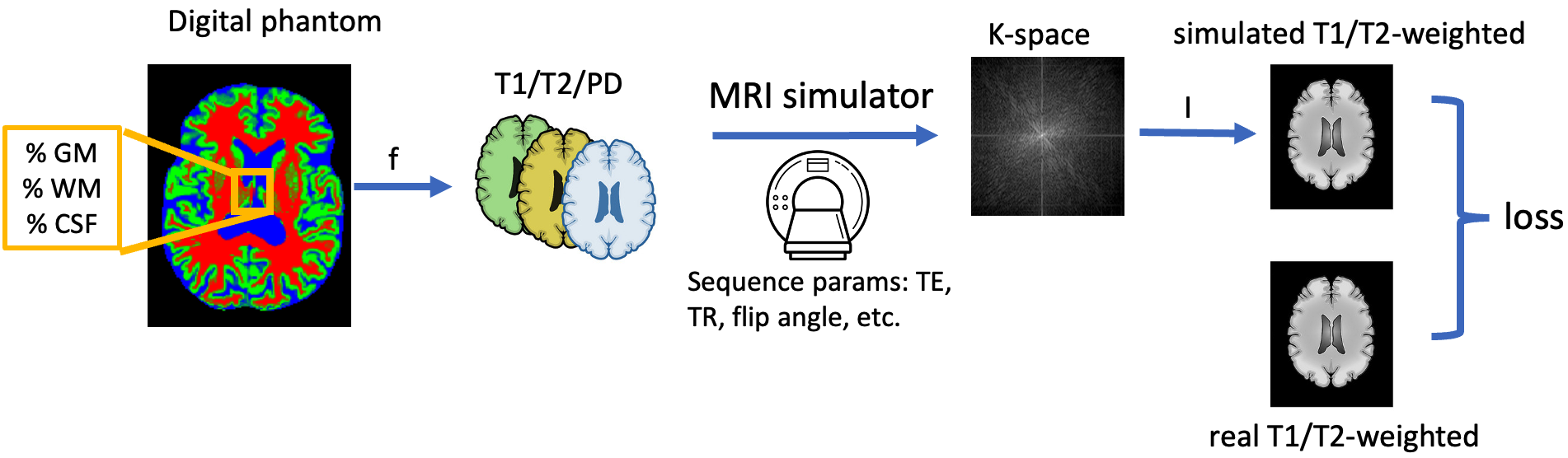
Reconstructing digital brain phantoms in the form of voxel-based, multi-channeled tissue probability maps for individual subjects is essential for capturing brain anatomical variability, understanding neurological diseases, as well as for testing image processing methods. We demonstrate the first framework that estimates brain tissue probability maps (Grey Matter - GM, White Matter - WM, and Cerebrospinal fluid - CSF) with the help of a Physics-based differentiable MRI simulator that models the magnetization signal at each voxel in the volume. Given an observed $T_1$/$T_2$-weighted MRI scan, the corresponding clinical MRI sequence, and the MRI differentiable simulator, we estimate the simulator's input probability maps by back-propagating the L2 loss between the simulator's output and the $T_1$/$T_2$-weighted scan. This approach has the significant advantage of not relying on any training data and instead uses the strong inductive bias of the MRI simulator. We tested the model on 20 scans from the BrainWeb database and demonstrated a highly accurate reconstruction of GM, WM, and CSF. Our source code is available online: https://github.com/BioMedAI-UCSC/BMapEst.
Read more7/2/2024

0
qMRI Diffusor: Quantitative T1 Mapping of the Brain using a Denoising Diffusion Probabilistic Model
Shishuai Wang, Hua Ma, Juan A. Hernandez-Tamames, Stefan Klein, Dirk H. J. Poot
Quantitative MRI (qMRI) offers significant advantages over weighted images by providing objective parameters related to tissue properties. Deep learning-based methods have demonstrated effectiveness in estimating quantitative maps from series of weighted images. In this study, we present qMRI Diffusor, a novel approach to qMRI utilising deep generative models. Specifically, we implemented denoising diffusion probabilistic models (DDPM) for T1 quantification in the brain, framing the estimation of quantitative maps as a conditional generation task. The proposed method is compared with the residual neural network (ResNet) and the recurrent inference machine (RIM) on both phantom and in vivo data. The results indicate that our method achieves improved accuracy and precision in parameter estimation, along with superior visual performance. Moreover, our method inherently incorporates stochasticity, enabling straightforward quantification of uncertainty. Hence, the proposed method holds significant promise for quantitative MR mapping.
Read more7/24/2024

0
Detailed delineation of the fetal brain in diffusion MRI via multi-task learning
Davood Karimi, Camilo Calixto, Haykel Snoussi, Maria Camila Cortes-Albornoz, Clemente Velasco-Annis, Caitlin Rollins, Camilo Jaimes, Ali Gholipour, Simon K. Warfield
Diffusion-weighted MRI is increasingly used to study the normal and abnormal development of fetal brain in-utero. Recent studies have shown that dMRI can offer invaluable insights into the neurodevelopmental processes in the fetal stage. However, because of the low data quality and rapid brain development, reliable analysis of fetal dMRI data requires dedicated computational methods that are currently unavailable. The lack of automated methods for fast, accurate, and reproducible data analysis has seriously limited our ability to tap the potential of fetal brain dMRI for medical and scientific applications. In this work, we developed and validated a unified computational framework to (1) segment the brain tissue into white matter, cortical/subcortical gray matter, and cerebrospinal fluid, (2) segment 31 distinct white matter tracts, and (3) parcellate the brain's cortex and delineate the deep gray nuclei and white matter structures into 96 anatomically meaningful regions. We utilized a set of manual, semi-automatic, and automatic approaches to annotate 97 fetal brains. Using these labels, we developed and validated a multi-task deep learning method to perform the three computations. Our evaluations show that the new method can accurately carry out all three tasks, achieving a mean Dice similarity coefficient of 0.865 on tissue segmentation, 0.825 on white matter tract segmentation, and 0.819 on parcellation. The proposed method can greatly advance the field of fetal neuroimaging as it can lead to substantial improvements in fetal brain tractography, tract-specific analysis, and structural connectivity assessment.
Read more9/14/2024
0
White Matter Geometry-Guided Score-Based Diffusion Model for Tissue Microstructure Imputation in Tractography Imaging
Yui Lo, Yuqian Chen, Fan Zhang, Dongnan Liu, Leo Zekelman, Suheyla Cetin-Karayumak, Yogesh Rathi, Weidong Cai, Lauren J. O'Donnell
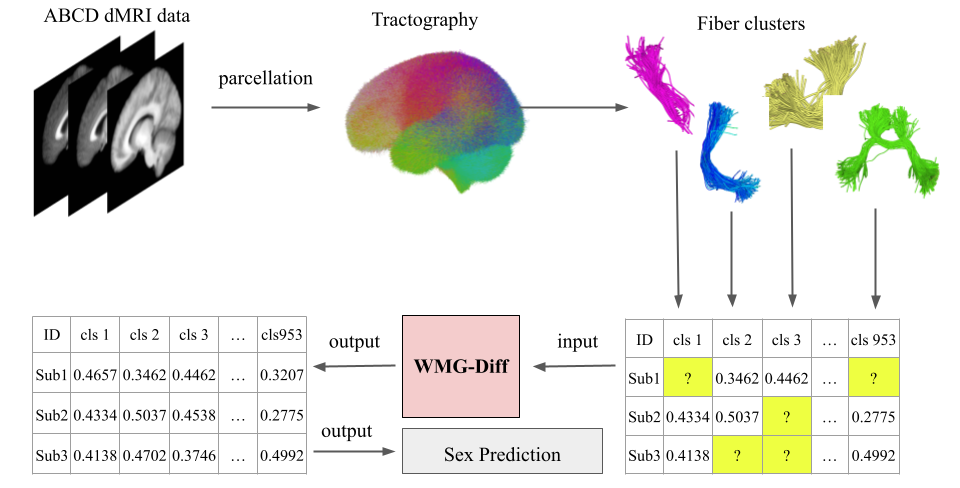
Parcellation of white matter tractography provides anatomical features for disease prediction, anatomical tract segmentation, surgical brain mapping, and non-imaging phenotype classifications. However, parcellation does not always reach 100% accuracy due to various factors, including inter-individual anatomical variability and the quality of neuroimaging scan data. The failure to identify parcels causes a problem of missing microstructure data values, which is especially challenging for downstream tasks that analyze large brain datasets. In this work, we propose a novel deep-learning model to impute tissue microstructure: the White Matter Geometry-guided Diffusion (WMG-Diff) model. Specifically, we first propose a deep score-based guided diffusion model to impute tissue microstructure for diffusion magnetic resonance imaging (dMRI) tractography fiber clusters. Second, we propose a white matter atlas geometric relationship-guided denoising function to guide the reverse denoising process at the subject-specific level. Third, we train and evaluate our model on a large dataset with 9342 subjects. Comprehensive experiments for tissue microstructure imputation and a downstream non-imaging phenotype prediction task demonstrate that our proposed WMG-Diff outperforms state-of-the-art methods.
Read more7/30/2024