World of Forms: Deformable Geometric Templates for One-Shot Surface Meshing in Coronary CT Angiography

0

Sign in to get full access
Overview
- The paper proposes a novel method for generating 3D surface meshes from coronary CT angiography (CCTA) data using deformable geometric templates.
- The approach allows for rapid, one-shot surface mesh generation, overcoming limitations of traditional time-consuming mesh generation techniques.
- The method is evaluated on a large dataset of CCTA scans, demonstrating its effectiveness and accuracy in capturing the complex topology of coronary arteries.
Plain English Explanation
The paper introduces a new way to create 3D models of coronary arteries from CT scans. Traditionally, generating these 3D models has been a slow and laborious process. The researchers have developed a "deformable geometric template" approach that can quickly and automatically create accurate 3D models of the arteries.
The key idea is to start with a pre-designed 3D template shape that roughly matches the expected structure of the coronary arteries. This template can then be "deformed" or adjusted to precisely match the actual shape of the arteries in each individual CT scan. This "one-shot" process allows the 3D model to be generated very quickly, in contrast to more time-consuming traditional methods.
The researchers tested their approach on a large dataset of CT scans and found that it was able to accurately capture the complex, branching structure of the coronary arteries. This rapid 3D modeling capability could be valuable for clinical applications, such as planning surgical procedures or monitoring changes in the arteries over time.
Technical Explanation
The paper introduces a "World of Forms" approach for rapid, one-shot surface meshing of coronary arteries from coronary CT angiography (CCTA) data. The key innovation is the use of deformable geometric templates that can be efficiently optimized to fit the target anatomy.
The method works by first constructing a reference template mesh that captures the general topology of the coronary arteries. This template is then deformed using a differentiable spatial transformer network to fit the specific geometry of the arteries in each CCTA scan. The deformation is driven by a novel "dynamic displacement field" that allows for local and global shape adjustments.
The deformation is optimized in an end-to-end manner using a combination of geometric and image-based loss functions. This one-shot optimization process allows the final 3D mesh to be generated very efficiently, in contrast to traditional approaches that require complex, time-consuming segmentation and meshing steps.
The authors evaluate their method on a large dataset of CCTA scans, demonstrating its ability to accurately capture the complex branching structure of the coronary arteries. They also show that the generated meshes can be used for downstream applications such as computational fluid dynamics analysis.
Critical Analysis
The "World of Forms" approach represents an innovative solution to the challenge of rapid 3D mesh generation from CCTA data. By leveraging deformable geometric templates, the method is able to overcome limitations of traditional mesh generation techniques, which can be computationally intensive and require significant user intervention.
One notable strength of the approach is its end-to-end optimization strategy, which allows the deformation process to be driven by a combination of geometric and image-based cues. This helps to ensure that the final mesh not only fits the target anatomy, but also captures the underlying vascular structure as observed in the CCTA data.
However, the paper does not fully address potential limitations or edge cases of the method. For example, it is not clear how the approach would perform on CCTA scans with significant anatomical variation or pathological changes to the coronary arteries. Additionally, the authors do not discuss the robustness of the method to factors such as image noise, resolution, or contrast agent distribution.
Further research is needed to explore the broader applicability of the "World of Forms" approach and to investigate potential refinements or extensions, such as the incorporation of additional anatomical priors or the use of more sophisticated deformation models. Nonetheless, this work represents an important step forward in the field of cardiac mesh generation and could have significant implications for clinical applications involving CCTA data analysis.
Conclusion
The "World of Forms" method presented in this paper offers a novel and efficient approach to 3D surface meshing of coronary arteries from CCTA data. By leveraging deformable geometric templates, the technique enables rapid, one-shot generation of accurate 3D models, overcoming the limitations of traditional mesh generation techniques.
The successful evaluation of the method on a large CCTA dataset demonstrates its potential for practical applications, such as surgical planning, computational fluid dynamics analysis, and monitoring of vascular changes over time. While further research is needed to explore the broader capabilities and limitations of the approach, this work represents an important contribution to the field of cardiac imaging and modeling.
This summary was produced with help from an AI and may contain inaccuracies - check out the links to read the original source documents!
Related Papers
0
New!World of Forms: Deformable Geometric Templates for One-Shot Surface Meshing in Coronary CT Angiography
Rudolf L. M. van Herten, Ioannis Lagogiannis, Jelmer M. Wolterink, Steffen Bruns, Eva R. Meulendijks, Damini Dey, Joris R. de Groot, Jos'e P. Henriques, R. Nils Planken, Simone Saitta, Ivana Iv{s}gum
Deep learning-based medical image segmentation and surface mesh generation typically involve a sequential pipeline from image to segmentation to meshes, often requiring large training datasets while making limited use of prior geometric knowledge. This may lead to topological inconsistencies and suboptimal performance in low-data regimes. To address these challenges, we propose a data-efficient deep learning method for direct 3D anatomical object surface meshing using geometric priors. Our approach employs a multi-resolution graph neural network that operates on a prior geometric template which is deformed to fit object boundaries of interest. We show how different templates may be used for the different surface meshing targets, and introduce a novel masked autoencoder pretraining strategy for 3D spherical data. The proposed method outperforms nnUNet in a one-shot setting for segmentation of the pericardium, left ventricle (LV) cavity and the LV myocardium. Similarly, the method outperforms other lumen segmentation operating on multi-planar reformatted images. Results further indicate that mesh quality is on par with or improves upon marching cubes post-processing of voxel mask predictions, while remaining flexible in the choice of mesh triangulation prior, thus paving the way for more accurate and topologically consistent 3D medical object surface meshing.
Read more9/19/2024
0
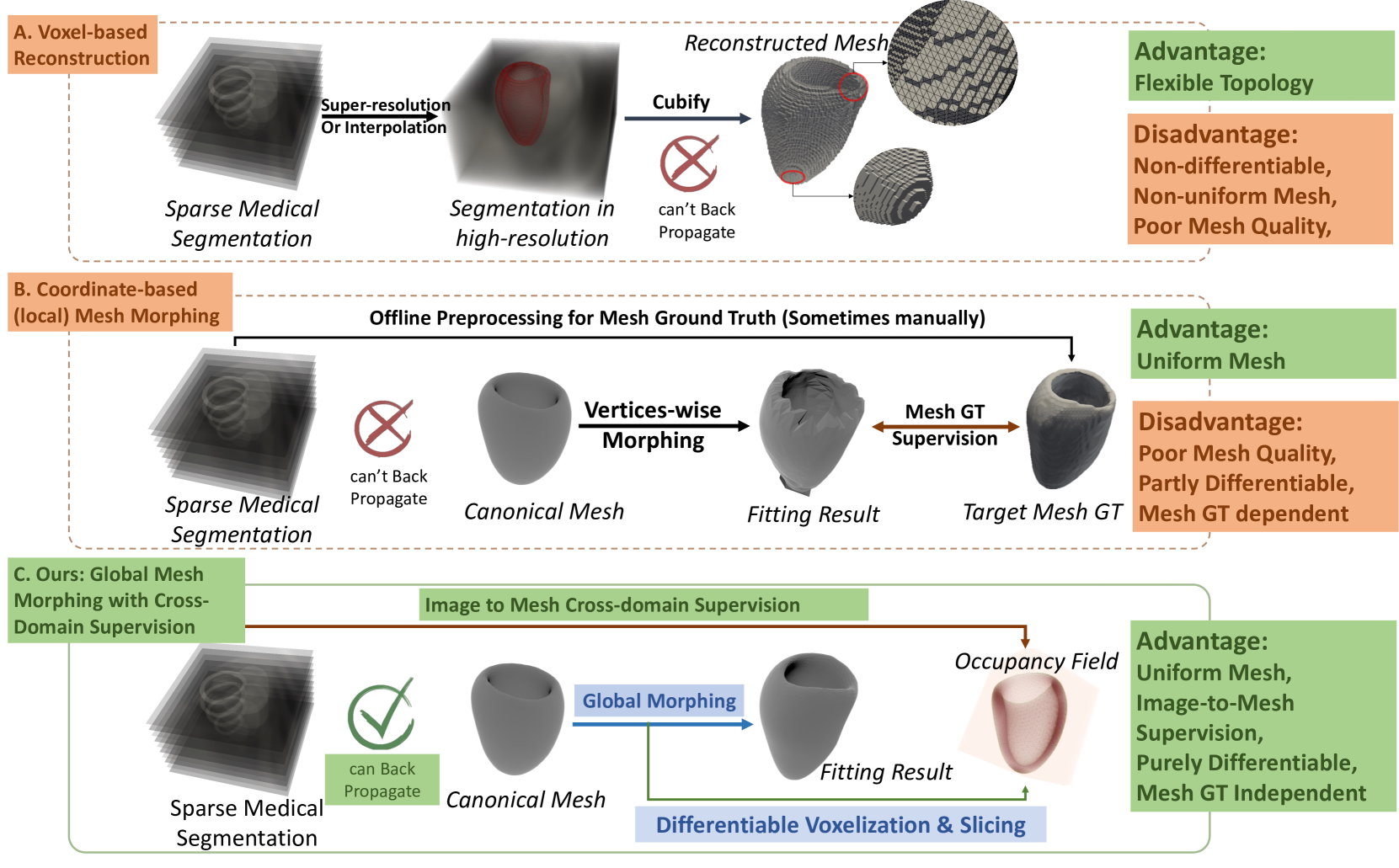
Explicit Differentiable Slicing and Global Deformation for Cardiac Mesh Reconstruction
Yihao Luo, Dario Sesia, Fanwen Wang, Yinzhe Wu, Wenhao Ding, Jiahao Huang, Fadong Shi Anoop Shah, Amit Kaural, Jamil Mayet, Guang Yang, ChoonHwai Yap
Mesh reconstruction of the cardiac anatomy from medical images is useful for shape and motion measurements and biophysics simulations to facilitate the assessment of cardiac function and health. However, 3D medical images are often acquired as 2D slices that are sparsely sampled and noisy, and mesh reconstruction on such data is a challenging task. Traditional voxel-based approaches rely on pre- and post-processing that compromises image fidelity, while mesh-level deep learning approaches require mesh annotations that are difficult to get. Therefore, direct cross-domain supervision from 2D images to meshes is a key technique for advancing 3D learning in medical imaging, but it has not been well-developed. While there have been attempts to approximate the optimized meshes' slicing, few existing methods directly use 2D slices to supervise mesh reconstruction in a differentiable manner. Here, we propose a novel explicit differentiable voxelization and slicing (DVS) algorithm that allows gradient backpropagation to a mesh from its slices, facilitating refined mesh optimization directly supervised by the losses defined on 2D images. Further, we propose an innovative framework for extracting patient-specific left ventricle (LV) meshes from medical images by coupling DVS with a graph harmonic deformation (GHD) mesh morphing descriptor of cardiac shape that naturally preserves mesh quality and smoothness during optimization. Experimental results demonstrate that our method achieves state-of-the-art performance in cardiac mesh reconstruction tasks from CT and MRI, with an overall Dice score of 90% on multi-datasets, outperforming existing approaches. The proposed method can further quantify clinically useful parameters such as ejection fraction and global myocardial strains, closely matching the ground truth and surpassing the traditional voxel-based approach in sparse images.
Read more9/4/2024

0
Multi-view Hybrid Graph Convolutional Network for Volume-to-mesh Reconstruction in Cardiovascular MRI
Nicol'as Gaggion, Benjamin A. Matheson, Yan Xia, Rodrigo Bonazzola, Nishant Ravikumar, Zeike A. Taylor, Diego H. Milone, Alejandro F. Frangi, Enzo Ferrante
Cardiovascular magnetic resonance imaging is emerging as a crucial tool to examine cardiac morphology and function. Essential to this endeavour are anatomical 3D surface and volumetric meshes derived from CMR images, which facilitate computational anatomy studies, biomarker discovery, and in-silico simulations. However, conventional surface mesh generation methods, such as active shape models and multi-atlas segmentation, are highly time-consuming and require complex processing pipelines to generate simulation-ready 3D meshes. In response, we introduce HybridVNet, a novel architecture for direct image-to-mesh extraction seamlessly integrating standard convolutional neural networks with graph convolutions, which we prove can efficiently handle surface and volumetric meshes by encoding them as graph structures. To further enhance accuracy, we propose a multiview HybridVNet architecture which processes both long axis and short axis CMR, showing that it can increase the performance of cardiac MR mesh generation. Our model combines traditional convolutional networks with variational graph generative models, deep supervision and mesh-specific regularisation. Experiments on a comprehensive dataset from the UK Biobank confirm the potential of HybridVNet to significantly advance cardiac imaging and computational cardiology by efficiently generating high-fidelity and simulation ready meshes from CMR images.
Read more8/15/2024
🤿
0
DCSM 2.0: Deep Conditional Shape Models for Data Efficient Segmentation
Athira J Jacob, Puneet Sharma, Daniel Rueckert
Segmentation is often the first step in many medical image analyses workflows. Deep learning approaches, while giving state-of-the-art accuracies, are data intensive and do not scale well to low data regimes. We introduce Deep Conditional Shape Models 2.0, which uses an edge detector, along with an implicit shape function conditioned on edge maps, to leverage cross-modality shape information. The shape function is trained exclusively on a source domain (contrasted CT) and applied to the target domain of interest (3D echocardiography). We demonstrate data efficiency in the target domain by varying the amounts of training data used in the edge detection stage. We observe that DCSM 2.0 outperforms the baseline at all data levels in terms of Hausdorff distances, and while using 50% or less of the training data in terms of average mesh distance, and at 10% or less of the data with the dice coefficient. The method scales well to low data regimes, with gains of up to 5% in dice coefficient, 2.58 mm in average surface distance and 21.02 mm in Hausdorff distance when using just 2% (22 volumes) of the training data.
Read more7/2/2024