Wound Tissue Segmentation in Diabetic Foot Ulcer Images Using Deep Learning: A Pilot Study

0
🤿
Sign in to get full access
Overview
- Researchers created a dataset called DFUTissue containing labeled and unlabeled images of diabetic foot ulcers (DFUs) to help evaluate wound tissue segmentation algorithms.
- They conducted a pilot study using deep learning to segment wound characteristics like fibrin, granulation, and callus.
- The researchers' framework combined supervised learning (SL) and semi-supervised learning (SSL) approaches due to the limited annotated data.
- The SL model uses a hybrid architecture with a Mix Transformer encoder and a CNN decoder, along with a spatial-channel squeeze-and-excitation module.
- The SSL phase employs pseudo-labeling to iteratively incorporate valuable unlabeled images and improve segmentation.
- The researchers compare their approach to state-of-the-art methods and achieve high performance on both SL and SSL tasks.
Plain English Explanation
Identifying different types of tissue in images of diabetic foot ulcers (DFUs) is a challenging task that has not been extensively studied, largely due to the lack of a comprehensive dataset. To address this gap, the researchers created the DFUTissue dataset, which contains 110 labeled images and 600 unlabeled images of DFUs.
Using this dataset, the researchers conducted a study to automatically segment key wound characteristics, such as fibrin, granulation, and callus, using deep learning. Due to the limited amount of labeled data, the researchers used a combination of supervised learning (SL) and semi-supervised learning (SSL) approaches.
The SL model features a hybrid architecture that combines a Mix Transformer encoder and a convolutional neural network (CNN) decoder, with an additional module to enhance the accuracy of boundary detection. The SSL phase then uses a pseudo-labeling technique to iteratively incorporate valuable unlabeled images and further improve the segmentation performance.
The researchers compare their approach to state-of-the-art methods and achieve impressive results, with the SL model reaching a Dice Similarity Coefficient (DSC) of 84.89% and the SSL model improving this to 87.64%. Additionally, when tested on a separate dataset, their hybrid model outperformed other methods in binary segmentation of DFU wound areas, achieving a DSC of 92.99%.
Technical Explanation
The researchers created the DFUTissue dataset to address the lack of available clinical image data for evaluating wound tissue segmentation algorithms. The dataset includes 110 images with tissues labeled by wound experts, as well as 600 unlabeled images.
For the supervised learning (SL) phase, the researchers proposed a hybrid deep learning model that combines a Mix Transformer (MiT-b3) encoder and a CNN decoder. This architecture is further enhanced by the integration of a parallel spatial and channel squeeze-and-excitation (P-scSE) module, which is known to improve boundary accuracy.
Due to the limited amount of annotated data, the researchers also implemented a semi-supervised learning (SSL) approach using pseudo-labeling. This technique iteratively identifies and incorporates valuable unlabeled images to enhance the overall segmentation performance.
The researchers conducted comparative evaluations of their SL and SSL models against state-of-the-art methods. The SL model achieved a Dice Similarity Coefficient (DSC) of 84.89%, which was then improved to 87.64% in the SSL phase. Additionally, the hybrid model outperformed other approaches with a 92.99% DSC when performing binary segmentation of DFU wound areas on the Chronic Wound dataset.
Critical Analysis
The researchers acknowledge the limited availability of annotated clinical data as a key challenge in this field, which motivated their use of semi-supervised learning techniques. While the DFUTissue dataset represents a valuable contribution, the relatively small size of the labeled subset may limit the generalizability of the findings.
Additionally, the paper does not provide detailed information on the clinical significance of the segmented wound characteristics or how the results could impact patient care and wound management. Further research is needed to evaluate the clinical utility of the proposed approach and its potential impact on healthcare outcomes.
It would also be interesting to see the researchers explore alternative semi-supervised learning methods, such as generative adversarial networks or cross-consistency training, and compare their performance to the pseudo-labeling approach used in this study.
Conclusion
This research represents an important step towards automating the analysis of diabetic foot ulcer images, a critical task that has received limited attention due to the lack of comprehensive datasets. The creation of the DFUTissue dataset and the development of a hybrid deep learning model that combines supervised and semi-supervised learning techniques demonstrate the researchers' efforts to address this challenge.
The high performance achieved by the researchers' approach, particularly in the SSL phase, highlights the potential of these methods to assist healthcare professionals in the accurate and efficient assessment of wound characteristics, which could ultimately lead to improved patient outcomes. As the field of medical image analysis continues to evolve, this work serves as a valuable contribution and a foundation for future research in the automated segmentation and analysis of diabetic foot ulcers.
This summary was produced with help from an AI and may contain inaccuracies - check out the links to read the original source documents!
Related Papers
🤿
0
Wound Tissue Segmentation in Diabetic Foot Ulcer Images Using Deep Learning: A Pilot Study
Mrinal Kanti Dhar, Chuanbo Wang, Yash Patel, Taiyu Zhang, Jeffrey Niezgoda, Sandeep Gopalakrishnan, Keke Chen, Zeyun Yu
Identifying individual tissues, so-called tissue segmentation, in diabetic foot ulcer (DFU) images is a challenging task and little work has been published, largely due to the limited availability of a clinical image dataset. To address this gap, we have created a DFUTissue dataset for the research community to evaluate wound tissue segmentation algorithms. The dataset contains 110 images with tissues labeled by wound experts and 600 unlabeled images. Additionally, we conducted a pilot study on segmenting wound characteristics including fibrin, granulation, and callus using deep learning. Due to the limited amount of annotated data, our framework consists of both supervised learning (SL) and semi-supervised learning (SSL) phases. In the SL phase, we propose a hybrid model featuring a Mix Transformer (MiT-b3) in the encoder and a CNN in the decoder, enhanced by the integration of a parallel spatial and channel squeeze-and-excitation (P-scSE) module known for its efficacy in improving boundary accuracy. The SSL phase employs a pseudo-labeling-based approach, iteratively identifying and incorporating valuable unlabeled images to enhance overall segmentation performance. Comparative evaluations with state-of-the-art methods are conducted for both SL and SSL phases. The SL achieves a Dice Similarity Coefficient (DSC) of 84.89%, which has been improved to 87.64% in the SSL phase. Furthermore, the results are benchmarked against two widely used SSL approaches: Generative Adversarial Networks and Cross-Consistency Training. Additionally, our hybrid model outperforms the state-of-the-art methods with a 92.99% DSC in performing binary segmentation of DFU wound areas when tested on the Chronic Wound dataset. Codes and data are available at https://github.com/uwm-bigdata/DFUTissueSegNet.
Read more6/26/2024

0
Deep Learning for Automated Wound Classification And Segmentation
Md. Zihad Bin Jahangir, Sumaiya Akter, MD Abdullah Al Nasim, Kishor Datta Gupta, Roy George
Wounds, such as foot ulcers, pressure ulcers, leg ulcers, and infected wounds, come up with substantial problems for healthcare professionals. Prompt and accurate segmentation is crucial for effective treatment. However, contemporary methods need an exhaustive model that is qualified for both classification and segmentation, especially lightweight ones. In this work, we tackle this issue by presenting a new architecture that incorporates U-Net, which is optimized for both wound classification and effective segmentation. We curated four extensive and diverse collections of wound images, utilizing the publicly available Medetec Dataset, and supplemented with additional data sourced from the Internet. Our model performed exceptionally well, with an F1 score of 0.929, a Dice score of 0.931 in segmentation, and an accuracy of 0.915 in classification, proving its effectiveness in both classification and segmentation work. This accomplishment highlights the potential of our approach to automating wound care management.
Read more8/22/2024

0
Early Explorations of Lightweight Models for Wound Segmentation on Mobile Devices
Vanessa Borst, Timo Dittus, Konstantin Muller, Samuel Kounev
The aging population poses numerous challenges to healthcare, including the increase in chronic wounds in the elderly. The current approach to wound assessment by therapists based on photographic documentation is subjective, highlighting the need for computer-aided wound recognition from smartphone photos. This offers objective and convenient therapy monitoring, while being accessible to patients from their home at any time. However, despite research in mobile image segmentation, there is a lack of focus on mobile wound segmentation. To address this gap, we conduct initial research on three lightweight architectures to investigate their suitability for smartphone-based wound segmentation. Using public datasets and UNet as a baseline, our results are promising, with both ENet and TopFormer, as well as the larger UNeXt variant, showing comparable performance to UNet. Furthermore, we deploy the models into a smartphone app for visual assessment of live segmentation, where results demonstrate the effectiveness of TopFormer in distinguishing wounds from wound-coloured objects. While our study highlights the potential of transformer models for mobile wound segmentation, future work should aim to further improve the mask contours.
Read more9/2/2024
0
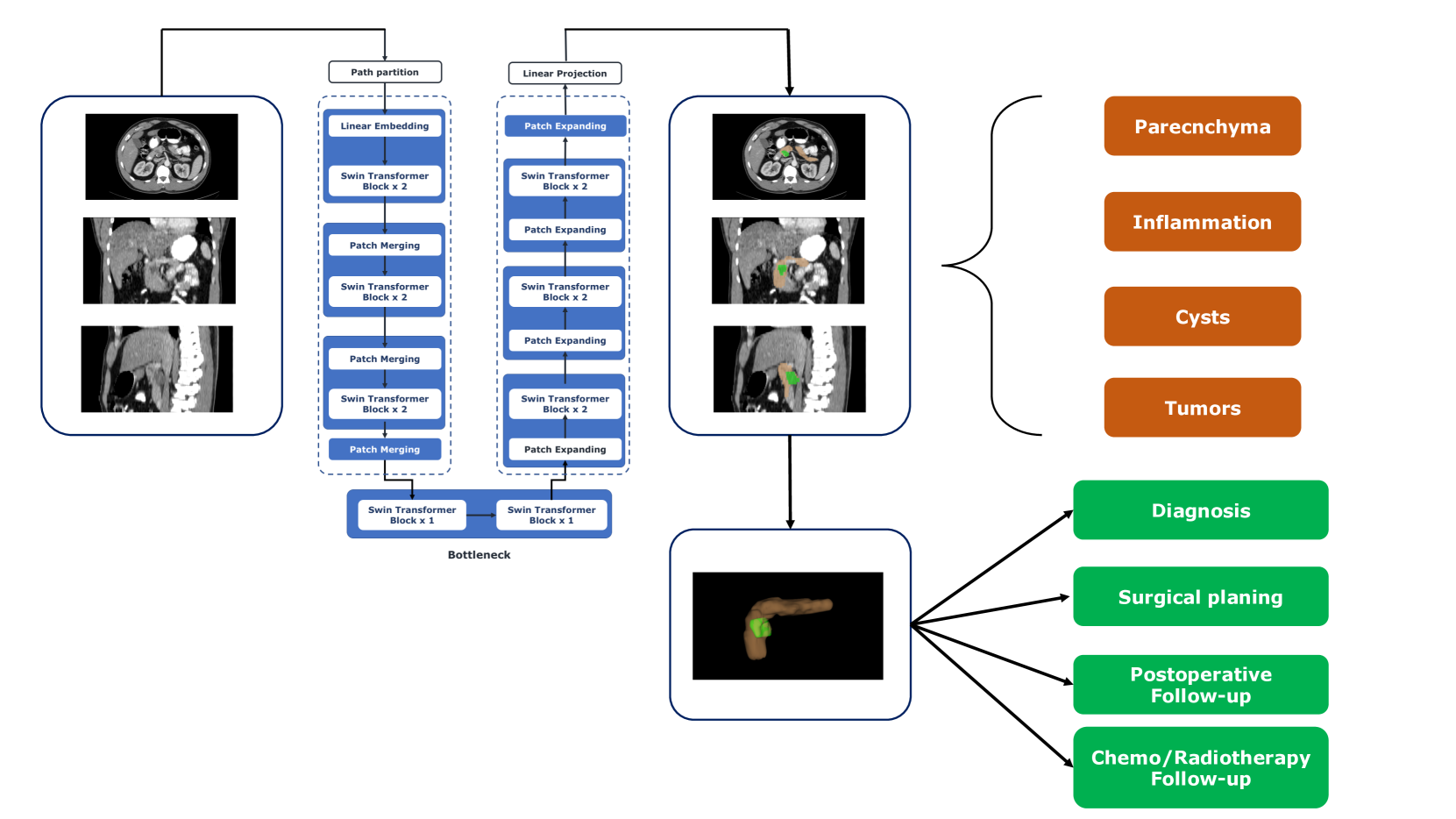
Deep Learning for Pancreas Segmentation: a Systematic Review
Andrea Moglia, Matteo Cavicchioli, Luca Mainardi, Pietro Cerveri
Pancreas segmentation has been traditionally challenging due to its small size in computed tomography abdominal volumes, high variability of shape and positions among patients, and blurred boundaries due to low contrast between the pancreas and surrounding organs. Many deep learning models for pancreas segmentation have been proposed in the past few years. We present a thorough systematic review based on the Preferred Reporting Items for Systematic Reviews and Meta-analyses (PRISMA) statement. The literature search was conducted on PubMed, Web of Science, Scopus, and IEEE Xplore on original studies published in peer-reviewed journals from 2013 to 2023. Overall, 130 studies were retrieved. We initially provided an overview of the technical background of the most common network architectures and publicly available datasets. Then, the analysis of the studies combining visual presentation in tabular form and text description was reported. The tables grouped the studies specifying the application, dataset size, design (model architecture, learning strategy, and loss function), results, and main contributions. We first analyzed the studies focusing on parenchyma segmentation using coarse-to-fine approaches, multi-organ segmentation, semi-supervised learning, and unsupervised learning, followed by those studies on generalization to other datasets and those concerning the design of new loss functions. Then, we analyzed the studies on segmentation of tumors, cysts, and inflammation reporting multi-stage methods, semi-supervised learning, generalization to other datasets, and design of new loss functions. Finally, we provided a critical discussion on the subject based on the published evidence underlining current issues that need to be addressed before clinical translation.
Read more7/24/2024