Adaptive Fusion of Radiomics and Deep Features for Lung Adenocarcinoma Subtype Recognition

0
🤿
Sign in to get full access
Overview
- Lung adenocarcinoma (LUAD) is the most common type of lung cancer.
- LUAD can be classified as pre-invasive (Pre-IAs) or invasive (IAs).
- Pre-IAs only require regular follow-up care, while IAs need immediate treatment.
- Prior research has focused on classifying Pre-IAs vs. IAs, but techniques for distinguishing different IA subtypes are lacking.
- This study proposes a multi-head attentional feature fusion (MHA-FF) model to distinguish Pre-IAs from IAs, as well as different IA subtypes.
- The model leverages both radiomics and deep features extracted from computed tomography (CT) images.
Plain English Explanation
Lung cancer is a serious health issue, and the most common type is called lung adenocarcinoma (LUAD). LUAD can be divided into two main categories: pre-invasive (Pre-IAs) and invasive (IAs). Pre-IAs cancers are less aggressive and only require regular check-ups, while IAs cancers are more serious and need immediate treatment, often involving surgery to remove the tumor.
Previous research has focused on identifying whether a lung tumor is Pre-IAs or IAs, but has struggled to accurately distinguish between the different subtypes of IAs. This is an important issue because the treatment approach needs to be tailored to the specific type of cancer.
To address this, the researchers in this study developed a new machine learning model called multi-head attentional feature fusion (MHA-FF). This model is able to not only tell if a lung tumor is Pre-IAs or IAs, but can also identify the specific subtype of IAs.
The key innovation of the MHA-FF model is that it combines two types of information extracted from CT scans of the lungs: radiomics features (mathematical properties of the tumor) and deep features (patterns identified by a deep learning algorithm). By fusing these features in an adaptive way, the model can learn the most important characteristics to accurately predict the LUAD subtype.
Technical Explanation
The researchers propose a multi-head attentional feature fusion (MHA-FF) model to address the challenge of distinguishing between different subtypes of invasive lung adenocarcinoma (IAs).
The model takes computed tomography (CT) images of lung nodules as input and leverages both radiomics features (mathematical properties of the tumor) and deep features (patterns identified by a deep learning algorithm) to make predictions. An adaptive fusion module is used to aggregate these complementary features, allowing the model to learn attention-based discriminative features that are most relevant for accurately predicting the LUAD subtype.
The utility of the MHA-FF model is demonstrated on a real-world, multi-center dataset of lung nodules. By incorporating both radiomics and deep features, the model is able to not only distinguish between pre-invasive (Pre-IAs) and invasive (IAs) LUAD, but also accurately classify the different subtypes of IAs. This is a significant advancement over previous approaches that have primarily focused on the Pre-IAs vs. IAs distinction.
Critical Analysis
The researchers acknowledge several limitations of their study. First, the dataset, while drawn from multiple centers, may not be fully representative of the broader population of lung cancer patients. Additionally, the study does not explore the clinical implications of accurately predicting LUAD subtypes, such as how this information could guide treatment decisions.
Another potential concern is the interpretability of the MHA-FF model. As a complex machine learning model, it may be difficult to understand the specific features and decision-making processes that lead to its predictions. Increased model interpretability could be valuable for building trust in the clinical application of such techniques.
Further research is also needed to validate the generalizability of the MHA-FF model on larger and more diverse datasets. Exploring the model's performance on different imaging modalities, such as X-ray or PET-CT, could also provide valuable insights.
Conclusion
This study presents a novel multi-head attentional feature fusion (MHA-FF) model that can accurately distinguish between pre-invasive and invasive lung adenocarcinoma, as well as identify the specific subtypes of invasive disease. By leveraging both radiomics and deep features extracted from CT images, the MHA-FF model demonstrates a significant advancement in the ability to diagnose and characterize this common form of lung cancer.
The accurate classification of LUAD subtypes has important implications for clinical practice, as it can help guide treatment decisions and improve patient outcomes. While further research is needed to address the limitations of this study, the MHA-FF model represents a promising step forward in the development of more sophisticated AI-based lung cancer diagnosis and analysis tools.
This summary was produced with help from an AI and may contain inaccuracies - check out the links to read the original source documents!
Related Papers
🤿
0
Adaptive Fusion of Radiomics and Deep Features for Lung Adenocarcinoma Subtype Recognition
Jing Zhou, Xiaotong Fu, Xirong Li, Ying Ji
The most common type of lung cancer, lung adenocarcinoma (LUAD), has been increasingly detected since the advent of low-dose computed tomography screening technology. In clinical practice, pre-invasive LUAD (Pre-IAs) should only require regular follow-up care, while invasive LUAD (IAs) should receive immediate treatment with appropriate lung cancer resection, based on the cancer subtype. However, prior research on diagnosing LUAD has mainly focused on classifying Pre-IAs/IAs, as techniques for distinguishing different subtypes of IAs have been lacking. In this study, we proposed a multi-head attentional feature fusion (MHA-FF) model for not only distinguishing IAs from Pre-IAs, but also for distinguishing the different subtypes of IAs. To predict the subtype of each nodule accurately, we leveraged both radiomics and deep features extracted from computed tomography images. Furthermore, those features were aggregated through an adaptive fusion module that can learn attention-based discriminative features. The utility of our proposed method is demonstrated here by means of real-world data collected from a multi-center cohort.
Read more8/28/2024
🔎
0
Domain Adaptive Lung Nodule Detection in X-ray Image
Haifeng Zhao, Lixiang Jiang, Leilei Ma, Dengdi Sun, Yanping Fu
Medical images from different healthcare centers exhibit varied data distributions, posing significant challenges for adapting lung nodule detection due to the domain shift between training and application phases. Traditional unsupervised domain adaptive detection methods often struggle with this shift, leading to suboptimal outcomes. To overcome these challenges, we introduce a novel domain adaptive approach for lung nodule detection that leverages mean teacher self-training and contrastive learning. First, we propose a hierarchical contrastive learning strategy to refine nodule representations and enhance the distinction between nodules and background. Second, we introduce a nodule-level domain-invariant feature learning (NDL) module to capture domain-invariant features through adversarial learning across different domains. Additionally, we propose a new annotated dataset of X-ray images to aid in advancing lung nodule detection research. Extensive experiments conducted on multiple X-ray datasets demonstrate the efficacy of our approach in mitigating domain shift impacts.
Read more8/6/2024
0
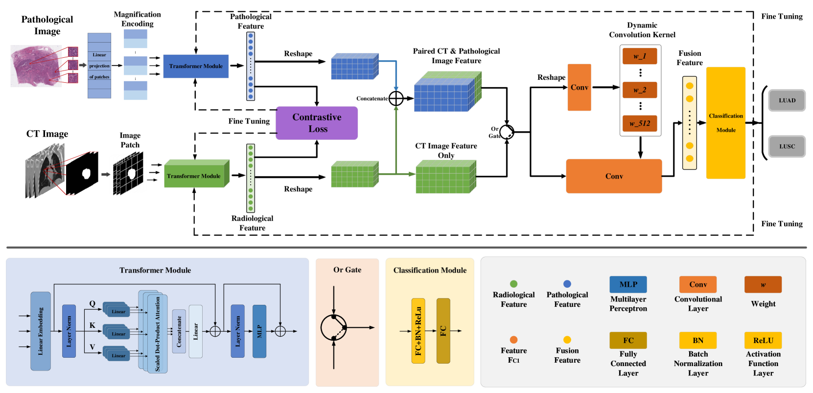
CC-DCNet: Dynamic Convolutional Neural Network with Contrastive Constraints for Identifying Lung Cancer Subtypes on Multi-modality Images
Yuan Jin, Gege Ma, Geng Chen, Tianling Lyu, Jan Egger, Junhui Lyu, Shaoting Zhang, Wentao Zhu
The accurate diagnosis of pathological subtypes of lung cancer is of paramount importance for follow-up treatments and prognosis managements. Assessment methods utilizing deep learning technologies have introduced novel approaches for clinical diagnosis. However, the majority of existing models rely solely on single-modality image input, leading to limited diagnostic accuracy. To this end, we propose a novel deep learning network designed to accurately classify lung cancer subtype with multi-dimensional and multi-modality images, i.e., CT and pathological images. The strength of the proposed model lies in its ability to dynamically process both paired CT-pathological image sets as well as independent CT image sets, and consequently optimize the pathology-related feature extractions from CT images. This adaptive learning approach enhances the flexibility in processing multi-dimensional and multi-modality datasets and results in performance elevating in the model testing phase. We also develop a contrastive constraint module, which quantitatively maps the cross-modality associations through network training, and thereby helps to explore the gold standard pathological information from the corresponding CT scans. To evaluate the effectiveness, adaptability, and generalization ability of our model, we conducted extensive experiments on a large-scale multi-center dataset and compared our model with a series of state-of-the-art classification models. The experimental results demonstrated the superiority of our model for lung cancer subtype classification, showcasing significant improvements in accuracy metrics such as ACC, AUC, and F1-score.
Read more7/19/2024
➖
0
A Lung Nodule Dataset with Histopathology-based Cancer Type Annotation
Muwei Jian, Hongyu Chen, Zaiyong Zhang, Nan Yang, Haorang Zhang, Lifu Ma, Wenjing Xu, Huixiang Zhi
Recently, Computer-Aided Diagnosis (CAD) systems have emerged as indispensable tools in clinical diagnostic workflows, significantly alleviating the burden on radiologists. Nevertheless, despite their integration into clinical settings, CAD systems encounter limitations. Specifically, while CAD systems can achieve high performance in the detection of lung nodules, they face challenges in accurately predicting multiple cancer types. This limitation can be attributed to the scarcity of publicly available datasets annotated with expert-level cancer type information. This research aims to bridge this gap by providing publicly accessible datasets and reliable tools for medical diagnosis, facilitating a finer categorization of different types of lung diseases so as to offer precise treatment recommendations. To achieve this objective, we curated a diverse dataset of lung Computed Tomography (CT) images, comprising 330 annotated nodules (nodules are labeled as bounding boxes) from 95 distinct patients. The quality of the dataset was evaluated using a variety of classical classification and detection models, and these promising results demonstrate that the dataset has a feasible application and further facilitate intelligent auxiliary diagnosis.
Read more6/27/2024