A Lung Nodule Dataset with Histopathology-based Cancer Type Annotation

0
➖
Sign in to get full access
Overview
- Researchers have developed a dataset of lung CT images with annotated lung nodules to improve the performance of computer-aided diagnosis (CAD) systems.
- The dataset aims to facilitate more accurate categorization of different lung diseases and provide better treatment recommendations.
- The dataset was evaluated using various classification and detection models, and the results demonstrate its feasibility for intelligent auxiliary diagnosis.
Plain English Explanation
Doctors often use computer-aided diagnosis (CAD) systems to help them analyze medical images and make diagnoses. These CAD systems have become essential tools in clinical settings, as they can assist radiologists and reduce their workload. However, current CAD systems still have some limitations, particularly in accurately predicting multiple types of lung cancer.
This research addresses this limitation by providing a publicly available dataset of lung CT images with detailed annotations. The dataset includes 330 annotated lung nodules (which are essentially abnormal growths) from 95 different patients. The researchers evaluated the quality of this dataset using various machine learning models, and the promising results show that it can be a valuable tool for improving the accuracy of lung disease diagnosis.
By having access to a high-quality dataset with detailed annotations, CAD systems can be trained to better distinguish between different types of lung diseases. This can lead to more precise treatment recommendations, ultimately benefiting patients. The researchers have made this dataset publicly available, which will help advance the field of explainable AI for lung health and improve the overall performance of CAD systems.
Technical Explanation
The researchers curated a diverse dataset of 330 annotated lung nodules from 95 distinct patients, with the goal of improving the performance of computer-aided diagnosis (CAD) systems in accurately predicting multiple cancer types. This is a crucial challenge, as current CAD systems often struggle to accurately categorize different types of lung diseases, despite their high performance in detecting lung nodules.
To address this limitation, the researchers created a publicly accessible dataset of lung Computed Tomography (CT) images, with each nodule labeled as a bounding box. This dataset was then evaluated using a variety of classical classification and detection models, and the promising results demonstrate the feasibility of using this dataset for intelligent auxiliary diagnosis.
The researchers chose to focus on lung nodules, as they are often the first indication of lung cancer or other lung diseases. By providing a well-annotated dataset, the researchers aim to facilitate the development of more accurate and precise CAD systems, which can ultimately lead to better treatment recommendations for patients.
Critical Analysis
The researchers have made a valuable contribution by providing a publicly accessible dataset of lung CT images with detailed annotations. This dataset can be a valuable resource for the development and evaluation of CAD systems, which are essential tools in clinical diagnostic workflows.
However, the researchers acknowledge that the dataset is limited in size, with only 95 distinct patients represented. While the results demonstrate the feasibility of using this dataset, it will be important to continue expanding the dataset with more diverse samples to further improve the performance and generalization of CAD systems.
Additionally, the researchers do not address the potential ethical considerations around the use of such datasets, such as the need for patient privacy and the potential for bias in the data collection and annotation processes. These are important factors that should be carefully considered as the research in this area continues to evolve.
Overall, the researchers have taken an important step forward in addressing the limitations of current CAD systems, and their work highlights the need for high-quality, publicly available datasets to drive progress in the field of explainable AI for lung health. As the research continues, it will be crucial to address the remaining challenges and ensure that the development of these systems is conducted in an ethical and responsible manner.
Conclusion
This research presents a valuable dataset of lung CT images with annotated lung nodules, aimed at improving the performance of computer-aided diagnosis (CAD) systems in accurately predicting multiple types of lung diseases. By providing a well-curated and publicly accessible dataset, the researchers have taken an important step towards advancing the field of AI-powered lung health diagnosis.
The promising results of the evaluation using various classification and detection models demonstrate the feasibility of this dataset for intelligent auxiliary diagnosis. This can ultimately lead to more precise treatment recommendations for patients, as CAD systems become better equipped to differentiate between different lung diseases.
While the dataset has limitations in terms of sample size, the researchers have made a significant contribution by addressing a crucial challenge in the development of CAD systems. As the research in this area continues to evolve, it will be important to expand the dataset, address ethical considerations, and ensure that these systems are developed and deployed in a responsible manner, ultimately benefiting patients and the healthcare community as a whole.
This summary was produced with help from an AI and may contain inaccuracies - check out the links to read the original source documents!
Related Papers
➖
0
A Lung Nodule Dataset with Histopathology-based Cancer Type Annotation
Muwei Jian, Hongyu Chen, Zaiyong Zhang, Nan Yang, Haorang Zhang, Lifu Ma, Wenjing Xu, Huixiang Zhi
Recently, Computer-Aided Diagnosis (CAD) systems have emerged as indispensable tools in clinical diagnostic workflows, significantly alleviating the burden on radiologists. Nevertheless, despite their integration into clinical settings, CAD systems encounter limitations. Specifically, while CAD systems can achieve high performance in the detection of lung nodules, they face challenges in accurately predicting multiple cancer types. This limitation can be attributed to the scarcity of publicly available datasets annotated with expert-level cancer type information. This research aims to bridge this gap by providing publicly accessible datasets and reliable tools for medical diagnosis, facilitating a finer categorization of different types of lung diseases so as to offer precise treatment recommendations. To achieve this objective, we curated a diverse dataset of lung Computed Tomography (CT) images, comprising 330 annotated nodules (nodules are labeled as bounding boxes) from 95 distinct patients. The quality of the dataset was evaluated using a variety of classical classification and detection models, and these promising results demonstrate that the dataset has a feasible application and further facilitate intelligent auxiliary diagnosis.
Read more6/27/2024
0
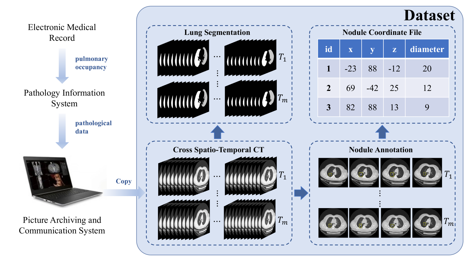
A Cross Spatio-Temporal Pathology-based Lung Nodule Dataset
Muwei Jian, Haoran Zhang, Mingju Shao, Hongyu Chen, Huihui Huang, Yanjie Zhong, Changlei Zhang, Bin Wang, Penghui Gao
Recently, intelligent analysis of lung nodules with the assistant of computer aided detection (CAD) techniques can improve the accuracy rate of lung cancer diagnosis. However, existing CAD systems and pulmonary datasets mainly focus on Computed Tomography (CT) images from one single period, while ignoring the cross spatio-temporal features associated with the progression of nodules contained in imaging data from various captured periods of lung cancer. If the evolution patterns of nodules across various periods in the patients' CT sequences can be explored, it will play a crucial role in guiding the precise screening identification of lung cancer. Therefore, a cross spatio-temporal lung nodule dataset with pathological information for nodule identification and diagnosis is constructed, which contains 328 CT sequences and 362 annotated nodules from 109 patients. This comprehensive database is intended to drive research in the field of CAD towards more practical and robust methods, and also contribute to the further exploration of precision medicine related field. To ensure patient confidentiality, we have removed sensitive information from the dataset.
Read more6/27/2024
0
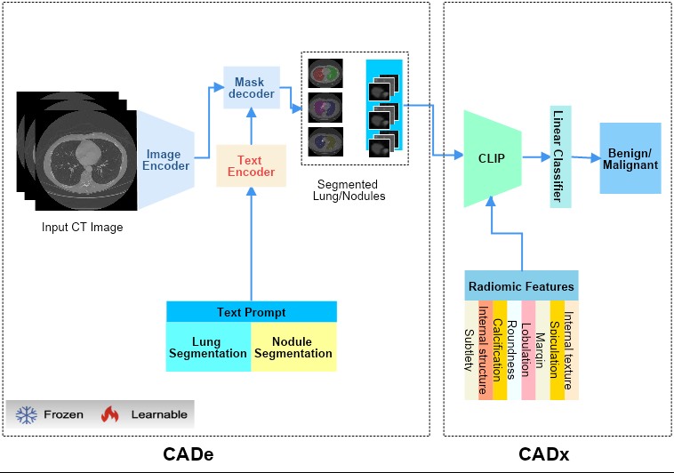
Lung-CADex: Fully automatic Zero-Shot Detection and Classification of Lung Nodules in Thoracic CT Images
Furqan Shaukat, Syed Muhammad Anwar, Abhijeet Parida, Van Khanh Lam, Marius George Linguraru, Mubarak Shah
Lung cancer has been one of the major threats to human life for decades. Computer-aided diagnosis can help with early lung nodul detection and facilitate subsequent nodule characterization. Large Visual Language models (VLMs) have been found effective for multiple downstream medical tasks that rely on both imaging and text data. However, lesion level detection and subsequent diagnosis using VLMs have not been explored yet. We propose CADe, for segmenting lung nodules in a zero-shot manner using a variant of the Segment Anything Model called MedSAM. CADe trains on a prompt suite on input computed tomography (CT) scans by using the CLIP text encoder through prefix tuning. We also propose, CADx, a method for the nodule characterization as benign/malignant by making a gallery of radiomic features and aligning image-feature pairs through contrastive learning. Training and validation of CADe and CADx have been done using one of the largest publicly available datasets, called LIDC. To check the generalization ability of the model, it is also evaluated on a challenging dataset, LUNGx. Our experimental results show that the proposed methods achieve a sensitivity of 0.86 compared to 0.76 that of other fully supervised methods.The source code, datasets and pre-processed data can be accessed using the link:
Read more7/4/2024
0
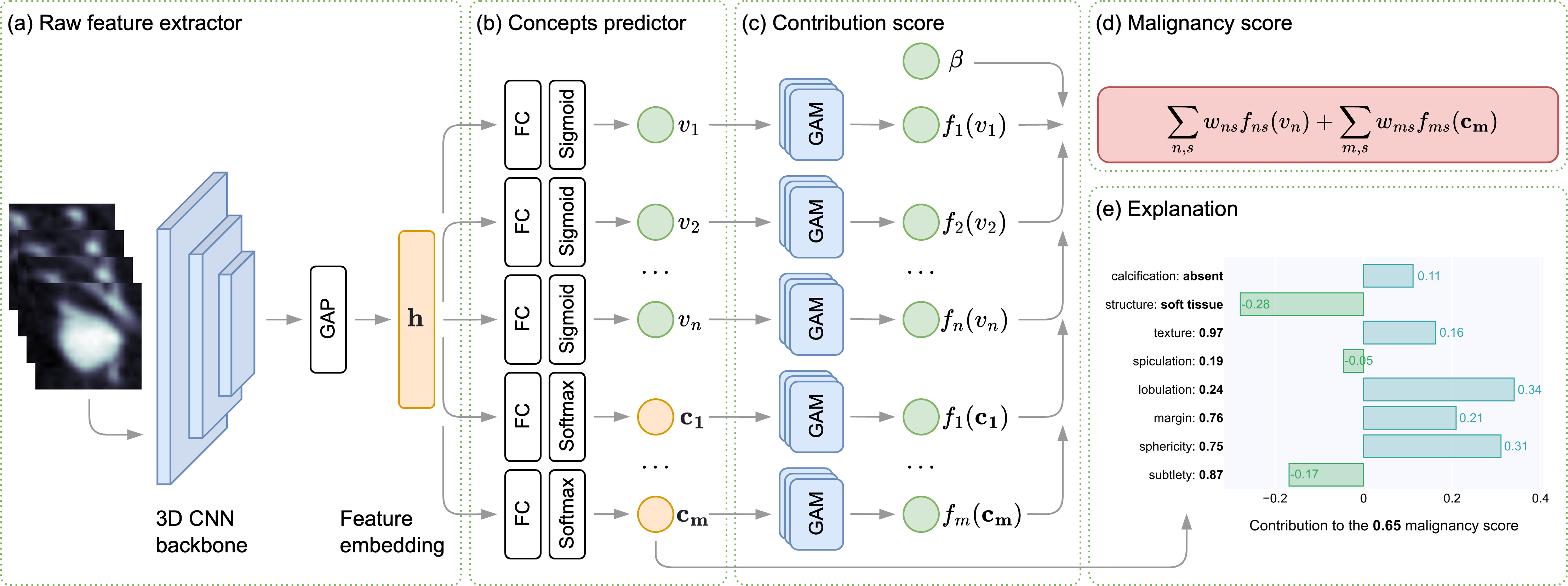
Concept-based Explainable Malignancy Scoring on Pulmonary Nodules in CT Images
Rinat I. Dumaev, Sergei A. Molodyakov, Lev V. Utkin
To increase the transparency of modern computer-aided diagnosis (CAD) systems for assessing the malignancy of lung nodules, an interpretable model based on applying the generalized additive models and the concept-based learning is proposed. The model detects a set of clinically significant attributes in addition to the final malignancy regression score and learns the association between the lung nodule attributes and a final diagnosis decision as well as their contributions into the decision. The proposed concept-based learning framework provides human-readable explanations in terms of different concepts (numerical and categorical), their values, and their contribution to the final prediction. Numerical experiments with the LIDC-IDRI dataset demonstrate that the diagnosis results obtained using the proposed model, which explicitly explores internal relationships, are in line with similar patterns observed in clinical practice. Additionally, the proposed model shows the competitive classification and the nodule attribute scoring performance, highlighting its potential for effective decision-making in the lung nodule diagnosis.
Read more5/29/2024