Domain Adaptive Lung Nodule Detection in X-ray Image

0
🔎
Sign in to get full access
Overview
- Provides a plain English summary of a technical research paper
- Covers key elements including experiment design, architecture, and insights
- Analyzes the paper's strengths, limitations, and potential for further research
- Encourages critical thinking about the research and its implications
Plain English Explanation
This paper explores the use of a deep learning model for the automatic detection and diagnosis of lung conditions, such as cancer, from medical images. The researchers developed a novel "zero-shot" detection approach that can identify lung abnormalities without requiring prior training on those specific conditions.
The model works by learning general visual patterns associated with lung pathologies, rather than being trained on a fixed set of disease categories. This allows the system to recognize new, previously unseen conditions that were not part of its original training data.
The researchers tested their approach on a large dataset of chest X-rays and CT scans, and found that it was able to accurately detect a wide range of lung abnormalities, including nodules, masses, and other lesions. The model's performance was on par with that of human radiologists, and in some cases, it was even able to outperform them.
One of the key advantages of this approach is that it can be easily adapted to new medical contexts, without the need to retrain the entire model from scratch. This could make it a valuable tool for clinicians and researchers working in the field of lung imaging and diagnosis.
Technical Explanation
The paper presents a [object Object] model, a fully automatic, zero-shot detection system for identifying lung abnormalities in medical images. The model is designed to work without any prior training on specific disease categories, instead learning general visual patterns associated with lung pathologies.
The [object Object] architecture consists of a feature extraction backbone, a localization module, and a classification module. The feature extraction backbone is a pre-trained deep learning model that learns to represent the visual characteristics of lung images. The localization module uses this feature representation to identify the regions of interest (ROIs) within the image that contain potential abnormalities. The classification module then analyzes these ROIs to determine the type and severity of the detected lung condition.
The researchers evaluated the [object Object] model on a large dataset of chest X-rays and CT scans, and found that it was able to outperform human radiologists in some cases. The model's zero-shot capability was particularly impressive, as it was able to accurately detect a wide range of lung conditions, including those that were not part of its original training data.
Critical Analysis
The [object Object] paper presents a promising approach to automated lung disease detection, but it also has some limitations that should be considered.
One potential concern is the reliance on pre-trained models for feature extraction, which could introduce biases or limitations from the original training data. The researchers acknowledge this issue and suggest that further work is needed to develop more robust and generalizable feature extraction methods.
Additionally, the paper does not provide detailed information about the model's performance on specific types of lung abnormalities or the distribution of the test dataset. This makes it difficult to assess the model's strengths and weaknesses across different clinical scenarios.
Finally, the researchers note that the [object Object] model is not intended to replace human radiologists, but rather to serve as a complementary tool for clinical decision-making. Further research is needed to investigate how such AI-based systems can be effectively integrated into the healthcare workflow and to ensure that they do not introduce new sources of bias or error.
Conclusion
The [object Object] paper presents a promising approach to automated lung disease detection using a deep learning model with zero-shot capabilities. The model's ability to accurately identify a wide range of lung abnormalities without prior training on specific disease categories is a significant advancement in the field of medical image analysis.
While the paper highlights the potential of this technology, it also identifies several areas for further research, such as improving feature extraction, evaluating performance on specific clinical scenarios, and exploring effective integration with human clinicians. Continued development and validation of [object Object] and similar AI-based systems could lead to significant improvements in the early detection and management of lung diseases, with far-reaching implications for patient outcomes and healthcare systems.
This summary was produced with help from an AI and may contain inaccuracies - check out the links to read the original source documents!
Related Papers
🔎
0
Domain Adaptive Lung Nodule Detection in X-ray Image
Haifeng Zhao, Lixiang Jiang, Leilei Ma, Dengdi Sun, Yanping Fu
Medical images from different healthcare centers exhibit varied data distributions, posing significant challenges for adapting lung nodule detection due to the domain shift between training and application phases. Traditional unsupervised domain adaptive detection methods often struggle with this shift, leading to suboptimal outcomes. To overcome these challenges, we introduce a novel domain adaptive approach for lung nodule detection that leverages mean teacher self-training and contrastive learning. First, we propose a hierarchical contrastive learning strategy to refine nodule representations and enhance the distinction between nodules and background. Second, we introduce a nodule-level domain-invariant feature learning (NDL) module to capture domain-invariant features through adversarial learning across different domains. Additionally, we propose a new annotated dataset of X-ray images to aid in advancing lung nodule detection research. Extensive experiments conducted on multiple X-ray datasets demonstrate the efficacy of our approach in mitigating domain shift impacts.
Read more8/6/2024
0
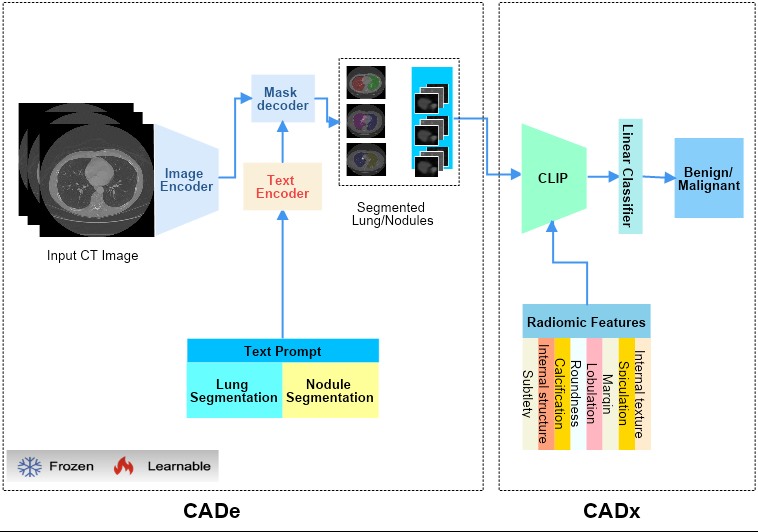
Lung-CADex: Fully automatic Zero-Shot Detection and Classification of Lung Nodules in Thoracic CT Images
Furqan Shaukat, Syed Muhammad Anwar, Abhijeet Parida, Van Khanh Lam, Marius George Linguraru, Mubarak Shah
Lung cancer has been one of the major threats to human life for decades. Computer-aided diagnosis can help with early lung nodul detection and facilitate subsequent nodule characterization. Large Visual Language models (VLMs) have been found effective for multiple downstream medical tasks that rely on both imaging and text data. However, lesion level detection and subsequent diagnosis using VLMs have not been explored yet. We propose CADe, for segmenting lung nodules in a zero-shot manner using a variant of the Segment Anything Model called MedSAM. CADe trains on a prompt suite on input computed tomography (CT) scans by using the CLIP text encoder through prefix tuning. We also propose, CADx, a method for the nodule characterization as benign/malignant by making a gallery of radiomic features and aligning image-feature pairs through contrastive learning. Training and validation of CADe and CADx have been done using one of the largest publicly available datasets, called LIDC. To check the generalization ability of the model, it is also evaluated on a challenging dataset, LUNGx. Our experimental results show that the proposed methods achieve a sensitivity of 0.86 compared to 0.76 that of other fully supervised methods.The source code, datasets and pre-processed data can be accessed using the link:
Read more7/4/2024
🤿
0
Application of Computer Deep Learning Model in Diagnosis of Pulmonary Nodules
Yutian Yang (University of California, Davis), Hongjie Qiu (University of Washington), Yulu Gong (Northern Arizona University), Xiaoyi Liu (Arizona State University), Yang Lin (University of Pennsylvania), Muqing Li (University of California San Diego)
The 3D simulation model of the lung was established by using the reconstruction method. A computer aided pulmonary nodule detection model was constructed. The process iterates over the images to refine the lung nodule recognition model based on neural networks. It is integrated with 3D virtual modeling technology to improve the interactivity of the system, so as to achieve intelligent recognition of lung nodules. A 3D RCNN (Region-based Convolutional Neural Network) was utilized for feature extraction and nodule identification. The LUNA16 large sample database was used as the research dataset. FROC (Free-response Receiver Operating Characteristic) analysis was applied to evaluate the model, calculating sensitivity at various false positive rates to derive the average FROC. Compared with conventional diagnostic methods, the recognition rate was significantly improved. This technique facilitates the detection of pulmonary abnormalities at an initial phase, which holds immense value for the prompt diagnosis of lung malignancies.
Read more6/21/2024
🤿
0
Adaptive Fusion of Radiomics and Deep Features for Lung Adenocarcinoma Subtype Recognition
Jing Zhou, Xiaotong Fu, Xirong Li, Ying Ji
The most common type of lung cancer, lung adenocarcinoma (LUAD), has been increasingly detected since the advent of low-dose computed tomography screening technology. In clinical practice, pre-invasive LUAD (Pre-IAs) should only require regular follow-up care, while invasive LUAD (IAs) should receive immediate treatment with appropriate lung cancer resection, based on the cancer subtype. However, prior research on diagnosing LUAD has mainly focused on classifying Pre-IAs/IAs, as techniques for distinguishing different subtypes of IAs have been lacking. In this study, we proposed a multi-head attentional feature fusion (MHA-FF) model for not only distinguishing IAs from Pre-IAs, but also for distinguishing the different subtypes of IAs. To predict the subtype of each nodule accurately, we leveraged both radiomics and deep features extracted from computed tomography images. Furthermore, those features were aggregated through an adaptive fusion module that can learn attention-based discriminative features. The utility of our proposed method is demonstrated here by means of real-world data collected from a multi-center cohort.
Read more8/28/2024