Application of Computer Deep Learning Model in Diagnosis of Pulmonary Nodules

0
🤿
Sign in to get full access
Overview
- The researchers developed a 3D simulation model of the lung using a reconstruction method.
- They built a computer-aided pulmonary nodule detection model using neural networks.
- The model integrates 3D virtual modeling technology to improve the interactivity of the system and enable intelligent recognition of lung nodules.
- A 3D RCNN (Region-based Convolutional Neural Network) was used for feature extraction and nodule identification.
- The LUNA16 large sample database was used as the research dataset.
- FROC (Free-response Receiver Operating Characteristic) analysis was applied to evaluate the model.
- Compared to conventional diagnostic methods, the recognition rate was significantly improved.
- This technique enables the early detection of pulmonary abnormalities, which is valuable for the prompt diagnosis of lung cancer.
Plain English Explanation
The researchers created a 3D computer model of the lungs using a special reconstruction technique. They then built a computer program that can automatically detect small growths called nodules in the lungs. This program uses a type of artificial intelligence called neural networks to identify the nodules.
To make the system more interactive and intelligent, the researchers integrated 3D virtual modeling technology. This allows users to better visualize and interact with the lung model.
The key part of the detection system is a type of neural network called a 3D RCNN. This network is trained to analyze the 3D lung images and identify any suspicious nodules or growths. The researchers used a large dataset of lung images, called LUNA16, to train and test the model.
To evaluate the performance of the model, the researchers used a metric called FROC analysis. This looks at how accurately the model can detect nodules, while also considering the number of false positives (incorrect detections).
Compared to traditional diagnostic methods, this AI-powered lung nodule detection system showed a significantly higher recognition rate. This is important because early detection of lung abnormalities, like nodules, is crucial for the prompt diagnosis and treatment of lung cancer.
By integrating 3D modeling and advanced AI techniques, this research demonstrates a powerful tool for improving lung cancer screening and diagnosis. This technique facilitates the detection of pulmonary abnormalities at an initial phase, which holds immense value for the prompt diagnosis of lung malignancies.
Technical Explanation
The researchers established a 3D simulation model of the lung using a reconstruction method. They then constructed a computer-aided pulmonary nodule detection model based on neural networks. The process iterates over the images to refine the lung nodule recognition model.
To improve the interactivity and intelligence of the system, the researchers integrated 3D virtual modeling technology. This allows for more intuitive visualization and interaction with the lung model.
A 3D RCNN (Region-based Convolutional Neural Network) was utilized for feature extraction and nodule identification. The LUNA16 large sample database was used as the research dataset.
FROC (Free-response Receiver Operating Characteristic) analysis was applied to evaluate the model's performance. This calculates the sensitivity (true positive rate) at various false positive rates to derive the average FROC.
Compared to conventional diagnostic methods, the researchers found that their recognition rate was significantly improved. This technique facilitates the detection of pulmonary abnormalities at an initial phase, which holds immense value for the prompt diagnosis of lung malignancies.
Critical Analysis
The paper demonstrates a promising approach for improving lung cancer screening and diagnosis through the integration of 3D modeling and advanced AI techniques. However, the researchers do not provide much detail on the specific architectural details or hyperparameters of the 3D RCNN model.
Additionally, while the FROC analysis shows improved performance compared to traditional methods, the paper does not quantify the extent of this improvement or provide a direct comparison. Further research is needed to benchmark the performance of this approach against other state-of-the-art lung nodule detection models.
The paper also does not discuss potential limitations or challenges of the proposed approach, such as the computational resources required, the robustness of the model to variations in image quality or nodule characteristics, or the generalizability of the approach to different patient populations.
Overall, this research demonstrates a promising direction for utilizing 3D virtual modeling and deep learning techniques to enhance lung cancer screening and early detection. However, further validation and refinement of the approach will be necessary before it can be widely adopted in clinical practice.
Conclusion
This research paper presents a novel approach for lung nodule detection that integrates 3D virtual modeling and deep learning techniques. The researchers developed a computer-aided pulmonary nodule detection model using a 3D RCNN neural network and the LUNA16 dataset.
Compared to conventional diagnostic methods, the proposed system showed significantly improved recognition rates for lung nodules. This is an important advancement, as early detection of pulmonary abnormalities is crucial for the prompt diagnosis and treatment of lung cancer.
By leveraging 3D virtual modeling and interactive visualization, the researchers have created a more intuitive and intelligent system for lung cancer screening. This work demonstrates the potential of AI-powered techniques to enhance medical imaging analysis and improve patient outcomes.
Further research is needed to fully validate the performance and robustness of this approach, but the results presented in this paper are a promising step towards more accurate and accessible lung cancer detection.
This summary was produced with help from an AI and may contain inaccuracies - check out the links to read the original source documents!
Related Papers
🤿
0
Application of Computer Deep Learning Model in Diagnosis of Pulmonary Nodules
Yutian Yang (University of California, Davis), Hongjie Qiu (University of Washington), Yulu Gong (Northern Arizona University), Xiaoyi Liu (Arizona State University), Yang Lin (University of Pennsylvania), Muqing Li (University of California San Diego)
The 3D simulation model of the lung was established by using the reconstruction method. A computer aided pulmonary nodule detection model was constructed. The process iterates over the images to refine the lung nodule recognition model based on neural networks. It is integrated with 3D virtual modeling technology to improve the interactivity of the system, so as to achieve intelligent recognition of lung nodules. A 3D RCNN (Region-based Convolutional Neural Network) was utilized for feature extraction and nodule identification. The LUNA16 large sample database was used as the research dataset. FROC (Free-response Receiver Operating Characteristic) analysis was applied to evaluate the model, calculating sensitivity at various false positive rates to derive the average FROC. Compared with conventional diagnostic methods, the recognition rate was significantly improved. This technique facilitates the detection of pulmonary abnormalities at an initial phase, which holds immense value for the prompt diagnosis of lung malignancies.
Read more6/21/2024
🧠
0
Convolutional Neural Networks for Predictive Modeling of Lung Disease
Yingbin Liang, Xiqing Liu, Haohao Xia, Yiru Cang, Zitao Zheng, Yuanfang Yang
In this paper, Pro-HRnet-CNN, an innovative model combining HRNet and void-convolution techniques, is proposed for disease prediction under lung imaging. Through the experimental comparison on the authoritative LIDC-IDRI dataset, we found that compared with the traditional ResNet-50, Pro-HRnet-CNN showed better performance in the feature extraction and recognition of small-size nodules, significantly improving the detection accuracy. Particularly within the domain of detecting smaller targets, the model has exhibited a remarkable enhancement in accuracy, thereby pioneering an innovative avenue for the early identification and prognostication of pulmonary conditions.
Read more8/26/2024
0
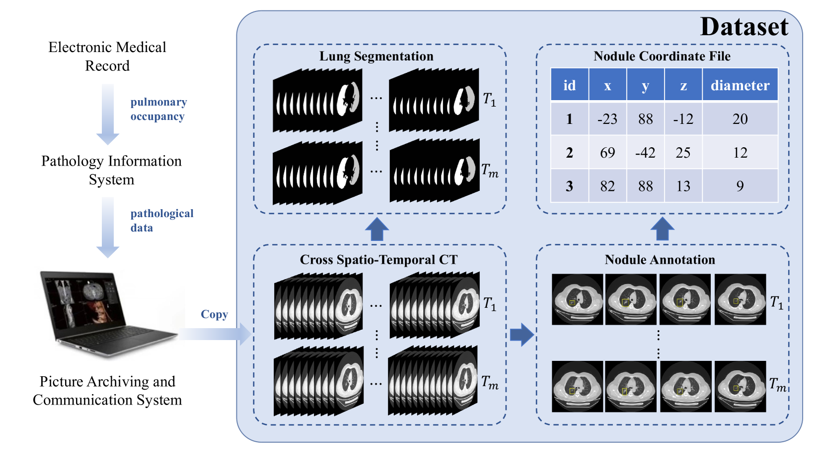
A Cross Spatio-Temporal Pathology-based Lung Nodule Dataset
Muwei Jian, Haoran Zhang, Mingju Shao, Hongyu Chen, Huihui Huang, Yanjie Zhong, Changlei Zhang, Bin Wang, Penghui Gao
Recently, intelligent analysis of lung nodules with the assistant of computer aided detection (CAD) techniques can improve the accuracy rate of lung cancer diagnosis. However, existing CAD systems and pulmonary datasets mainly focus on Computed Tomography (CT) images from one single period, while ignoring the cross spatio-temporal features associated with the progression of nodules contained in imaging data from various captured periods of lung cancer. If the evolution patterns of nodules across various periods in the patients' CT sequences can be explored, it will play a crucial role in guiding the precise screening identification of lung cancer. Therefore, a cross spatio-temporal lung nodule dataset with pathological information for nodule identification and diagnosis is constructed, which contains 328 CT sequences and 362 annotated nodules from 109 patients. This comprehensive database is intended to drive research in the field of CAD towards more practical and robust methods, and also contribute to the further exploration of precision medicine related field. To ensure patient confidentiality, we have removed sensitive information from the dataset.
Read more6/27/2024
0
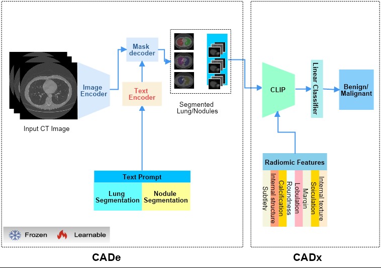
Lung-CADex: Fully automatic Zero-Shot Detection and Classification of Lung Nodules in Thoracic CT Images
Furqan Shaukat, Syed Muhammad Anwar, Abhijeet Parida, Van Khanh Lam, Marius George Linguraru, Mubarak Shah
Lung cancer has been one of the major threats to human life for decades. Computer-aided diagnosis can help with early lung nodul detection and facilitate subsequent nodule characterization. Large Visual Language models (VLMs) have been found effective for multiple downstream medical tasks that rely on both imaging and text data. However, lesion level detection and subsequent diagnosis using VLMs have not been explored yet. We propose CADe, for segmenting lung nodules in a zero-shot manner using a variant of the Segment Anything Model called MedSAM. CADe trains on a prompt suite on input computed tomography (CT) scans by using the CLIP text encoder through prefix tuning. We also propose, CADx, a method for the nodule characterization as benign/malignant by making a gallery of radiomic features and aligning image-feature pairs through contrastive learning. Training and validation of CADe and CADx have been done using one of the largest publicly available datasets, called LIDC. To check the generalization ability of the model, it is also evaluated on a challenging dataset, LUNGx. Our experimental results show that the proposed methods achieve a sensitivity of 0.86 compared to 0.76 that of other fully supervised methods.The source code, datasets and pre-processed data can be accessed using the link:
Read more7/4/2024