Concept-based Explainable Malignancy Scoring on Pulmonary Nodules in CT Images

0
Sign in to get full access
Overview
- This paper proposes a concept-based explainable AI system for scoring the malignancy of pulmonary nodules in CT images.
- The system uses a generalized additive model and a neural network to predict malignancy, while also providing explanations for the predictions based on learned concepts.
- The research aims to improve the interpretability and trustworthiness of AI systems for medical image analysis.
Plain English Explanation
The researchers developed an AI system to analyze CT scans of lung nodules and determine how likely they are to be cancerous. Typically, AI systems like this can make accurate predictions, but it's hard to understand how they arrive at those conclusions. This can make doctors and patients hesitant to trust the system's recommendations.
To address this, the researchers created an AI model that not only predicts malignancy, but also explains its reasoning. The model learns to recognize specific "concepts" in the CT scans, such as the shape, texture, or location of the nodule. When the model makes a prediction, it can point to which concepts it relied on most heavily, similar to how a human doctor might explain their thought process.
By making the AI's decision-making more transparent, the researchers hope to build trust in the technology and help doctors make more informed decisions about patient care. This type of concept-based explainable AI could also have applications in other medical imaging tasks, like detecting and diagnosing conditions from X-rays or MRI scans.
Technical Explanation
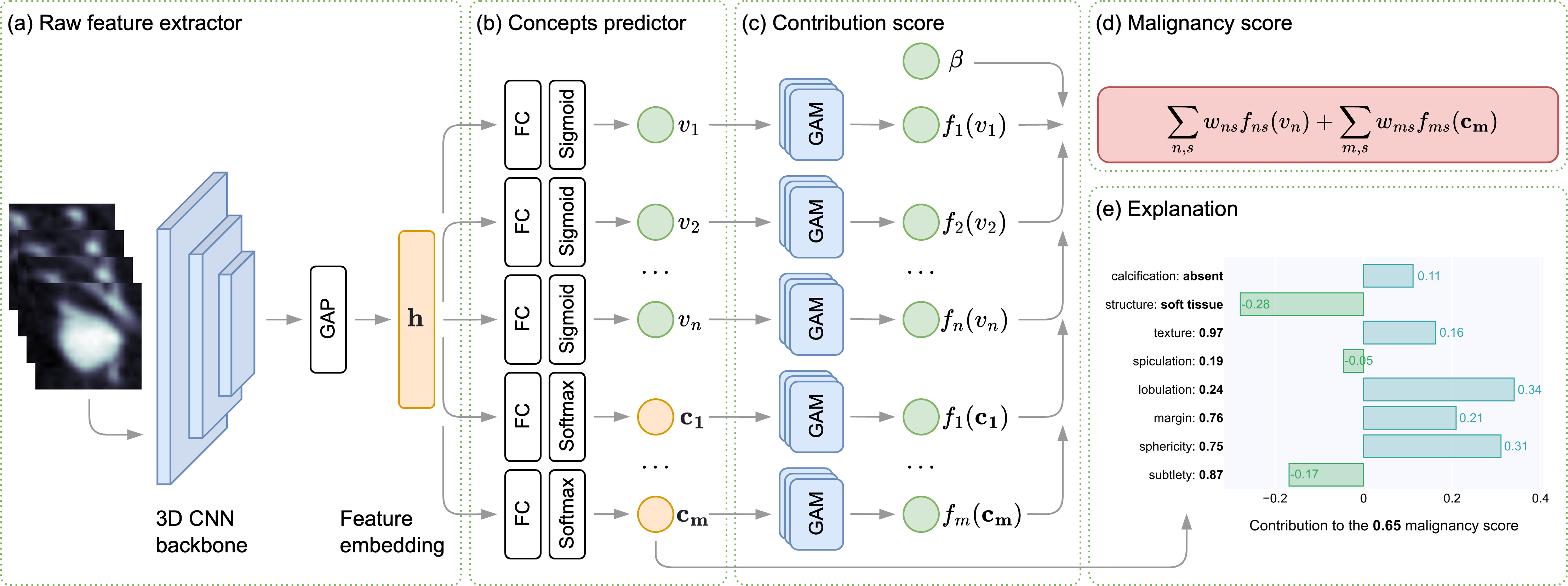
The proposed system uses a two-stage approach. First, it employs a generalized additive model (GAM) to predict the malignancy score of a pulmonary nodule based on a set of low-level visual features extracted from the CT image. The GAM model allows the system to explain its predictions by highlighting the relative importance of each feature.
Second, the system uses a neural network that learns a set of high-level "concepts" directly from the CT image data. These concepts capture more abstract visual patterns associated with malignancy, such as nodule shape, texture, or location. The neural network then uses these learned concepts to make its own malignancy prediction, which can be explained by showing which concepts contributed most to the final score.
By combining the interpretable GAM model with the more powerful concept-based neural network, the researchers aimed to create an AI system that achieves high predictive performance while also providing transparent, human-understandable explanations for its decisions. This aligns with the growing interest in explainable AI techniques that can improve the trust and adoption of AI in sensitive domains like healthcare.
Critical Analysis
The researchers acknowledge several limitations of their approach. First, the concept-based neural network requires a large amount of annotated training data to learn meaningful visual concepts, which may be difficult to obtain in some medical imaging domains. Additionally, the explanations provided by the system, while more interpretable than a "black box" neural network, may still be challenging for non-expert users to fully comprehend.
Another potential concern is the reliance on low-level visual features in the GAM model, which may miss important high-level semantic information about the nodules. While the concept-based neural network addresses this to some degree, further research is needed to determine the optimal balance between interpretability and predictive performance.
Overall, the concept-based explainable malignancy scoring system represents an interesting and promising approach to improving the transparency and trustworthiness of AI in medical image analysis. However, as with any new technology, continued refinement and validation will be necessary before widespread clinical adoption.
Conclusion
This paper presents a novel concept-based explainable AI system for predicting the malignancy of pulmonary nodules in CT images. By combining an interpretable generalized additive model with a more powerful concept-learning neural network, the researchers have created a system that can both accurately predict malignancy and provide human-understandable explanations for its decisions.
The proposed approach aligns with the growing emphasis on explainable AI in healthcare, where trust and transparency are critical for adoption and clinical use. While the system has some limitations, it represents an important step towards developing AI tools that can assist and augment human medical experts in a way that is reliable and understandable.
This summary was produced with help from an AI and may contain inaccuracies - check out the links to read the original source documents!
Related Papers
0
Concept-based Explainable Malignancy Scoring on Pulmonary Nodules in CT Images
Rinat I. Dumaev, Sergei A. Molodyakov, Lev V. Utkin
To increase the transparency of modern computer-aided diagnosis (CAD) systems for assessing the malignancy of lung nodules, an interpretable model based on applying the generalized additive models and the concept-based learning is proposed. The model detects a set of clinically significant attributes in addition to the final malignancy regression score and learns the association between the lung nodule attributes and a final diagnosis decision as well as their contributions into the decision. The proposed concept-based learning framework provides human-readable explanations in terms of different concepts (numerical and categorical), their values, and their contribution to the final prediction. Numerical experiments with the LIDC-IDRI dataset demonstrate that the diagnosis results obtained using the proposed model, which explicitly explores internal relationships, are in line with similar patterns observed in clinical practice. Additionally, the proposed model shows the competitive classification and the nodule attribute scoring performance, highlighting its potential for effective decision-making in the lung nodule diagnosis.
Read more5/29/2024
➖
0
A Lung Nodule Dataset with Histopathology-based Cancer Type Annotation
Muwei Jian, Hongyu Chen, Zaiyong Zhang, Nan Yang, Haorang Zhang, Lifu Ma, Wenjing Xu, Huixiang Zhi
Recently, Computer-Aided Diagnosis (CAD) systems have emerged as indispensable tools in clinical diagnostic workflows, significantly alleviating the burden on radiologists. Nevertheless, despite their integration into clinical settings, CAD systems encounter limitations. Specifically, while CAD systems can achieve high performance in the detection of lung nodules, they face challenges in accurately predicting multiple cancer types. This limitation can be attributed to the scarcity of publicly available datasets annotated with expert-level cancer type information. This research aims to bridge this gap by providing publicly accessible datasets and reliable tools for medical diagnosis, facilitating a finer categorization of different types of lung diseases so as to offer precise treatment recommendations. To achieve this objective, we curated a diverse dataset of lung Computed Tomography (CT) images, comprising 330 annotated nodules (nodules are labeled as bounding boxes) from 95 distinct patients. The quality of the dataset was evaluated using a variety of classical classification and detection models, and these promising results demonstrate that the dataset has a feasible application and further facilitate intelligent auxiliary diagnosis.
Read more6/27/2024
🤿
0
Application of Computer Deep Learning Model in Diagnosis of Pulmonary Nodules
Yutian Yang (University of California, Davis), Hongjie Qiu (University of Washington), Yulu Gong (Northern Arizona University), Xiaoyi Liu (Arizona State University), Yang Lin (University of Pennsylvania), Muqing Li (University of California San Diego)
The 3D simulation model of the lung was established by using the reconstruction method. A computer aided pulmonary nodule detection model was constructed. The process iterates over the images to refine the lung nodule recognition model based on neural networks. It is integrated with 3D virtual modeling technology to improve the interactivity of the system, so as to achieve intelligent recognition of lung nodules. A 3D RCNN (Region-based Convolutional Neural Network) was utilized for feature extraction and nodule identification. The LUNA16 large sample database was used as the research dataset. FROC (Free-response Receiver Operating Characteristic) analysis was applied to evaluate the model, calculating sensitivity at various false positive rates to derive the average FROC. Compared with conventional diagnostic methods, the recognition rate was significantly improved. This technique facilitates the detection of pulmonary abnormalities at an initial phase, which holds immense value for the prompt diagnosis of lung malignancies.
Read more6/21/2024
0
Lung-CADex: Fully automatic Zero-Shot Detection and Classification of Lung Nodules in Thoracic CT Images
Furqan Shaukat, Syed Muhammad Anwar, Abhijeet Parida, Van Khanh Lam, Marius George Linguraru, Mubarak Shah
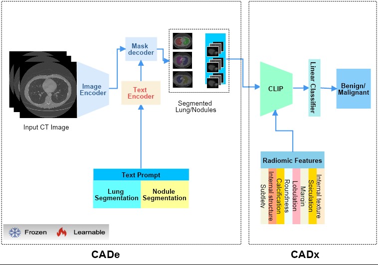
Lung cancer has been one of the major threats to human life for decades. Computer-aided diagnosis can help with early lung nodul detection and facilitate subsequent nodule characterization. Large Visual Language models (VLMs) have been found effective for multiple downstream medical tasks that rely on both imaging and text data. However, lesion level detection and subsequent diagnosis using VLMs have not been explored yet. We propose CADe, for segmenting lung nodules in a zero-shot manner using a variant of the Segment Anything Model called MedSAM. CADe trains on a prompt suite on input computed tomography (CT) scans by using the CLIP text encoder through prefix tuning. We also propose, CADx, a method for the nodule characterization as benign/malignant by making a gallery of radiomic features and aligning image-feature pairs through contrastive learning. Training and validation of CADe and CADx have been done using one of the largest publicly available datasets, called LIDC. To check the generalization ability of the model, it is also evaluated on a challenging dataset, LUNGx. Our experimental results show that the proposed methods achieve a sensitivity of 0.86 compared to 0.76 that of other fully supervised methods.The source code, datasets and pre-processed data can be accessed using the link:
Read more7/4/2024