CXR-Agent: Vision-language models for chest X-ray interpretation with uncertainty aware radiology reporting

0
Sign in to get full access
Overview
- The paper presents a new radiological assistant model called d-RAX that leverages large language models and multimodal techniques to assist radiologists in their tasks.
- The model is designed to enhance human-computer interaction in the radiology domain, building on prior work such as MedXChat and MAIRA-1.
- The paper also explores the use of vision-language generative models to generate view-specific chest X-ray images.
Plain English Explanation
The researchers have developed a new artificial intelligence (AI) system called d-RAX that is designed to help radiologists, or doctors who specialize in medical imaging, with their work. The system uses large language models and multimodal techniques, which means it can work with both text and images.
The goal of d-RAX is to make it easier for radiologists to do their jobs. Radiologists often have to look at many medical images, like X-rays or MRI scans, and try to identify problems or abnormalities. The d-RAX system is meant to assist them in this process, for example by providing relevant information or suggestions.
The researchers built on previous work in this area, including systems called MedXChat and MAIRA-1, which also aimed to help radiologists. They also explored using a special type of AI model that can generate new images, specifically view-specific chest X-ray images.
Overall, the d-RAX system is designed to improve the way radiologists and computers work together, making the radiologists' jobs easier and more efficient.
Technical Explanation
The paper presents a new radiological assistant model called d-RAX (domain-specific Radiologic Assistant leveraging X) that leverages large language models and multimodal techniques to assist radiologists in their tasks. The model is built upon prior work in the field, such as MedXChat, a unified multimodal large language model framework, and MAIRA-1, a specialized large multimodal model for radiology.
The key contributions of the d-RAX system include:
- Developing a domain-specific radiological assistant that can understand and generate natural language responses to assist radiologists in their workflow.
- Incorporating multimodal capabilities to enable the model to understand and process both text and medical images, such as X-rays and MRI scans.
- Exploring the use of vision-language generative models to generate view-specific chest X-ray images, which can be used to enhance human-computer interaction in the radiology domain.
The paper describes the architecture and training of the d-RAX model, as well as the experimental setup and evaluation of its performance on various tasks, including image-text retrieval, image captioning, and question answering.
Critical Analysis
The paper provides a comprehensive overview of the d-RAX system and its key contributions to the field of radiological assistants. However, the authors acknowledge several limitations and areas for further research:
- The model is trained on a limited dataset of radiology-specific data, which may limit its generalization to a broader range of medical imaging modalities and tasks.
- The generation of view-specific chest X-ray images, while promising, still has room for improvement in terms of realism and medical accuracy.
- The long-term impact of such AI-powered radiological assistants on the workflow and decision-making processes of radiologists is not fully explored and may require further investigation.
Additionally, it would be valuable to see more detailed analysis on the potential biases or ethical considerations that may arise from the use of such a system, particularly in terms of its impact on patient care and the potential for misdiagnosis or over-reliance on the model's recommendations.
Conclusion
The d-RAX system represents a significant advancement in the field of radiological assistants, leveraging large language models and multimodal techniques to enhance the interaction between radiologists and computers. By enabling natural language processing and generation, as well as the integration of medical images, the d-RAX model has the potential to streamline the radiologist's workflow and improve the overall efficiency of medical imaging analysis.
The exploration of vision-language generative models for view-specific chest X-ray image generation is an intriguing area of research that could lead to further advancements in human-computer interaction within the radiology domain.
While the paper acknowledges some limitations and areas for further study, the d-RAX system demonstrates the growing capabilities of AI-powered assistants in the healthcare field and highlights the importance of continued research and development in this space.
This summary was produced with help from an AI and may contain inaccuracies - check out the links to read the original source documents!
Related Papers
0
CXR-Agent: Vision-language models for chest X-ray interpretation with uncertainty aware radiology reporting
Naman Sharma
Recently large vision-language models have shown potential when interpreting complex images and generating natural language descriptions using advanced reasoning. Medicine's inherently multimodal nature incorporating scans and text-based medical histories to write reports makes it conducive to benefit from these leaps in AI capabilities. We evaluate the publicly available, state of the art, foundational vision-language models for chest X-ray interpretation across several datasets and benchmarks. We use linear probes to evaluate the performance of various components including CheXagent's vision transformer and Q-former, which outperform the industry-standard Torch X-ray Vision models across many different datasets showing robust generalisation capabilities. Importantly, we find that vision-language models often hallucinate with confident language, which slows down clinical interpretation. Based on these findings, we develop an agent-based vision-language approach for report generation using CheXagent's linear probes and BioViL-T's phrase grounding tools to generate uncertainty-aware radiology reports with pathologies localised and described based on their likelihood. We thoroughly evaluate our vision-language agents using NLP metrics, chest X-ray benchmarks and clinical evaluations by developing an evaluation platform to perform a user study with respiratory specialists. Our results show considerable improvements in accuracy, interpretability and safety of the AI-generated reports. We stress the importance of analysing results for normal and abnormal scans separately. Finally, we emphasise the need for larger paired (scan and report) datasets alongside data augmentation to tackle overfitting seen in these large vision-language models.
Read more7/15/2024
0
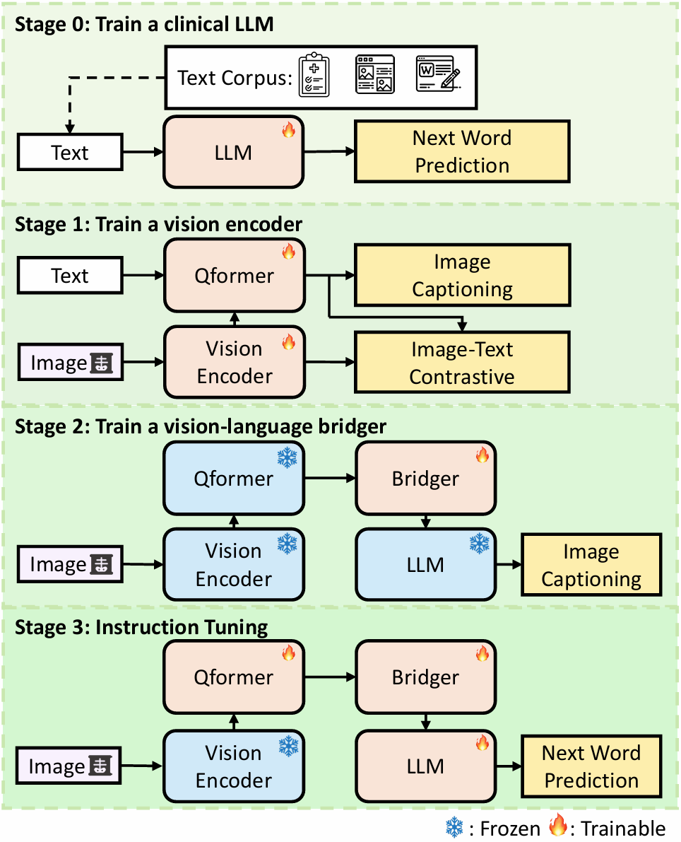
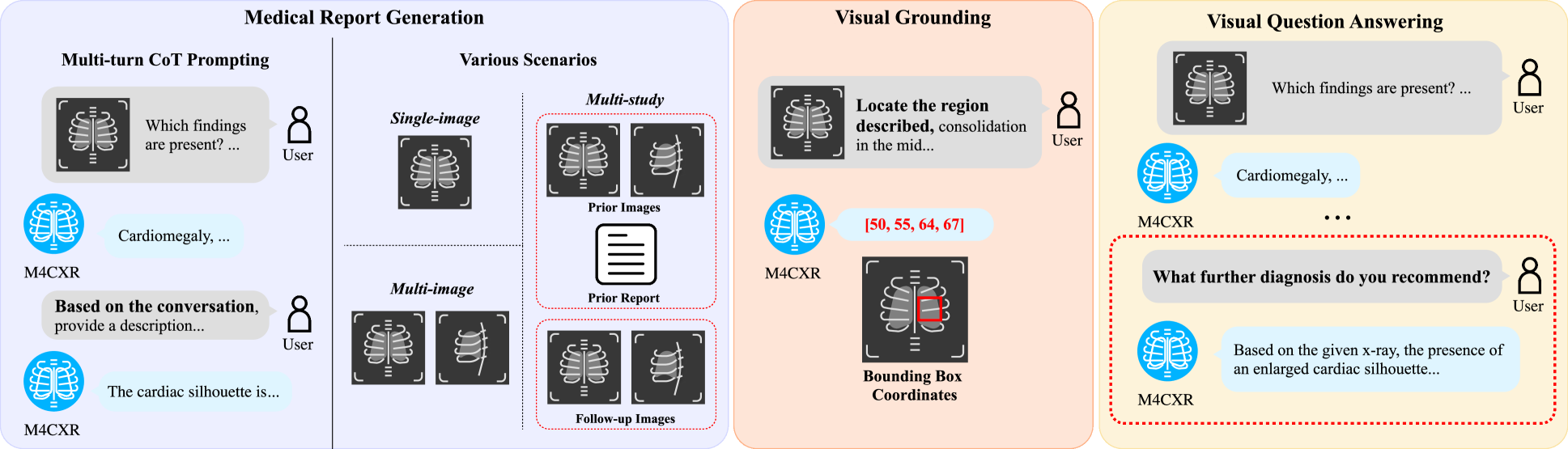
M4CXR: Exploring Multi-task Potentials of Multi-modal Large Language Models for Chest X-ray Interpretation
Jonggwon Park, Soobum Kim, Byungmu Yoon, Jihun Hyun, Kyoyun Choi
The rapid evolution of artificial intelligence, especially in large language models (LLMs), has significantly impacted various domains, including healthcare. In chest X-ray (CXR) analysis, previous studies have employed LLMs, but with limitations: either underutilizing the multi-tasking capabilities of LLMs or lacking clinical accuracy. This paper presents M4CXR, a multi-modal LLM designed to enhance CXR interpretation. The model is trained on a visual instruction-following dataset that integrates various task-specific datasets in a conversational format. As a result, the model supports multiple tasks such as medical report generation (MRG), visual grounding, and visual question answering (VQA). M4CXR achieves state-of-the-art clinical accuracy in MRG by employing a chain-of-thought prompting strategy, in which it identifies findings in CXR images and subsequently generates corresponding reports. The model is adaptable to various MRG scenarios depending on the available inputs, such as single-image, multi-image, and multi-study contexts. In addition to MRG, M4CXR performs visual grounding at a level comparable to specialized models and also demonstrates outstanding performance in VQA. Both quantitative and qualitative assessments reveal M4CXR's versatility in MRG, visual grounding, and VQA, while consistently maintaining clinical accuracy.
Read more8/30/2024
0
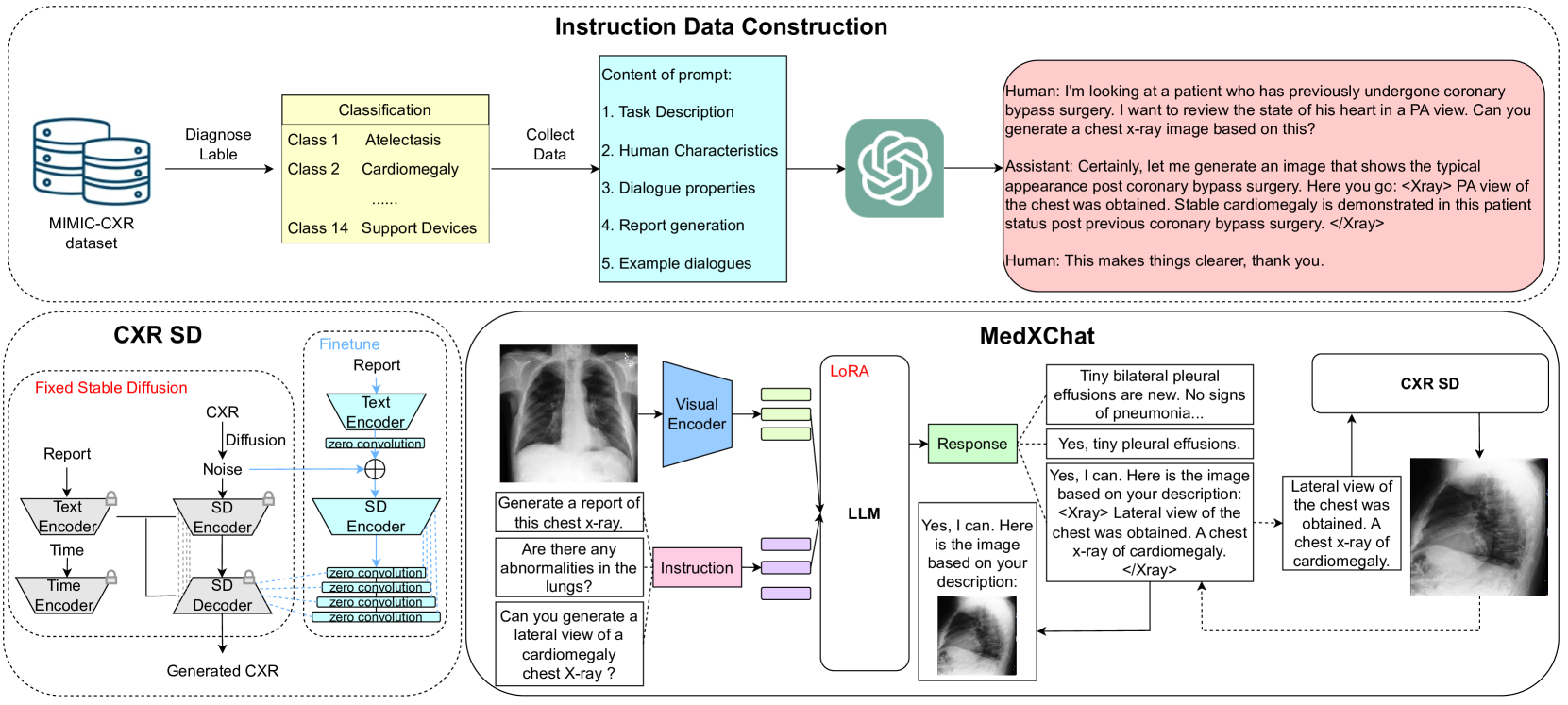
MedXChat: A Unified Multimodal Large Language Model Framework towards CXRs Understanding and Generation
Ling Yang, Zhanyu Wang, Zhenghao Chen, Xinyu Liang, Luping Zhou
Multimodal Large Language Models (MLLMs) have shown success in various general image processing tasks, yet their application in medical imaging is nascent, lacking tailored models. This study investigates the potential of MLLMs in improving the understanding and generation of Chest X-Rays (CXRs). We introduce MedXChat, a unified framework facilitating seamless interactions between medical assistants and users for diverse CXR tasks, including text report generation, visual question-answering (VQA), and Text-to-CXR generation. Our MLLMs using natural language as the input breaks task boundaries, maximally simplifying medical professional training by allowing diverse tasks within a single environment. For CXR understanding, we leverage powerful off-the-shelf visual encoders (e.g., ViT) and LLMs (e.g., mPLUG-Owl) to convert medical imagery into language-like features, and subsequently fine-tune our large pre-trained models for medical applications using a visual adapter network and a delta-tuning approach. For CXR generation, we introduce an innovative synthesis approach that utilizes instruction-following capabilities within the Stable Diffusion (SD) architecture. This technique integrates smoothly with the existing model framework, requiring no extra parameters, thereby maintaining the SD's generative strength while also bestowing upon it the capacity to render fine-grained medical images with high fidelity. Through comprehensive experiments, our model demonstrates exceptional cross-task adaptability, displaying adeptness across all three defined tasks. Our MedXChat model and the instruction dataset utilized in this research will be made publicly available to encourage further exploration in the field.
Read more5/13/2024

0
D-Rax: Domain-specific Radiologic assistant leveraging multi-modal data and eXpert model predictions
Hareem Nisar, Syed Muhammad Anwar, Zhifan Jiang, Abhijeet Parida, Ramon Sanchez-Jacob, Vishwesh Nath, Holger R. Roth, Marius George Linguraru
Large vision language models (VLMs) have progressed incredibly from research to applicability for general-purpose use cases. LLaVA-Med, a pioneering large language and vision assistant for biomedicine, can perform multi-modal biomedical image and data analysis to provide a natural language interface for radiologists. While it is highly generalizable and works with multi-modal data, it is currently limited by well-known challenges that exist in the large language model space. Hallucinations and imprecision in responses can lead to misdiagnosis which currently hinder the clinical adaptability of VLMs. To create precise, user-friendly models in healthcare, we propose D-Rax -- a domain-specific, conversational, radiologic assistance tool that can be used to gain insights about a particular radiologic image. In this study, we enhance the conversational analysis of chest X-ray (CXR) images to support radiological reporting, offering comprehensive insights from medical imaging and aiding in the formulation of accurate diagnosis. D-Rax is achieved by fine-tuning the LLaVA-Med architecture on our curated enhanced instruction-following data, comprising of images, instructions, as well as disease diagnosis and demographic predictions derived from MIMIC-CXR imaging data, CXR-related visual question answer (VQA) pairs, and predictive outcomes from multiple expert AI models. We observe statistically significant improvement in responses when evaluated for both open and close-ended conversations. Leveraging the power of state-of-the-art diagnostic models combined with VLMs, D-Rax empowers clinicians to interact with medical images using natural language, which could potentially streamline their decision-making process, enhance diagnostic accuracy, and conserve their time.
Read more8/6/2024