Enhancing super-resolution ultrasound localisation through multi-frame deconvolution exploiting spatiotemporal coherence

0
👨🏫
Sign in to get full access
Overview
- This paper presents a novel method called Multi-Frame Deconvolution (MF-Decon) to enhance the localization of microbubbles (MBs) in contrast-enhanced ultrasound (CEUS) images for super-resolution ultrasound imaging.
- Super-resolution ultrasound imaging through MB localization and tracking, also known as ultrasound localization microscopy, allows non-invasive high-resolution imaging of microvasculature in animals and humans.
- The quality of the resulting super-resolution images depends on the number of MBs localized and the localization precision, which can be challenging due to noise in the CEUS images.
- The proposed MF-Decon framework exploits the spatiotemporal coherence in the CEUS data using new spatial and temporal regularizers based on total variation (TV) and regularization by denoising (RED).
Plain English Explanation
Ultrasound imaging is a widely used medical technique that uses sound waves to create images of the inside of the body. Super-resolution ultrasound imaging is a more advanced technique that can produce even higher-quality images, particularly of small blood vessels.
This technique works by tracking the movement of tiny bubbles, called microbubbles, that are injected into the body. As the microbubbles flow through the blood vessels, they can be detected by the ultrasound system, and their positions can be used to create a very detailed image of the blood vessel network.
However, the quality of these super-resolution images can be affected by noise in the original ultrasound data, which can make it difficult to accurately track the microbubbles. To address this, the researchers developed a new method called Multi-Frame Deconvolution (MF-Decon) that uses advanced mathematical techniques to enhance the localization of the microbubbles and improve the quality of the super-resolution images.
The MF-Decon method takes advantage of the fact that the microbubbles move in a coherent way over time, and it uses this information to better distinguish the microbubbles from the background noise. The researchers also developed new mathematical tools, based on concepts like total variation and regularization by denoising, to further improve the microbubble localization and the quality of the final images.
The researchers tested their MF-Decon method using computer simulations and real-world data from experiments on rats, and they found that it outperformed other commonly used methods in terms of both the precision and the recall of the microbubble localization. The super-resolution images generated using the MF-Decon method also showed less noise, better contrast, and more detailed blood vessel structures compared to other techniques.
Technical Explanation
The proposed MF-Decon framework exploits the spatiotemporal coherence inherent in the CEUS data to enhance the localization of microbubbles (MBs). The framework introduces new spatial and temporal regularizers designed based on total variation (TV) and regularization by denoising (RED).
Specifically, the researchers introduce two novel methods:
- MF-Decon with spatial and temporal TVs (MF-Decon+3DTV): This method uses 3D TV regularization to capture both the spatial and temporal information in the CEUS data.
- MF-Decon with spatial RED and temporal TV (MF-Decon+RED+TV): This method combines spatial regularization using RED, which leverages the inherent structure of the CEUS images, with temporal TV regularization.
The performance of these MF-Decon methods is evaluated using in silico simulations and compared to two widely used techniques: deconvolution and normalized cross-correlation. The results show that the proposed methods outperform the baseline approaches across various evaluation metrics, including precision, recall, F1 score, and localization error.
In particular, the MF-Decon methods improve MB localization precision by up to 39% and recall by up to 12%. The super-resolution microvasculature maps generated using the MF-Decon methods on a publicly available in vivo rat brain dataset exhibit less noise, better contrast, higher resolution, and more vessel structures compared to the other techniques.
Critical Analysis
The researchers have presented a promising approach to enhance microbubble localization in super-resolution ultrasound imaging. The key strengths of their work include the novel spatial and temporal regularizers based on TV and RED, which effectively leverage the inherent structure and coherence in the CEUS data.
However, the paper does not provide a comprehensive analysis of the limitations of the proposed methods. For example, it would be valuable to understand the computational complexity of the MF-Decon framework and its implications for real-time or clinical applications. Additionally, the performance of the methods on more diverse datasets, such as human clinical data, should be evaluated to assess their generalizability.
Furthermore, the paper does not discuss potential sources of bias or artifacts that may arise from the MF-Decon framework. It would be important to investigate the robustness of the methods to factors such as variations in microbubble concentration, tissue heterogeneity, or imaging depth.
Future research could also explore the integration of the MF-Decon methods with other advanced techniques, such as inverse problem approaches to aberration correction or transformer-based local feature matching, to further enhance the quality and reliability of super-resolution ultrasound imaging.
Conclusion
The proposed MF-Decon framework represents a significant contribution to the field of super-resolution ultrasound imaging. By leveraging the spatiotemporal coherence in CEUS data and introducing novel regularization techniques, the researchers have demonstrated improved microbubble localization and the generation of higher-quality super-resolution microvasculature maps.
The potential clinical impact of this work is substantial, as it could lead to more accurate and detailed visualization of the microvascular network in both animal and human subjects. This improved imaging capability may enable earlier diagnosis, better treatment monitoring, and a deeper understanding of vascular physiology and pathology.
While the paper highlights the strengths of the MF-Decon methods, further research is needed to address the limitations and explore the integration of this approach with other state-of-the-art techniques in the field of high-resolution power Doppler imaging and edge-guided cross-scale feature fusion. By continuing to push the boundaries of super-resolution ultrasound imaging, researchers can unlock new possibilities for non-invasive, high-resolution visualization of the human body.
This summary was produced with help from an AI and may contain inaccuracies - check out the links to read the original source documents!
Related Papers
👨🏫
0
Enhancing super-resolution ultrasound localisation through multi-frame deconvolution exploiting spatiotemporal coherence
Su Yan, Clotilde Vi'e, Marcelo Lerendegui, Herman Verinaz-Jadan, Jipeng Yan, Martina Tashkova, James Burn, Bingxue Wang, Gary Frost, Kevin G. Murphy, Meng-Xing Tang
Super-resolution ultrasound imaging through microbubble (MB) localisation and tracking, also known as ultrasound localisation microscopy, allows non-invasive sub-diffraction resolution imaging of microvasculature in animals and humans. The number of MBs localised from the acquired contrast-enhanced ultrasound (CEUS) images and the localisation precision directly influence the quality of the resulting super-resolution microvasculature images. However, non-negligible noise present in the CEUS images can make localising MBs challenging. To enhance the MB localisation performance, we propose a Multi-Frame Deconvolution (MF-Decon) framework that can exploit the spatiotemporal coherence inherent in the CEUS data, with new spatial and temporal regularisers designed based on total variation (TV) and regularisation by denoising (RED). Based on the MF-Decon framework, we introduce two novel methods: MF-Decon with spatial and temporal TVs (MF-Decon+3DTV) and MF-Decon with spatial RED and temporal TV (MF-Decon+RED+TV). Results from in silico simulations indicate that our methods outperform two widely used methods using deconvolution or normalised cross-correlation across all evaluation metrics, including precision, recall, $F_1$ score, mean and standard localisation errors. In particular, our methods improve MB localisation precision by up to 39% and recall by up to 12%. Super-resolution microvasculature maps generated with our methods on a publicly available in vivo rat brain dataset show less noise, better contrast, higher resolution and more vessel structures.
Read more7/10/2024
🤿
0
Deep Learning for Super-resolution Ultrasound Imaging with Spatiotemporal Data
Arthur David Redfern, Katherine G. Brown
Super-resolution ultrasound imaging (SRUS) is an active area of research as it brings up to a ten-fold improvement in the resolution of microvascular structures. The limitations to the clinical adoption of SRUS include long acquisition times and long image processing times. Both these limitations can be alleviated with deep learning approaches to the processing of SRUS images. In this study we propose an optimized architecture based on modern improvements to convolutional neural networks from the ConvNeXt architecture and further customize the choice of features to improve performance on the specific tasks of both MB detection and localization within a single network. We employ a spatiotemporal input of up to five successive image frames to increase the number of MBs detected. The output structure produces three classifications: a MB detection Boolean for each pixel in the central image frame, as well as x and z offsets at 4-fold subpixel resolution for each MB detected. Ultrasound simulations generated images based on the L22-14v transducer (Verasonics) for training and testing of the proposed SRUS-ConvNeXt network. In vivo image data of a mouse brain was used as further validation of the architecture. The proposed network had the highest performance as measured by F1 score when configured for a 3-frame spatiotemporal input. The smallest localization error of {lambda}/22 was achieved when the network was configured for a single input frame. The flexibility of the proposed architecture allows extension to 10-fold upscaling for SRUS images with a much lower impact to number of parameters and subsequent increase in inference time than typical U-Net style approaches. This network is promising in the quest to develop a SRUS deep network architecture for real time image formation.
Read more8/5/2024
0
New!Online 4D Ultrasound-Guided Robotic Tracking Enables 3D Ultrasound Localisation Microscopy with Large Tissue Displacements
Jipeng Yan, Shusei Kawara, Qingyuan Tan, Jingwen Zhu, Bingxue Wang, Matthieu Toulemonde, Honghai Liu, Ying Tan, Meng-Xing Tang
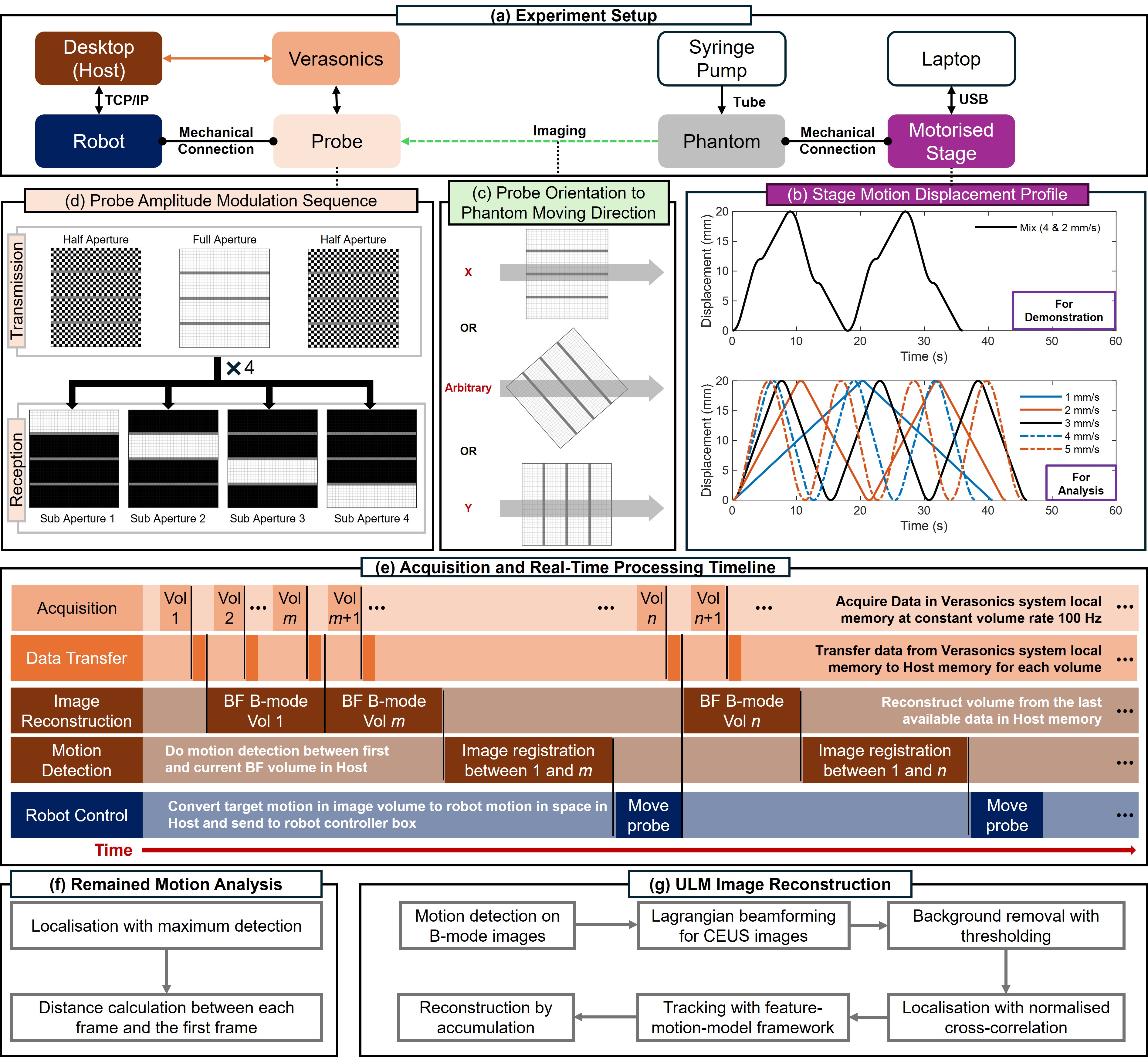
Super-Resolution Ultrasound (SRUS) imaging through localising and tracking microbubbles, also known as Ultrasound Localisation Microscopy (ULM), has demonstrated significant potential for reconstructing microvasculature and flows with sub-diffraction resolution in clinical diagnostics. However, imaging organs with large tissue movements, such as those caused by respiration, presents substantial challenges. Existing methods often require breath holding to maintain accumulation accuracy, which limits data acquisition time and ULM image saturation. To improve image quality in the presence of large tissue movements, this study introduces an approach integrating high-frame-rate ultrasound with online precise robotic probe control. Tested on a microvasculature phantom with translation motions up to 20 mm, twice the aperture size of the matrix array used, our method achieved real-time tracking of the moving phantom and imaging volume rate at 85 Hz, keeping majority of the target volume in the imaging field of view. ULM images of the moving cross channels in the phantom were successfully reconstructed in post-processing, demonstrating the feasibility of super-resolution imaging under large tissue motions. This represents a significant step towards ULM imaging of organs with large motion.
Read more9/18/2024

0
PHOCUS: Physics-Based Deconvolution for Ultrasound Resolution Enhancement
Felix Duelmer, Walter Simson, Mohammad Farid Azampour, Magdalena Wysocki, Angelos Karlas, Nassir Navab
Ultrasound is widely used in medical diagnostics allowing for accessible and powerful imaging but suffers from resolution limitations due to diffraction and the finite aperture of the imaging system, which restricts diagnostic use. The impulse function of an ultrasound imaging system is called the point spread function (PSF), which is convolved with the spatial distribution of reflectors in the image formation process. Recovering high-resolution reflector distributions by removing image distortions induced by the convolution process improves image clarity and detail. Conventionally, deconvolution techniques attempt to rectify the imaging system's dependent PSF, working directly on the radio-frequency (RF) data. However, RF data is often not readily accessible. Therefore, we introduce a physics-based deconvolution process using a modeled PSF, working directly on the more commonly available B-mode images. By leveraging Implicit Neural Representations (INRs), we learn a continuous mapping from spatial locations to their respective echogenicity values, effectively compensating for the discretized image space. Our contribution consists of a novel methodology for retrieving a continuous echogenicity map directly from a B-mode image through a differentiable physics-based rendering pipeline for ultrasound resolution enhancement. We qualitatively and quantitatively evaluate our approach on synthetic data, demonstrating improvements over traditional methods in metrics such as PSNR and SSIM. Furthermore, we show qualitative enhancements on an ultrasound phantom and an in-vivo acquisition of a carotid artery.
Read more8/9/2024