A kinetic approach to consensus-based segmentation of biomedical images

0
🗣️
Sign in to get full access
Overview
- Applies a kinetic version of a bounded confidence consensus model to biomedical segmentation problems
- Microscopic model includes particle/pixel position and a feature representing the gray level of each pixel
- Derives a kinetic formulation and uses a Fokker-Planck approach to compute the large time behavior of the system
- Exploits the computational efficiency of direct simulation Monte Carlo methods for parameter identification tasks
- Minimizes a segmentation metric for 2D gray-scale images, with applications in various biomedical imaging contexts
Plain English Explanation
This research applies a type of motion-informed needle segmentation in ultrasound images model to problems in biomedical image segmentation. The key idea is to represent each pixel in the image as a "particle" that has both a physical location and a feature (in this case, the gray level or brightness of that pixel).
The researchers then derive a mathematical model that describes how these particles/pixels interact and evolve over time. This model is based on the concept of "bounded confidence consensus," where particles/pixels tend to converge towards a common state if they are sufficiently similar to each other.
To make this model computationally efficient, the researchers use a technique called the "Fokker-Planck approach" to simplify the calculations. They also leverage a method called "direct simulation Monte Carlo" to quickly identify the best set of parameters for the model.
By minimizing a metric that compares the model's segmentation output to the ground truth, the researchers are able to apply this approach to a variety of 2D biomedical imaging problems, such as consistency-based medical image segmentation and failure detection in medical imaging. The core idea is to use the motion and appearance of pixels to guide the segmentation process, rather than relying solely on static image information.
Technical Explanation
The researchers apply a kinetic version of a bounded confidence consensus model to biomedical segmentation problems. The microscopic state of each particle/pixel includes its spatial position and a feature representing the gray level of the pixel. From this microscopic model, the researchers derive a kinetic formulation, which they then compute the large-time behavior of using a surrogate Fokker-Planck approach.
To efficiently solve the resulting Boltzmann-type description of the problem, the researchers exploit the computational advantages of direct simulation Monte Carlo methods. They use these methods to perform parameter identification tasks, minimizing a suitable loss function that measures the distance between the ground truth segmentation mask and the evaluated mask. The applications of this approach focus on various biomedical imaging contexts, leveraging the kinetics perspective in predicting optical flow to guide the segmentation process.
Critical Analysis
The paper presents a novel application of a kinetic consensus model to biomedical image segmentation, which is an important and challenging problem in the field. The use of the Fokker-Planck approach and direct simulation Monte Carlo methods appears to be a computationally efficient way to solve the model and perform parameter identification.
However, the paper does not provide much detail on the specific biomedical imaging applications or the performance of the method compared to other state-of-the-art segmentation techniques. It would be helpful to see more comparative benchmarking of failure detection methods in medical imaging to better understand the strengths and limitations of this approach.
Additionally, the paper does not discuss the potential limitations or challenges of applying a consensus-based model to biomedical segmentation, such as the sensitivity to initialization or the ability to handle complex, heterogeneous image structures. Further research may be needed to adaptively generalize the approach to different MRI imaging modalities.
Overall, the work presents an interesting and potentially useful approach to biomedical image segmentation, but more thorough evaluation and discussion of its practical implications would be beneficial.
Conclusion
This research applies a kinetic version of a bounded confidence consensus model to the problem of biomedical image segmentation. By representing each pixel as a particle with both spatial and appearance-based features, the researchers derive a computationally efficient method for performing segmentation tasks.
The use of the Fokker-Planck approach and direct simulation Monte Carlo techniques allows for effective parameter identification and optimization of the segmentation model. While the paper demonstrates the potential of this approach, further research is needed to fully understand its capabilities and limitations in real-world biomedical imaging applications.
This summary was produced with help from an AI and may contain inaccuracies - check out the links to read the original source documents!
Related Papers
🗣️
0
A kinetic approach to consensus-based segmentation of biomedical images
Raffaella Fiamma Cabini, Anna Pichiecchio, Alessandro Lascialfari, Silvia Figini, Mattia Zanella
In this work, we apply a kinetic version of a bounded confidence consensus model to biomedical segmentation problems. In the presented approach, time-dependent information on the microscopic state of each particle/pixel includes its space position and a feature representing a static characteristic of the system, i.e. the gray level of each pixel. From the introduced microscopic model we derive a kinetic formulation of the model. The large time behavior of the system is then computed with the aid of a surrogate Fokker-Planck approach that can be obtained in the quasi-invariant scaling. We exploit the computational efficiency of direct simulation Monte Carlo methods for the obtained Boltzmann-type description of the problem for parameter identification tasks. Based on a suitable loss function measuring the distance between the ground truth segmentation mask and the evaluated mask, we minimize the introduced segmentation metric for a relevant set of 2D gray-scale images. Applications to biomedical segmentation concentrate on different imaging research contexts.
Read more6/17/2024
🧠
0
Motion Informed Needle Segmentation in Ultrasound Images
Raghavv Goel, Cecilia Morales, Manpreet Singh, Artur Dubrawski, John Galeotti, Howie Choset
Segmenting a moving needle in ultrasound images is challenging due to the presence of artifacts, noise, and needle occlusion. This task becomes even more demanding in scenarios where data availability is limited. In this paper, we present a novel approach for needle segmentation for 2D ultrasound that combines classical Kalman Filter (KF) techniques with data-driven learning, incorporating both needle features and needle motion. Our method offers three key contributions. First, we propose a compatible framework that seamlessly integrates into commonly used encoder-decoder style architectures. Second, we demonstrate superior performance compared to recent state-of-the-art needle segmentation models using our novel convolutional neural network (CNN) based KF-inspired block, achieving a 15% reduction in pixel-wise needle tip error and an 8% reduction in length error. Third, to our knowledge we are the first to implement a learnable filter to incorporate non-linear needle motion for improving needle segmentation.
Read more5/7/2024
0
CTS: A Consistency-Based Medical Image Segmentation Model
Kejia Zhang, Lan Zhang, Haiwei Pan, Baolong Yu
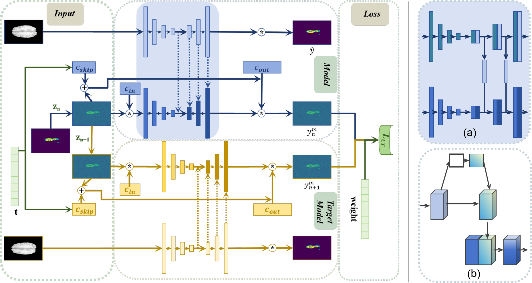
In medical image segmentation tasks, diffusion models have shown significant potential. However, mainstream diffusion models suffer from drawbacks such as multiple sampling times and slow prediction results. Recently, consistency models, as a standalone generative network, have resolved this issue. Compared to diffusion models, consistency models can reduce the sampling times to once, not only achieving similar generative effects but also significantly speeding up training and prediction. However, they are not suitable for image segmentation tasks, and their application in the medical imaging field has not yet been explored. Therefore, this paper applies the consistency model to medical image segmentation tasks, designing multi-scale feature signal supervision modes and loss function guidance to achieve model convergence. Experiments have verified that the CTS model can obtain better medical image segmentation results with a single sampling during the test phase.
Read more5/16/2024

0
Comparative Benchmarking of Failure Detection Methods in Medical Image Segmentation: Unveiling the Role of Confidence Aggregation
Maximilian Zenk, David Zimmerer, Fabian Isensee, Jeremias Traub, Tobias Norajitra, Paul F. Jager, Klaus Maier-Hein
Semantic segmentation is an essential component of medical image analysis research, with recent deep learning algorithms offering out-of-the-box applicability across diverse datasets. Despite these advancements, segmentation failures remain a significant concern for real-world clinical applications, necessitating reliable detection mechanisms. This paper introduces a comprehensive benchmarking framework aimed at evaluating failure detection methodologies within medical image segmentation. Through our analysis, we identify the strengths and limitations of current failure detection metrics, advocating for the risk-coverage analysis as a holistic evaluation approach. Utilizing a collective dataset comprising five public 3D medical image collections, we assess the efficacy of various failure detection strategies under realistic test-time distribution shifts. Our findings highlight the importance of pixel confidence aggregation and we observe superior performance of the pairwise Dice score (Roy et al., 2019) between ensemble predictions, positioning it as a simple and robust baseline for failure detection in medical image segmentation. To promote ongoing research, we make the benchmarking framework available to the community.
Read more6/6/2024