Promptable Counterfactual Diffusion Model for Unified Brain Tumor Segmentation and Generation with MRIs

0

Sign in to get full access
Overview
- This paper proposes a Promptable Counterfactual Diffusion Model (PCDM) for unified brain tumor segmentation and generation from MRI scans.
- PCDM leverages diffusion models, which are a type of generative AI that can create new images by gradually adding noise to an existing image and then removing the noise.
- The model is "promptable," meaning it can be controlled and customized using text prompts, similar to how text-to-image models work.
- PCDM can perform both tumor segmentation (identifying the location and boundaries of brain tumors) and tumor generation (creating synthetic brain MRI scans with realistic tumors).
Plain English Explanation
The researchers have developed a new AI system that can work with brain MRI scans in two very useful ways. First, it can analyze an MRI scan and automatically identify the location and boundaries of any brain tumors present. This is called "segmentation" and is a crucial task for medical diagnosis and treatment planning.
Secondly, the system can also generate completely new, synthetic brain MRI scans that include realistic-looking brain tumors. This "tumor generation" capability could be used to create training data for other AI models or to test the performance of tumor segmentation algorithms.
What makes this system unique is that it is "promptable." This means the user can provide the AI with text instructions or prompts to control its behavior. For example, you could ask the system to generate a brain MRI with a specific type or size of tumor, or to segment a tumor with particular characteristics. This added flexibility and customization is a key innovation of this research.
The underlying technology behind PCDM is a type of generative AI called a diffusion model. Diffusion models work by gradually adding noise to an image and then learning how to reverse that process, allowing them to create new images from scratch. By combining diffusion modeling with the ability to follow text prompts, the researchers have developed a powerful tool for both brain tumor analysis and synthesis.
Technical Explanation
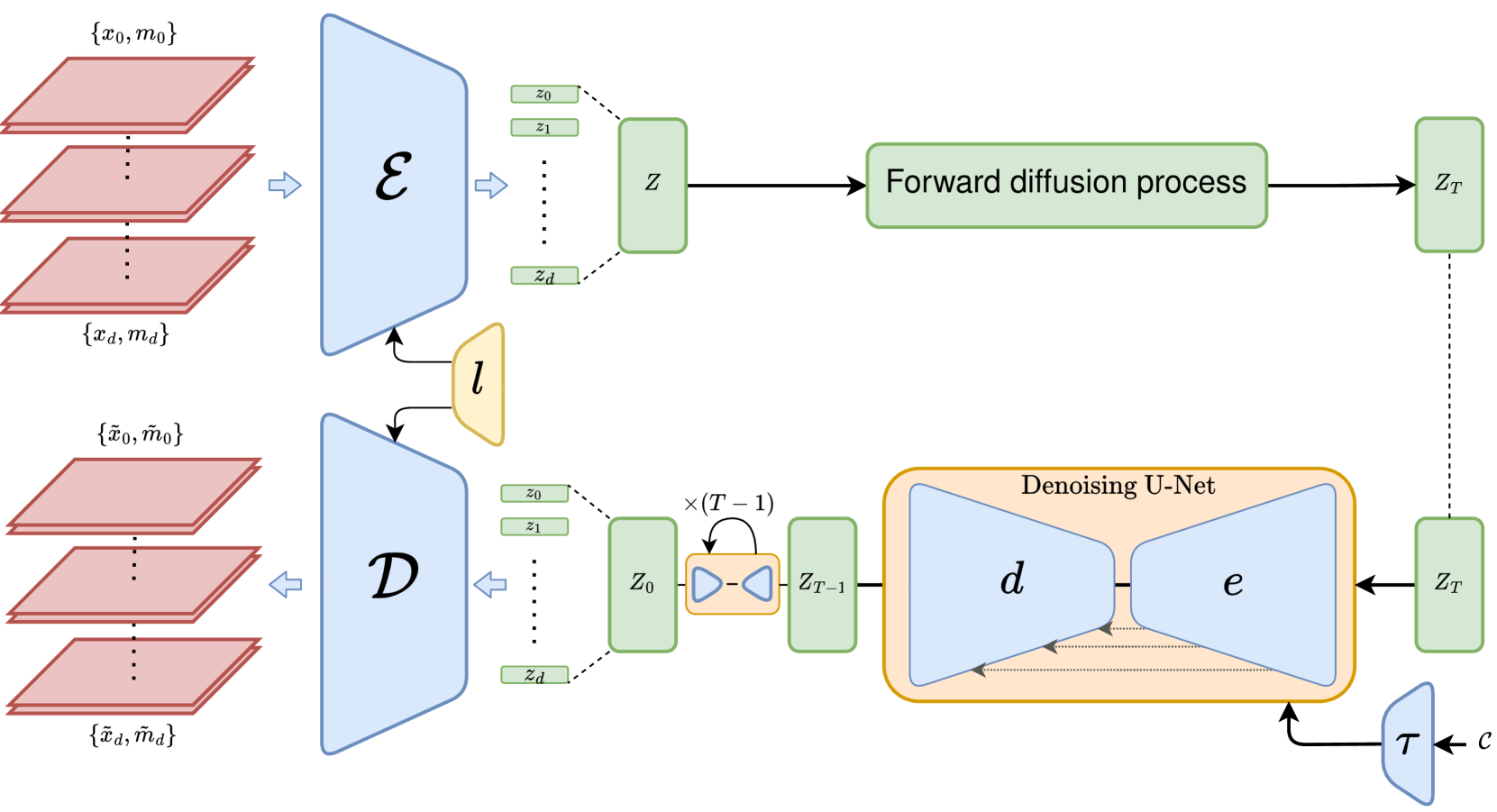
The Promptable Counterfactual Diffusion Model (PCDM) proposed in this paper leverages the flexibility and controllability of diffusion models for the tasks of brain tumor segmentation and generation from MRI scans. Diffusion models are a type of generative AI that can create new images by gradually adding noise to an existing image and then learning to reverse that process.
The key innovations of PCDM are:
-
Prompt-based Control: The model can be controlled and customized using text prompts, similar to how text-to-image models work. This allows the system to generate brain MRIs with specific tumor characteristics or to focus the segmentation on particular regions of interest.
-
Unified Segmentation and Generation: PCDM is capable of both segmenting brain tumors from existing MRI scans and generating new, synthetic MRI scans with realistic-looking tumors. This unified approach leverages the shared underlying representations learned by the model.
-
Counterfactual Reasoning: The model is trained to not only generate realistic brain MRIs, but also to reason about "counterfactual" scenarios - i.e., how the MRI would change if certain attributes (such as tumor size or location) were different. This provides additional flexibility and control over the generated outputs.
The PCDM architecture builds upon recent advances in diffusion models, such as Anatomically Controllable Medical Image Generation and Segmentation and Discrepancy-based Diffusion Models for Lesion Detection in Brain MRI. It integrates a prompt encoder, a diffusion model, and segmentation and generation heads to enable the unified task capabilities.
The researchers evaluate PCDM on brain MRI datasets, demonstrating its effectiveness in both tumor segmentation and generation. They also showcase the model's ability to perform counterfactual reasoning, such as generating MRIs with tumors of different sizes or in different locations.
Critical Analysis
The paper presents a compelling and technically sophisticated approach to brain tumor analysis and synthesis using diffusion models. The key strengths of the PCDM system are its flexibility, control, and unified capabilities for both segmentation and generation.
However, the paper also acknowledges several limitations and areas for further research:
-
Dataset Diversity: The experiments were conducted on relatively homogeneous datasets of brain MRI scans. Evaluating the model's performance on more diverse, real-world clinical data would be an important next step.
-
Computational Complexity: Diffusion models, while powerful, can be computationally intensive, particularly for high-resolution medical imaging tasks. Techniques to improve the efficiency of PCDM would be valuable.
-
Clinical Validation: While the paper demonstrates strong technical performance, further research is needed to validate the clinical utility and safety of using PCDM for tasks like tumor diagnosis and treatment planning.
-
Discrepancy-based Diffusion Models for Lesion Detection in Brain MRI and CT-based Brain Ventricle Segmentation via Diffusion are related works that could provide additional insights and baselines for evaluating PCDM's performance.
Overall, the Promptable Counterfactual Diffusion Model represents an exciting advancement in the field of medical image analysis and synthesis. With further refinement and validation, this type of technology could have significant implications for improving brain tumor diagnosis, treatment, and understanding.
Conclusion
The Promptable Counterfactual Diffusion Model (PCDM) proposed in this paper is a innovative AI system that can perform both brain tumor segmentation and generation from MRI scans. By leveraging diffusion models and enabling prompt-based control, PCDM provides a flexible and customizable tool for analyzing and synthesizing brain MRI data.
The key contributions of this research include the unified segmentation and generation capabilities, the ability to perform counterfactual reasoning, and the integration of prompt-based control. While the paper highlights several limitations that require further investigation, PCDM represents an important step forward in the development of advanced medical imaging technologies that can assist clinicians and researchers in understanding and treating brain tumors.
As diffusion models and other generative AI techniques continue to advance, we can expect to see even more powerful and versatile tools emerge for a wide range of medical imaging and analysis tasks. The work presented in this paper serves as a valuable example of how these cutting-edge AI capabilities can be harnessed to tackle critical challenges in the field of neuroscience and healthcare.
This summary was produced with help from an AI and may contain inaccuracies - check out the links to read the original source documents!
Related Papers
0
Promptable Counterfactual Diffusion Model for Unified Brain Tumor Segmentation and Generation with MRIs
Yiqing Shen, Guannan He, Mathias Unberath
Brain tumor analysis in Magnetic Resonance Imaging (MRI) is crucial for accurate diagnosis and treatment planning. However, the task remains challenging due to the complexity and variability of tumor appearances, as well as the scarcity of labeled data. Traditional approaches often address tumor segmentation and image generation separately, limiting their effectiveness in capturing the intricate relationships between healthy and pathological tissue structures. We introduce a novel promptable counterfactual diffusion model as a unified solution for brain tumor segmentation and generation in MRI. The key innovation lies in our mask-level prompting mechanism at the sampling stage, which enables guided generation and manipulation of specific healthy or unhealthy regions in MRI images. Specifically, the model's architecture allows for bidirectional inference, which can segment tumors in existing images and generate realistic tumor structures in healthy brain scans. Furthermore, we present a two-step approach for tumor generation and position transfer, showcasing the model's versatility in synthesizing realistic tumor structures. Experiments on the BRATS2021 dataset demonstrate that our method outperforms traditional counterfactual diffusion approaches, achieving a mean IoU of 0.653 and mean Dice score of 0.785 for tumor segmentation, outperforming the 0.344 and 0.475 of conventional counterfactual diffusion model. Our work contributes to improving brain tumor detection and segmentation accuracy, with potential implications for data augmentation and clinical decision support in neuro-oncology. The code is available at https://github.com/arcadelab/counterfactual_diffusion.
Read more7/18/2024

0
MedEdit: Counterfactual Diffusion-based Image Editing on Brain MRI
Malek Ben Alaya, Daniel M. Lang, Benedikt Wiestler, Julia A. Schnabel, Cosmin I. Bercea
Denoising diffusion probabilistic models enable high-fidelity image synthesis and editing. In biomedicine, these models facilitate counterfactual image editing, producing pairs of images where one is edited to simulate hypothetical conditions. For example, they can model the progression of specific diseases, such as stroke lesions. However, current image editing techniques often fail to generate realistic biomedical counterfactuals, either by inadequately modeling indirect pathological effects like brain atrophy or by excessively altering the scan, which disrupts correspondence to the original images. Here, we propose MedEdit, a conditional diffusion model for medical image editing. MedEdit induces pathology in specific areas while balancing the modeling of disease effects and preserving the integrity of the original scan. We evaluated MedEdit on the Atlas v2.0 stroke dataset using Frechet Inception Distance and Dice scores, outperforming state-of-the-art diffusion-based methods such as Palette (by 45%) and SDEdit (by 61%). Additionally, clinical evaluations by a board-certified neuroradiologist confirmed that MedEdit generated realistic stroke scans indistinguishable from real ones. We believe this work will enable counterfactual image editing research to further advance the development of realistic and clinically useful imaging tools.
Read more7/23/2024

0
A Weakly Supervised and Globally Explainable Learning Framework for Brain Tumor Segmentation
Ruitao Xie, Limai Jiang, Xiaoxi He, Yi Pan, Yunpeng Cai
Machine-based brain tumor segmentation can help doctors make better diagnoses. However, the complex structure of brain tumors and expensive pixel-level annotations present challenges for automatic tumor segmentation. In this paper, we propose a counterfactual generation framework that not only achieves exceptional brain tumor segmentation performance without the need for pixel-level annotations, but also provides explainability. Our framework effectively separates class-related features from class-unrelated features of the samples, and generate new samples that preserve identity features while altering class attributes by embedding different class-related features. We perform topological data analysis on the extracted class-related features and obtain a globally explainable manifold, and for each abnormal sample to be segmented, a meaningful normal sample could be effectively generated with the guidance of the rule-based paths designed within the manifold for comparison for identifying the tumor regions. We evaluate our proposed method on two datasets, which demonstrates superior performance of brain tumor segmentation. The code is available at https://github.com/xrt11/tumor-segmentation.
Read more8/6/2024
0
3D MRI Synthesis with Slice-Based Latent Diffusion Models: Improving Tumor Segmentation Tasks in Data-Scarce Regimes
Aghiles Kebaili, J'er^ome Lapuyade-Lahorgue, Pierre Vera, Su Ruan
Despite the increasing use of deep learning in medical image segmentation, the limited availability of annotated training data remains a major challenge due to the time-consuming data acquisition and privacy regulations. In the context of segmentation tasks, providing both medical images and their corresponding target masks is essential. However, conventional data augmentation approaches mainly focus on image synthesis. In this study, we propose a novel slice-based latent diffusion architecture designed to address the complexities of volumetric data generation in a slice-by-slice fashion. This approach extends the joint distribution modeling of medical images and their associated masks, allowing a simultaneous generation of both under data-scarce regimes. Our approach mitigates the computational complexity and memory expensiveness typically associated with diffusion models. Furthermore, our architecture can be conditioned by tumor characteristics, including size, shape, and relative position, thereby providing a diverse range of tumor variations. Experiments on a segmentation task using the BRATS2022 confirm the effectiveness of the synthesized volumes and masks for data augmentation.
Read more6/11/2024