SegStitch: Multidimensional Transformer for Robust and Efficient Medical Imaging Segmentation

0

Sign in to get full access
Overview
- The paper introduces a novel medical imaging segmentation model called SegStitch that uses a multidimensional transformer architecture.
- The key contributions include improved robustness, efficiency, and segmentation accuracy compared to existing approaches.
- The model is evaluated on various medical imaging tasks and datasets, demonstrating its versatility and effectiveness.
Plain English Explanation
The paper presents a new deep learning model called SegStitch that is designed for the task of medical imaging segmentation. Medical imaging segmentation is the process of automatically identifying and delineating different anatomical structures or regions of interest within medical images, such as MRI or CT scans.
The core innovation of SegStitch is its multidimensional transformer architecture. Transformers are a type of neural network that have become popular in recent years for their ability to effectively capture long-range dependencies in data. SegStitch extends this transformer-based approach to work with multidimensional medical images, allowing it to better understand the complex spatial relationships present in these types of data.
Compared to previous methods, the researchers claim that SegStitch demonstrates improved robustness to factors like noise, occlusions, and variations in the input data. It also shows increased efficiency, requiring fewer computational resources to achieve high segmentation accuracy.
The paper evaluates SegStitch on a variety of medical imaging tasks and datasets, showcasing its versatility and strong performance. The results suggest that SegStitch could be a valuable tool for automating and enhancing medical image analysis in clinical and research settings.
Technical Explanation
The SegStitch model uses a multidimensional transformer architecture that is designed to effectively process 2D, 3D, and higher-dimensional medical images. The core components include:
- A multidimensional attention module that allows the model to capture long-range spatial dependencies across multiple dimensions of the input data.
- A stitching module that combines information from different spatial scales to improve the model's understanding of the input.
- A lightweight decoder that efficiently generates the final segmentation output.
The researchers conducted extensive experiments on several medical imaging datasets, including tasks like organ segmentation, lesion segmentation, and tissue classification. They compared SegStitch to a range of baseline models and found that it consistently achieved state-of-the-art performance in terms of segmentation accuracy, robustness, and computational efficiency.
Critical Analysis
The paper provides a thorough evaluation of the SegStitch model and makes a compelling case for its advantages over existing approaches. However, a few potential limitations or areas for further research are worth noting:
- The paper does not delve into the theoretical underpinnings or interpretability of the multidimensional transformer architecture in depth. More insights into how this design choice enables the observed performance improvements would be valuable.
- While the model demonstrates strong robustness, the paper does not explore its sensitivity to factors like dataset bias or distribution shift. Evaluating SegStitch's performance in more diverse or challenging real-world scenarios could provide additional insights.
- The paper focuses on segmentation tasks but does not explore the potential of SegStitch for other medical imaging applications, such as disease detection or radiomics. Expanding the evaluation to a broader range of clinical use cases could further demonstrate the versatility of the approach.
Overall, the SegStitch model represents an innovative and promising contribution to the field of medical image analysis. The researchers have made a compelling case for the benefits of their multidimensional transformer-based approach, but continued exploration and validation will be important to fully realize its potential impact.
Conclusion
The SegStitch paper introduces a novel deep learning model for robust and efficient medical imaging segmentation. By leveraging a multidimensional transformer architecture, the researchers have demonstrated significant improvements in segmentation accuracy, robustness, and computational efficiency compared to existing methods.
The versatility and strong performance of SegStitch, as evidenced by the comprehensive evaluation across diverse medical imaging tasks and datasets, suggest that this approach could have a transformative impact on the field of automated medical image analysis. As healthcare systems increasingly rely on advanced imaging technologies, tools like SegStitch that can reliably and efficiently extract clinically relevant information from these data could play a crucial role in improving patient outcomes and accelerating medical research.
This summary was produced with help from an AI and may contain inaccuracies - check out the links to read the original source documents!
Related Papers
0
SegStitch: Multidimensional Transformer for Robust and Efficient Medical Imaging Segmentation
Shengbo Tan, Zeyu Zhang, Ying Cai, Daji Ergu, Lin Wu, Binbin Hu, Pengzhang Yu, Yang Zhao
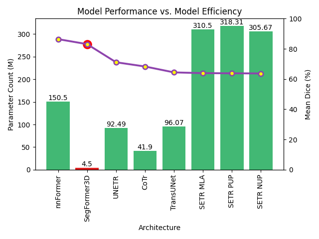
Medical imaging segmentation plays a significant role in the automatic recognition and analysis of lesions. State-of-the-art methods, particularly those utilizing transformers, have been prominently adopted in 3D semantic segmentation due to their superior performance in scalability and generalizability. However, plain vision transformers encounter challenges due to their neglect of local features and their high computational complexity. To address these challenges, we introduce three key contributions: Firstly, we proposed SegStitch, an innovative architecture that integrates transformers with denoising ODE blocks. Instead of taking whole 3D volumes as inputs, we adapt axial patches and customize patch-wise queries to ensure semantic consistency. Additionally, we conducted extensive experiments on the BTCV and ACDC datasets, achieving improvements up to 11.48% and 6.71% respectively in mDSC, compared to state-of-the-art methods. Lastly, our proposed method demonstrates outstanding efficiency, reducing the number of parameters by 36.7% and the number of FLOPS by 10.7% compared to UNETR. This advancement holds promising potential for adapting our method to real-world clinical practice. The code will be available at https://github.com/goblin327/SegStitch
Read more8/2/2024
0
SegFormer3D: an Efficient Transformer for 3D Medical Image Segmentation
Shehan Perera, Pouyan Navard, Alper Yilmaz
The adoption of Vision Transformers (ViTs) based architectures represents a significant advancement in 3D Medical Image (MI) segmentation, surpassing traditional Convolutional Neural Network (CNN) models by enhancing global contextual understanding. While this paradigm shift has significantly enhanced 3D segmentation performance, state-of-the-art architectures require extremely large and complex architectures with large scale computing resources for training and deployment. Furthermore, in the context of limited datasets, often encountered in medical imaging, larger models can present hurdles in both model generalization and convergence. In response to these challenges and to demonstrate that lightweight models are a valuable area of research in 3D medical imaging, we present SegFormer3D, a hierarchical Transformer that calculates attention across multiscale volumetric features. Additionally, SegFormer3D avoids complex decoders and uses an all-MLP decoder to aggregate local and global attention features to produce highly accurate segmentation masks. The proposed memory efficient Transformer preserves the performance characteristics of a significantly larger model in a compact design. SegFormer3D democratizes deep learning for 3D medical image segmentation by offering a model with 33x less parameters and a 13x reduction in GFLOPS compared to the current state-of-the-art (SOTA). We benchmark SegFormer3D against the current SOTA models on three widely used datasets Synapse, BRaTs, and ACDC, achieving competitive results. Code: https://github.com/OSUPCVLab/SegFormer3D.git
Read more4/17/2024
0
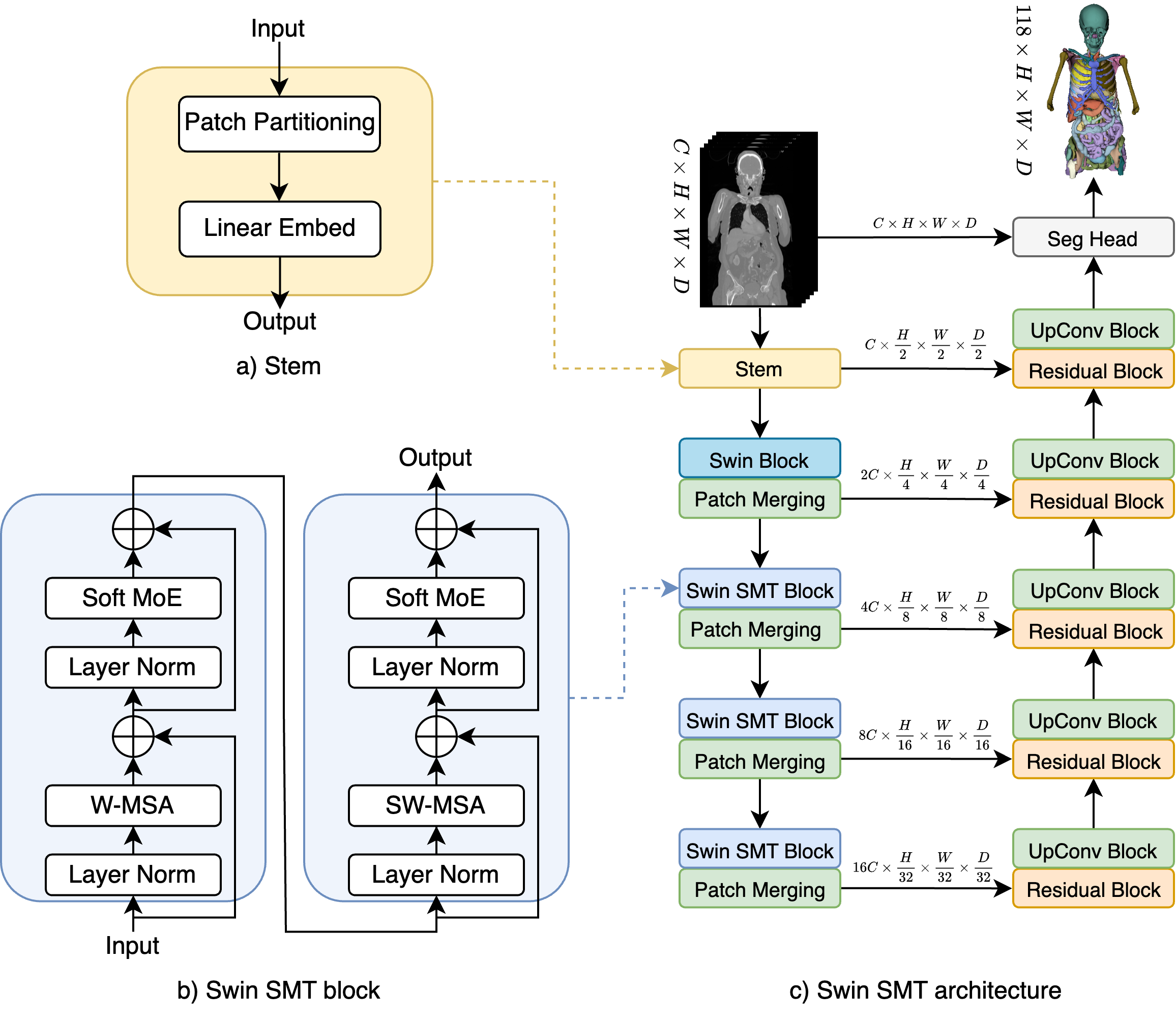
Swin SMT: Global Sequential Modeling in 3D Medical Image Segmentation
Szymon P{l}otka, Maciej Chrabaszcz, Przemyslaw Biecek
Recent advances in Vision Transformers (ViTs) have significantly enhanced medical image segmentation by facilitating the learning of global relationships. However, these methods face a notable challenge in capturing diverse local and global long-range sequential feature representations, particularly evident in whole-body CT (WBCT) scans. To overcome this limitation, we introduce Swin Soft Mixture Transformer (Swin SMT), a novel architecture based on Swin UNETR. This model incorporates a Soft Mixture-of-Experts (Soft MoE) to effectively handle complex and diverse long-range dependencies. The use of Soft MoE allows for scaling up model parameters maintaining a balance between computational complexity and segmentation performance in both training and inference modes. We evaluate Swin SMT on the publicly available TotalSegmentator-V2 dataset, which includes 117 major anatomical structures in WBCT images. Comprehensive experimental results demonstrate that Swin SMT outperforms several state-of-the-art methods in 3D anatomical structure segmentation, achieving an average Dice Similarity Coefficient of 85.09%. The code and pre-trained weights of Swin SMT are publicly available at https://github.com/MI2DataLab/SwinSMT.
Read more7/11/2024
🌐
0
Multi-Aperture Fusion of Transformer-Convolutional Network (MFTC-Net) for 3D Medical Image Segmentation and Visualization
Siyavash Shabani, Muhammad Sohaib, Sahar A. Mohammed, Bahram Parvin
Vision Transformers have shown superior performance to the traditional convolutional-based frameworks in many vision applications, including but not limited to the segmentation of 3D medical images. To further advance this area, this study introduces the Multi-Aperture Fusion of Transformer-Convolutional Network (MFTC-Net), which integrates the output of Swin Transformers and their corresponding convolutional blocks using 3D fusion blocks. The Multi-Aperture incorporates each image patch at its original resolutions with its pyramid representation to better preserve minute details. The proposed architecture has demonstrated a score of 89.73 and 7.31 for Dice and HD95, respectively, on the Synapse multi-organs dataset an improvement over the published results. The improved performance also comes with the added benefits of the reduced complexity of approximately 40 million parameters. Our code is available at https://github.com/Siyavashshabani/MFTC-Net
Read more6/26/2024