Attention-based Shape-Deformation Networks for Artifact-Free Geometry Reconstruction of Lumbar Spine from MR Images

0

Sign in to get full access
Overview
- Proposes a novel attention-based shape deformation network for reconstructing 3D geometry of the lumbar spine from MRI data
- Addresses the issue of artifacts in existing geometry reconstruction methods
- Leverages an attention mechanism to capture complex shape deformations and produce artifact-free reconstructions
Plain English Explanation
This paper presents a new deep learning approach for reconstructing the 3D shape of the lumbar spine from MRI scans. Existing methods for this task can sometimes produce reconstructions with artifacts, which are unwanted distortions or inaccuracies in the final 3D model.
The key innovation of this work is the use of an attention mechanism to better capture the complex deformations and variations in spine shape across different patients. By focusing the network's attention on the most relevant information, it can produce higher-quality, artifact-free reconstructions of the lumbar spine geometry.
This is an important advance, as accurate 3D models of the spine are crucial for various medical applications, such as surgical planning, biomechanical analysis, and patient monitoring. The proposed method could help improve the reliability and clinical utility of these applications by providing more precise and realistic 3D representations of the spine.
Technical Explanation
The authors propose an attention-based shape deformation network for reconstructing the 3D geometry of the lumbar spine from MRI data. The network takes as input a set of 2D MRI slices and outputs a detailed 3D mesh representation of the spine.
The key component of the architecture is the attention module, which allows the network to focus on the most relevant features for accurately deforming the initial 3D shape template to match the target anatomy. This attention mechanism learns to weight the contributions of different parts of the input, helping the network better capture complex shape variations.
The network is trained end-to-end on a dataset of MRI scans and corresponding 3D spine meshes. The authors compare their approach to several baseline methods and demonstrate that it outperforms them in terms of reconstruction accuracy and artifact reduction.
Critical Analysis
The paper provides a thorough evaluation of the proposed method, including comparisons to state-of-the-art alternatives and ablation studies to understand the contribution of different components. However, the authors do not discuss potential limitations or areas for further research in depth.
One aspect that could be explored further is the generalization of the method to other anatomical structures beyond the lumbar spine. The attention mechanism may be applicable to 3D reconstruction of other musculoskeletal regions, but this would require additional validation.
Additionally, the paper does not address how the reconstructed 3D models could be integrated into real-world clinical workflows or the potential challenges in deploying such a system in a practical setting. Discussing these practical considerations would help readers better understand the feasibility and impact of the proposed approach.
Conclusion
This paper presents a novel attention-based shape deformation network for reconstructing the 3D geometry of the lumbar spine from MRI data. By leveraging an attention mechanism, the method is able to produce accurate, artifact-free 3D models, which is a significant advancement over existing techniques.
The proposed approach has the potential to improve various medical applications that rely on detailed 3D representations of the spine, such as surgical planning and biomechanical analysis. While the paper provides a robust technical evaluation, further research is needed to explore the method's generalization to other anatomical structures and its practical integration into clinical workflows.
This summary was produced with help from an AI and may contain inaccuracies - check out the links to read the original source documents!
Related Papers
0
Attention-based Shape-Deformation Networks for Artifact-Free Geometry Reconstruction of Lumbar Spine from MR Images
Linchen Qian, Jiasong Chen, Linhai Ma, Timur Urakov, Weiyong Gu, Liang Liang
Lumbar disc degeneration, a progressive structural wear and tear of lumbar intervertebral disc, is regarded as an essential role on low back pain, a significant global health concern. Automated lumbar spine geometry reconstruction from MR images will enable fast measurement of medical parameters to evaluate the lumbar status, in order to determine a suitable treatment. Existing image segmentation-based techniques often generate erroneous segments or unstructured point clouds, unsuitable for medical parameter measurement. In this work, we present $textit{UNet-DeformSA}$ and $textit{TransDeformer}$: novel attention-based deep neural networks that reconstruct the geometry of the lumbar spine with high spatial accuracy and mesh correspondence across patients, and we also present a variant of $textit{TransDeformer}$ for error estimation. Specially, we devise new attention modules with a new attention formula, which integrate image features and tokenized contour features to predict the displacements of the points on a shape template without the need for image segmentation. The deformed template reveals the lumbar spine geometry in an image. Experiment results show that our networks generate artifact-free geometry outputs, and the variant of $textit{TransDeformer}$ can predict the errors of a reconstructed geometry. Our code is available at https://github.com/linchenq/TransDeformer-Mesh.
Read more5/2/2024

0
Panoptic Segmentation and Labelling of Lumbar Spine Vertebrae using Modified Attention Unet
Rikathi Pal, Priya Saha, Somoballi Ghoshal, Amlan Chakrabarti, Susmita Sur-Kolay
Segmentation and labeling of vertebrae in MRI images of the spine are critical for the diagnosis of illnesses and abnormalities. These steps are indispensable as MRI technology provides detailed information about the tissue structure of the spine. Both supervised and unsupervised segmentation methods exist, yet acquiring sufficient data remains challenging for achieving high accuracy. In this study, we propose an enhancing approach based on modified attention U-Net architecture for panoptic segmentation of 3D sliced MRI data of the lumbar spine. Our method achieves an impressive accuracy of 99.5% by incorporating novel masking logic, thus significantly advancing the state-of-the-art in vertebral segmentation and labeling. This contributes to more precise and reliable diagnosis and treatment planning.
Read more4/30/2024
0
GeoLRM: Geometry-Aware Large Reconstruction Model for High-Quality 3D Gaussian Generation
Chubin Zhang, Hongliang Song, Yi Wei, Yu Chen, Jiwen Lu, Yansong Tang
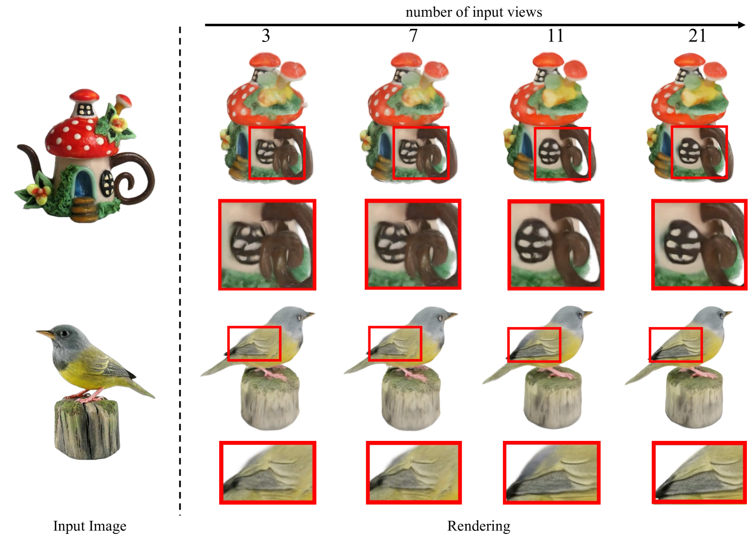
In this work, we introduce the Geometry-Aware Large Reconstruction Model (GeoLRM), an approach which can predict high-quality assets with 512k Gaussians and 21 input images in only 11 GB GPU memory. Previous works neglect the inherent sparsity of 3D structure and do not utilize explicit geometric relationships between 3D and 2D images. This limits these methods to a low-resolution representation and makes it difficult to scale up to the dense views for better quality. GeoLRM tackles these issues by incorporating a novel 3D-aware transformer structure that directly processes 3D points and uses deformable cross-attention mechanisms to effectively integrate image features into 3D representations. We implement this solution through a two-stage pipeline: initially, a lightweight proposal network generates a sparse set of 3D anchor points from the posed image inputs; subsequently, a specialized reconstruction transformer refines the geometry and retrieves textural details. Extensive experimental results demonstrate that GeoLRM significantly outperforms existing models, especially for dense view inputs. We also demonstrate the practical applicability of our model with 3D generation tasks, showcasing its versatility and potential for broader adoption in real-world applications.
Read more6/24/2024
🔗
0
New!World of Forms: Deformable Geometric Templates for One-Shot Surface Meshing in Coronary CT Angiography
Rudolf L. M. van Herten, Ioannis Lagogiannis, Jelmer M. Wolterink, Steffen Bruns, Eva R. Meulendijks, Damini Dey, Joris R. de Groot, Jos'e P. Henriques, R. Nils Planken, Simone Saitta, Ivana Iv{s}gum
Deep learning-based medical image segmentation and surface mesh generation typically involve a sequential pipeline from image to segmentation to meshes, often requiring large training datasets while making limited use of prior geometric knowledge. This may lead to topological inconsistencies and suboptimal performance in low-data regimes. To address these challenges, we propose a data-efficient deep learning method for direct 3D anatomical object surface meshing using geometric priors. Our approach employs a multi-resolution graph neural network that operates on a prior geometric template which is deformed to fit object boundaries of interest. We show how different templates may be used for the different surface meshing targets, and introduce a novel masked autoencoder pretraining strategy for 3D spherical data. The proposed method outperforms nnUNet in a one-shot setting for segmentation of the pericardium, left ventricle (LV) cavity and the LV myocardium. Similarly, the method outperforms other lumen segmentation operating on multi-planar reformatted images. Results further indicate that mesh quality is on par with or improves upon marching cubes post-processing of voxel mask predictions, while remaining flexible in the choice of mesh triangulation prior, thus paving the way for more accurate and topologically consistent 3D medical object surface meshing.
Read more9/19/2024