Correlating Stroke Risk with Non-Invasive Tracing of Brain Blood Dynamic via a Portable Speckle Contrast Optical Spectroscopy Laser Device

0
👨🏫
Sign in to get full access
Overview
- Stroke is a major global health problem, leading to significant morbidity and mortality.
- Current stroke risk assessment relies on markers like demographics, blood tests, and comorbidities.
- Measuring cerebral blood flow could provide a minimally invasive, cost-effective way to assess stroke risk.
- Physiological changes in the cerebral vascular system, such as during breath-holding, can offer insights into stroke risk.
- Existing methods for measuring cerebral perfusion reserve have limitations.
Plain English Explanation
The research paper discusses a non-invasive technique to measure brain blood flow and its potential for assessing stroke risk. Stroke is a major health problem that affects many people worldwide, often leading to serious complications or even death. Currently, doctors use factors like age, medical conditions, and blood tests to estimate a person's stroke risk.
The researchers propose using a technique called speckle contrast optical spectroscopy (SCOS) to directly measure changes in brain blood flow. This method is non-invasive, meaning it doesn't require any procedures that break the skin. By monitoring how brain blood flow changes when people hold their breath, the researchers hope to gain insights into a person's risk of having a stroke.
Existing ways of measuring blood flow in the brain have drawbacks, such as being too complex or expensive for widespread use. The researchers believe their SCOS-based approach could be a more practical and affordable way to screen for stroke risk in the general population. This could lead to earlier detection and better treatment of stroke risk factors.
Technical Explanation
The researchers conducted a study on 50 individuals, dividing them into two groups: those at low risk for stroke and those at higher risk. They used SCOS, a non-invasive optical imaging technique, to monitor regional changes in brain blood flow and volume while the participants held their breath.
Breath-holding was used as a way to induce physiological changes in the cerebral vascular system, such as changes in carbon dioxide and oxygen levels. These changes can provide insights into a person's stroke risk. The researchers found significant differences in the blood flow and volume changes during breath-holding between the low-risk and higher-risk groups.
The SCOS-based approach offers several advantages, including cost-effectiveness, scalability, portability, and simplicity. The researchers believe this laser-based tool has great potential for enhancing pre-screening and early intervention for stroke prevention in the general population.
Critical Analysis
The paper provides a promising non-invasive approach for assessing stroke risk, but there are some limitations and areas for further research:
- The study was relatively small, with only 50 participants. Larger-scale studies would be needed to validate the findings and establish the technique's clinical utility.
- The paper does not address the specific thresholds or criteria for determining stroke risk based on the observed blood flow and volume changes. More work is needed to develop a robust risk assessment framework.
- The long-term reliability and repeatability of the SCOS-based measurements over time need to be evaluated, as stroke risk can change over an individual's lifetime.
- The researchers mention the potential for this approach to enhance pre-screening and early intervention, but they do not provide details on how it could be integrated into existing clinical workflows or healthcare systems.
Overall, the research presents an interesting and innovative approach to stroke risk assessment, but further studies and refinements are necessary to fully realize its potential impact on stroke prevention and intervention.
Conclusion
The research paper explores a non-invasive, cost-effective technique using speckle contrast optical spectroscopy (SCOS) to monitor changes in brain blood flow and volume during breath-holding. The researchers found significant differences in these physiological responses between individuals at low risk and higher risk for stroke, providing a potential new avenue for stroke risk assessment.
The SCOS-based approach offers several practical advantages, including its simplicity, portability, and scalability. If further developed and validated, this technique could enable more widespread screening for stroke risk in the general population, leading to earlier detection and more effective interventions to prevent strokes and improve public health outcomes.
This summary was produced with help from an AI and may contain inaccuracies - check out the links to read the original source documents!
Related Papers
👨🏫
0
Correlating Stroke Risk with Non-Invasive Tracing of Brain Blood Dynamic via a Portable Speckle Contrast Optical Spectroscopy Laser Device
Yu Xi Huang, Simon Mahler, Aidin Abedi, Julian Michael Tyszka, Yu Tung Lo, Patrick D. Lyden, Jonathan Russin, Charles Liu, Changhuei Yang
Stroke poses a significant global health threat, with millions affected annually, leading to substantial morbidity and mortality. Current stroke risk assessment for the general population relies on markers such as demographics, blood tests, and comorbidities. A minimally invasive, clinically scalable, and cost-effective way to directly measure cerebral blood flow presents an opportunity. This opportunity has potential to positively impact effective stroke risk assessment prevention and intervention. Physiological changes in the cerebral vascular system, particularly in response to carbon dioxide level changes and oxygen deprivation, such as during breath-holding, can offer insights into stroke risk assessment. However, existing methods for measuring cerebral perfusion reserve, such as blood flow and blood volume changes, are limited by either invasiveness or impracticality. Here, we propose a transcranial approach using speckle contrast optical spectroscopy (SCOS) to non-invasively monitor regional changes in brain blood flow and volume during breath-holding. Our study, conducted on 50 individuals classified into two groups (low-risk and higher-risk for stroke), shows significant differences in blood dynamic changes during breath-holding between the two groups, providing physiological insights for stroke risk assessment using a non-invasive quantification paradigm. Given its cost-effectiveness, scalability, portability, and simplicity, this laser-centric tool has significant potential in enhancing the pre-screening of stroke and mitigating strokes in the general population through early diagnosis and intervention.
Read more7/24/2024
0
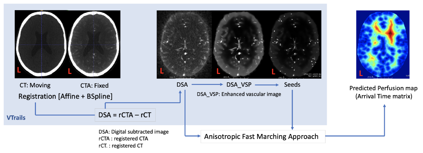
Framework to generate perfusion map from CT and CTA images in patients with acute ischemic stroke: A longitudinal and cross-sectional study
Chayanin Tangwiriyasakul, Pedro Borges, Stefano Moriconi, Paul Wright, Yee-Haur Mah, James Teo, Parashkev Nachev, Sebastien Ourselin, M. Jorge Cardoso
Stroke is a leading cause of disability and death. Effective treatment decisions require early and informative vascular imaging. 4D perfusion imaging is ideal but rarely available within the first hour after stroke, whereas plain CT and CTA usually are. Hence, we propose a framework to extract a predicted perfusion map (PPM) derived from CT and CTA images. In all eighteen patients, we found significantly high spatial similarity (with average Spearman's correlation = 0.7893) between our predicted perfusion map (PPM) and the T-max map derived from 4D-CTP. Voxelwise correlations between the PPM and National Institutes of Health Stroke Scale (NIHSS) subscores for L/R hand motor, gaze, and language on a large cohort of 2,110 subjects reliably mapped symptoms to expected infarct locations. Therefore our PPM could serve as an alternative for 4D perfusion imaging, if the latter is unavailable, to investigate blood perfusion in the first hours after hospital admission.
Read more4/8/2024
⛏️
0
Retinal arterial blood flow measured by real-time Doppler holography at 33,000 frames per second
Yann Fischer, Zacharie Auray, Olivier Martinache, Marius Dubosc, No'e Top'eza, Chlo'e Magnier, Maxime Boy-Arnould, Michael Atlan
This study presents a novel quantitative estimation method for total retinal arterial blood flow utilizing real-time Doppler holography at an unprecedented frame rate of 33,000 frames per second. This technique, leveraging high-speed digital holography, enables non-invasive angiographic imaging of the retina, providing detailed blood flow contrasts essential for assessing retinal health. The proposed quantitative analysis method consists of segmenting primary in-plane retinal arteries and calculating local blood velocity using Doppler frequency broadening. The analysis integrates a forward scattering model to achieve blood flow estimation. Our findings highlight the potential of Doppler holography as a powerful tool for diagnosing and monitoring the treatment of retinal vascular conditions, complementary to existing imaging methods.
Read more9/27/2024

0
Functional Assessment of Cerebral Capillaries using Single Capillary Reporters in Ultrasound Localization Microscopy
Stephen A Lee, Alexis Leconte, Alice Wu, Joshua Kinugasa, Jonathan Poree, Andreas Linninger, Jean Provost
The brain's microvascular cerebral capillary network plays a vital role in maintaining neuronal health, yet capillary dynamics are still not well understood due to limitations in existing imaging techniques. Here, we present Single Capillary Reporters (SCaRe) for transcranial Ultrasound Localization Microscopy (ULM), a novel approach enabling non-invasive, whole-brain mapping of single capillaries and estimates of their transit-time as a neurovascular biomarker. We accomplish this first through computational Monte Carlo and ultrasound simulations of microbubbles flowing through a fully-connected capillary network. We unveil distinct capillary flow behaviors which informs methodological changes to ULM acquisitions to better capture capillaries in vivo. Subsequently, applying SCaRe-ULM in vivo, we achieve unprecedented visualization of single capillary tracks across brain regions, analysis of layer-specific capillary heterogeneous transit times (CHT), and characterization of whole microbubble trajectories from arterioles to venules. Lastly, we evaluate capillary biomarkers using injected lipopolysaccharide to induce systemic neuroinflammation and track the increase in SCaRe-ULM CHT, demonstrating the capability to detect subtle capillary functional changes. SCaRe-ULM represents a significant advance in studying microvascular dynamics, offering novel avenues for investigating capillary patterns in neurological disorders and potential diagnostic applications.
Read more7/12/2024